Related Research Articles

Caenorhabditis elegans is a free-living transparent nematode about 1 mm in length that lives in temperate soil environments. It is the type species of its genus. The name is a blend of the Greek caeno- (recent), rhabditis (rod-like) and Latin elegans (elegant). In 1900, Maupas initially named it Rhabditides elegans. Osche placed it in the subgenus Caenorhabditis in 1952, and in 1955, Dougherty raised Caenorhabditis to the status of genus.

The Rickettsiales, informally called rickettsias, are an order of small Alphaproteobacteria. They are obligate intracellular parasites, and some are notable pathogens, including Rickettsia, which causes a variety of diseases in humans, and Ehrlichia, which causes diseases in livestock. Another genus of well-known Rickettsiales is the Wolbachia, which infect about two-thirds of all arthropods and nearly all filarial nematodes. Genetic studies support the endosymbiotic theory according to which mitochondria and related organelles developed from members of this group.
Nosema apis is a microsporidian, a small, unicellular parasite recently reclassified as a fungus that mainly affects honey bees. It causes nosemosis, also called nosema, which is the most common and widespread of adult honey bee diseases. The dormant stage of N. apis is a long-lived spore which is resistant to temperature extremes and dehydration, and cannot be killed by freezing the contaminated comb. Nosemosis is a listed disease with the Office International des Epizooties (OIE).

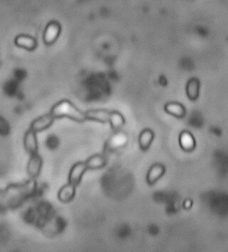
Microsporidia are a group of spore-forming unicellular parasites. These spores contain an extrusion apparatus that has a coiled polar tube ending in an anchoring disc at the apical part of the spore. They were once considered protozoans or protists, but are now known to be fungi, or a sister group to fungi. These fungal microbes are obligate eukaryotic parasites that use a unique mechanism to infect host cells. They have recently been discovered in a 2017 Cornell study to infect Coleoptera on a large scale. So far, about 1500 of the probably more than one million species are named. Microsporidia are restricted to animal hosts, and all major groups of animals host microsporidia. Most infect insects, but they are also responsible for common diseases of crustaceans and fish. The named species of microsporidia usually infect one host species or a group of closely related taxa. Approximately 10 percent of the species are parasites of vertebrates —several species, most of which are opportunistic, can infect humans, in whom they can cause microsporidiosis.
Intracellular parasites are microparasites that are capable of growing and reproducing inside the cells of a host. They are also called intracellular pathogens.
Microsporidiosis is an opportunistic intestinal infection that causes diarrhea and wasting in immunocompromised individuals. It results from different species of microsporidia, a group of microbial (unicellular) fungi.
A xenoma is a growth caused by various protists and fungi, most notably microsporidia. It can occur on numerous organisms; however is predominantly found on fish.
Enterocytozoon bieneusi is a species of the order Chytridiopsida which infects the intestinal epithelial cells. It is an obligate intracellular parasite.
Nosema ceranae is a microsporidian, a small, unicellular parasite that mainly affects Apis cerana, the Asiatic honey bee. Along with Nosema apis, it causes the disease nosemosis, the most widespread of the diseases of adult honey bees. N. ceranae can remain dormant as a long-lived spore which is resistant to temperature extremes and dehydration. This fungus has been shown to act in a synergistic fashion with diverse insecticides such as fipronil or neonicotinoids, by increasing the toxicity of pesticides for bees, leading to higher bee mortality. It may thus play an indirect role in colony collapse disorder. In addition, the interaction between fipronil and N. ceranae induces changes in male physiology leading to sterility.
Encephalitozoon intestinalis is a parasite. It can cause microsporidiosis.

Encephalitozoon cuniculi is a microsporidial parasite of mammals with world-wide distribution. An important cause of neurologic and renal disease in rabbits, E. cuniculi can also cause disease in immunocompromised people.

Theileria parva is a species of parasites, named in honour of Arnold Theiler, that causes East Coast fever (theileriosis) in cattle, a costly disease in Africa. The main vector for T. parva is the tick Rhipicephalus appendiculatus. Theiler found that East Coast fever was not the same as redwater, but caused by a different protozoan.
Loma salmonae is a species of microsporidian parasite, infecting Pacific salmon. L. salmonae is the causative agent of microsporidial gill disease of salmon. It is an intracellular parasite which induces respiratory distress, secondary infection, and increased mortality rates.

Caenorhabditis elegans- microbe interactions are defined as any interaction that encompasses the association with microbes that temporarily or permanently live in or on the nematode C. elegans. The microbes can engage in a commensal, mutualistic or pathogenic interaction with the host. These include bacterial, viral, unicellular eukaryotic, and fungal interactions. In nature C. elegans harbours a diverse set of microbes. In contrast, C. elegans strains that are cultivated in laboratories for research purposes have lost the natural associated microbial communities and are commonly maintained on a single bacterial strain, Escherichia coli OP50. However, E. coli OP50 does not allow for reverse genetic screens because RNAi libraries have only been generated in strain HT115. This limits the ability to study bacterial effects on host phenotypes. The host microbe interactions of C. elegans are closely studied because of their orthologs in humans. Therefore, the better we understand the host interactions of C. elegans the better we can understand the host interactions within the human body.
Ordospora colligata is an intracellular parasite belonging to the Microsporidia. It is an obligatory gut parasite with the crustacean Daphnia magna as its only host. So far it has been reported from Europe and Asia.

Hamiltosporidium is a genus of Microsporidia, which are intracellular and unicellular parasites. The genus, proposed by Haag et al. in 2010, contains two species; Hamiltosporidium tvaerminnensis, and Hamiltosporidium magnivora. Both species infect only the crustacean Daphnia magna (Waterflea).
Nematocida is a genus of Microsporidia fungi. One species, N. parisii, is found in wild isolates of Caenorhabditis elegans. It has been nicknamed the nematode-killer from Paris. This species replicates in the intestines of C. elegans.
Caenorhabditis tropicalis is a species of Caenorhabditis nematodes, belonging to the Elegans super-group and Elegans group within the genus. It is a close relative of C. wallacei.C. tropicalis is collected frequently in tropical South America, Caribbean islands, and various islands in the Indian and Pacific Oceans from rotting fruit, flowers and stems. C. tropicalis was referred to as “C. sp. 11” prior to 2014.

Steinernema carpocapsae is an entomopathogenic nematode and a member of the family Steinernematidae. It is a parasitic roundworm that has evolved an insect-killing symbiosis with bacteria, and kills its hosts within a few days of infection. This parasite releases its bacterial symbiont along with a variety of proteins into the host after infection, and together the bacteria and nematode overcome host immunity and kill the host quickly. As a consequence, S. carpocapsae has been widely adapted for use as a biological control agent in agriculture and pest control. S. carpocapsae is considered a generalist parasite and has been effectively used to control a variety of insects including: Webworms, cutworms, armyworms, girdlers, some weevils, and wood-borers. This species is an example of an "ambush" forager, standing on its tail in an upright position near the soil surface and attaching to passing hosts, even capable of jumping. As an ambush forager, S. carpocapsae is thought to be especially effective when applied against highly mobile surface-adapted insects. S. carpocapsae can sense carbon dioxide production, making the spiracles a key portal of entry into its insect hosts. It is most effective at temperatures ranging from 22–28 °C (72–82 °F).

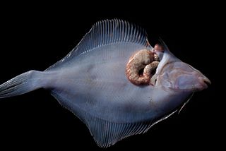
Enterospora nucleophila is a microsporidian infecting the gilt-head bream. It develops primarily within the nuclei of rodlet cells and enterocytes, at the intestinal epithelium. It can also be found in cytoplasmic position within other cell types, including phagocytes, at subepithelial layers. It is the causative agent of emaciative microsporidiosis of gilthead sea bream, a chronic condition manifested as a severe growth arrestment, normally accompanied by trickling mortality.
References
- ↑ Cuomo, C. A.; Desjardins, C. A.; Bakowski, M. A.; Goldberg, J.; Ma, A. T.; Becnel, J. J.; Didier, E. S.; Fan, L.; Heiman, D. I.; Levin, J. Z.; Young, S.; Zeng, Q.; Troemel, E. R. (2012-07-18). "Microsporidian genome analysis reveals evolutionary strategies for obligate intracellular growth". Genome Research. 22 (12). Cold Spring Harbor Laboratory: 2478–2488. doi: 10.1101/gr.142802.112 . ISSN 1088-9051. PMC 3514677 . PMID 22813931.
- 1 2 3 4 Moretto, Magali M.; Khan, Imtiaz A.; Weiss, Louis M. (2012-07-12). "Gastrointestinal Cell Mediated Immunity and the Microsporidia". PLOS Pathogens. 8 (7): e1002775. doi: 10.1371/journal.ppat.1002775 . ISSN 1553-7374. PMC 3395611 . PMID 22807673.
- ↑ Szumowski, Suzannah C.; Botts, Michael R.; Popovich, John J.; Smelkinson, Margery G.; Troemel, Emily R. (2014-06-03). "The small GTPase RAB-11 directs polarized exocytosis of the intracellular pathogen N. parisii for fecal-oral transmission from C. elegans". Proceedings of the National Academy of Sciences. 111 (22): 8215–8220. doi: 10.1073/pnas.1400696111 . ISSN 0027-8424. PMC 4050618 . PMID 24843160.
- 1 2 3 Troemel ER, Félix M, Whiteman NK, Barrière N, Ausubel FM (2008) Microsporidia are natural intracellular parasites of the nematode Caenorhabditis elegans. PLoS Biol 6(12): e309. doi : 10.1371/journal.pbio.0060309
- 1 2 3 4 Ardila-Garcia, A. M.; Fast, N. M. (2012-12-01). "Microsporidian Infection in a Free-Living Marine Nematode". Eukaryotic Cell. 11 (12): 1544–1551. doi: 10.1128/EC.00228-12 . ISSN 1535-9778. PMC 3536275 . PMID 23087371.
- ↑ Luallen, Robert J.; Reinke, Aaron W.; Tong, Linda; Botts, Michael R.; Félix, Marie-Anne; Troemel, Emily R. (2016-06-30). "Discovery of a Natural Microsporidian Pathogen with a Broad Tissue Tropism in Caenorhabditis elegans". PLOS Pathogens. 12 (6): e1005724. doi: 10.1371/journal.ppat.1005724 . PMC 4928854 . PMID 27362540.
| | This fungus-related article is a stub. You can help Wikipedia by expanding it. |
| | This article related to parasites is a stub. You can help Wikipedia by expanding it. |