Related Research Articles
Working memory is a cognitive system with a limited capacity that can hold information temporarily. It is important for reasoning and the guidance of decision-making and behavior. Working memory is often used synonymously with short-term memory, but some theorists consider the two forms of memory distinct, assuming that working memory allows for the manipulation of stored information, whereas short-term memory only refers to the short-term storage of information. Working memory is a theoretical concept central to cognitive psychology, neuropsychology, and neuroscience.
Human intelligence is the intellectual capability of humans, which is marked by complex cognitive feats and high levels of motivation and self-awareness. Using their intelligence, humans are able to learn, form concepts, understand, and apply logic and reason. Human intelligence is also thought to encompass our capacities to recognize patterns, plan, innovate, solve problems, make decisions, retain information, and use language to communicate.

In the human brain, the anterior cingulate cortex (ACC) is the frontal part of the cingulate cortex that resembles a "collar" surrounding the frontal part of the corpus callosum. It consists of Brodmann areas 24, 32, and 33.

Facial perception is an individual's understanding and interpretation of the face. Here, perception implies the presence of consciousness and hence excludes automated facial recognition systems. Although facial recognition is found in other species, this article focuses on facial perception in humans.
In the philosophy of mind, neuroscience, and cognitive science, a mental image is an experience that, on most occasions, significantly resembles the experience of "perceiving" some object, event, or scene but occurs when the relevant object, event, or scene is not actually present to the senses. There are sometimes episodes, particularly on falling asleep and waking up, when the mental imagery may be dynamic, phantasmagoric, and involuntary in character, repeatedly presenting identifiable objects or actions, spilling over from waking events, or defying perception, presenting a kaleidoscopic field, in which no distinct object can be discerned. Mental imagery can sometimes produce the same effects as would be produced by the behavior or experience imagined.
Neuroscience and intelligence refers to the various neurological factors that are partly responsible for the variation of intelligence within species or between different species. A large amount of research in this area has been focused on the neural basis of human intelligence. Historic approaches to studying the neuroscience of intelligence consisted of correlating external head parameters, for example head circumference, to intelligence. Post-mortem measures of brain weight and brain volume have also been used. More recent methodologies focus on examining correlates of intelligence within the living brain using techniques such as magnetic resonance imaging (MRI), functional MRI (fMRI), electroencephalography (EEG), positron emission tomography and other non-invasive measures of brain structure and activity.

The somatic marker hypothesis, formulated by Antonio Damasio and associated researchers, proposes that emotional processes guide behavior, particularly decision-making.
Affective neuroscience is the study of how the brain processes emotions. This field combines neuroscience with the psychological study of personality, emotion, and mood. The basis of emotions and what emotions are remains an issue of debate within the field of affective neuroscience.
Social neuroscience is an interdisciplinary field devoted to understanding the relationship between social experiences and biological systems. Humans are fundamentally a social species, rather than solitary. As such, Homo sapiens create emergent organizations beyond the individual—structures that range from dyads, families, and groups to cities, civilizations, and cultures. In this regard, studies indicate that various social influences, including life events, poverty, unemployment and loneliness can influence health related biomarkers. The term "social neuroscience" can be traced to a publication entitled "Social Neuroscience Bulletin" which was published quarterly between 1988 and 1994. The term was subsequently popularized in an article by John Cacioppo and Gary Berntson, published in the American Psychologist in 1992. Cacioppo and Berntson are considered as the legitimate fathers of social neuroscience. Still a young field, social neuroscience is closely related to affective neuroscience and cognitive neuroscience, focusing on how the brain mediates social interactions. The biological underpinnings of social cognition are investigated in social cognitive neuroscience.

The posterior cingulate cortex (PCC) is the caudal part of the cingulate cortex, located posterior to the anterior cingulate cortex. This is the upper part of the "limbic lobe". The cingulate cortex is made up of an area around the midline of the brain. Surrounding areas include the retrosplenial cortex and the precuneus.

Mental chronometry is the scientific study of processing speed or reaction time on cognitive tasks to infer the content, duration, and temporal sequencing of mental operations. Reaction time is measured by the elapsed time between stimulus onset and an individual's response on elementary cognitive tasks (ECTs), which are relatively simple perceptual-motor tasks typically administered in a laboratory setting. Mental chronometry is one of the core methodological paradigms of human experimental, cognitive, and differential psychology, but is also commonly analyzed in psychophysiology, cognitive neuroscience, and behavioral neuroscience to help elucidate the biological mechanisms underlying perception, attention, and decision-making in humans and other species.
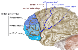
The dorsolateral prefrontal cortex is an area in the prefrontal cortex of the primate brain. It is one of the most recently derived parts of the human brain. It undergoes a prolonged period of maturation which lasts into adulthood. The DLPFC is not an anatomical structure, but rather a functional one. It lies in the middle frontal gyrus of humans. In macaque monkeys, it is around the principal sulcus. Other sources consider that DLPFC is attributed anatomically to BA 9 and 46 and BA 8, 9 and 10.
Marcel Just is D. O. Hebb Professor of Psychology at Carnegie Mellon University. His research uses brain imaging (fMRI) in high-level cognitive tasks to study the neuroarchitecture of cognition. Just's areas of expertise include psycholinguistics, object recognition, and autism, with particular attention to cognitive and neural substrates. Just directs the Center for Cognitive Brain Imaging and is a member of the Center for the Neural Basis of Cognition at CMU.
Cultural neuroscience is a field of research that focuses on the interrelation between a human's cultural environment and neurobiological systems. The field particularly incorporates ideas and perspectives from related domains like anthropology, psychology, and cognitive neuroscience to study sociocultural influences on human behaviors. Such impacts on behavior are often measured using various neuroimaging methods, through which cross-cultural variability in neural activity can be examined.
Neuroscience of multilingualism is the study of multilingualism within the field of neurology. These studies include the representation of different language systems in the brain, the effects of multilingualism on the brain's structural plasticity, aphasia in multilingual individuals, and bimodal bilinguals. Neurological studies of multilingualism are carried out with functional neuroimaging, electrophysiology, and through observation of people who have suffered brain damage.
Neuroimaging intelligence testing concerns the use of neuroimaging techniques to evaluate human intelligence. Neuroimaging technology has advanced such that scientists hope to use neuroimaging increasingly for investigations of brain function related to IQ.
A neurological look at race is multifaceted. The cross-race effect has been neurologically explained by there being differences in brain processing while viewing same-race and other-race faces. There is a debate over the cause of the cross-race effect.
The biological basis of personality is the collection of brain systems and mechanisms that underlie human personality. Human neurobiology, especially as it relates to complex traits and behaviors, is not well understood, but research into the neuroanatomical and functional underpinnings of personality are an active field of research. Animal models of behavior, molecular biology, and brain imaging techniques have provided some insight into human personality, especially trait theories.
The parieto-frontal integration theory (P-FIT) considers intelligence to relate to how well different brain regions integrate to form intelligent behaviors. The theory proposes that large scale brain networks connect brain regions, including regions within frontal, parietal, temporal, and cingulate cortices, underlie the biological basis of human intelligence. These regions, which overlap significantly with the task-positive network, allow the brain to communicate and exchange information efficiently with one another. Support for this theory is primarily based on neuroimaging evidence, with support from lesion studies. The P-FIT is influential in that it explains the majority of current neuroimaging findings, as well as increasing empirical support for cognition being the result of large-scale brain networks, rather than numerous domain-specific processes or modules. A 2010 review of the neuroscience of intelligence described P-FIT as "the best available answer to the question of where in the brain intelligence resides".
Sex differences in cognition are widely studied in the current scientific literature. Biological and genetic differences in combination with environment and culture have resulted in the cognitive differences among males and females. Among biological factors, hormones such as testosterone and estrogen may play some role mediating these differences. Among differences of diverse mental and cognitive abilities, the largest or most well known are those relating to spatial abilities, social cognition and verbal skills and abilities.
References
- 1 2 Dunst B, Benedek M, Jauk E, Bergner S, Koschutnig K, Sommer M, et al. (January 2014). "Neural efficiency as a function of task demands". Intelligence. 42 (100): 22–30. doi:10.1016/j.intell.2013.09.005. PMC 3907682 . PMID 24489416.
- ↑ Neubauer AC, Fink A (July 2009). "Intelligence and neural efficiency". Neuroscience and Biobehavioral Reviews. 33 (7): 1004–1023. doi:10.1016/j.neubiorev.2009.04.001. PMID 19580915. S2CID 7125675.
- ↑ Baumeister R, Vohs K (2007). Encyclopedia of Social Psychology. Thousand Oaks, California. doi:10.4135/9781412956253. ISBN 9781412916707.
{{cite book}}: CS1 maint: location missing publisher (link) - ↑ Brancucci A (July 2012). "Neural correlates of cognitive ability". Journal of Neuroscience Research. 90 (7): 1299–1309. doi:10.1002/jnr.23045. PMID 22422612. S2CID 16156840.
- ↑ Deary IJ, Penke L, Johnson W (March 2010). "The neuroscience of human intelligence differences" (PDF). Nature Reviews. Neuroscience. 11 (3): 201–211. doi:10.1038/nrn2793. PMID 20145623. S2CID 5136934.
- 1 2 Haier RJ, Siegel BV, Nuechterlein KH, Hazlett E, Wu JC, Paek J, Browning HL, Buchsbaum MS (1988). "Cortical glucose metabolic rate correlates of abstract reasoning and attention studied with positron emission tomography". Intelligence. 12 (2): 199–217. doi:10.1016/0160-2896(88)90016-5. ISSN 0160-2896.
- ↑ Sabharwal N, Arumugam P, Kelion A (2017). "Cardiac positron emission tomography (PET)". Oxford Medicine Online. doi:10.1093/med/9780198759942.003.0012.
- ↑ Haier RJ, Siegel B, Tang C, Abel L, Buchsbaum MS (1992). "Intelligence and changes in regional cerebral glucose metabolic rate following learning". Intelligence. 16 (3–4): 415–426. doi:10.1016/0160-2896(92)90018-m. ISSN 0160-2896.
- 1 2 Di Domenico SI, Rodrigo AH, Ayaz H, Fournier MA, Ruocco AC (April 2015). "Decision-making conflict and the neural efficiency hypothesis of intelligence: a functional near-infrared spectroscopy investigation". NeuroImage. 109: 307–317. doi:10.1016/j.neuroimage.2015.01.039. PMID 25625894. S2CID 19265562.
- ↑ Li L, Smith DM (2021-08-05). "Neural Efficiency in Athletes: A Systematic Review". Frontiers in Behavioral Neuroscience. 15: 698555. doi: 10.3389/fnbeh.2021.698555 . PMC 8374331 . PMID 34421553.
- ↑ Doppelmayr M, Klimesch W, Sauseng P, Hödlmoser K, Stadler W, Hanslmayr S (June 2005). "Intelligence related differences in EEG-bandpower". Neuroscience Letters. 381 (3): 309–313. doi:10.1016/j.neulet.2005.02.037. PMID 15896490. S2CID 42449563.
- ↑ Neubauer AC, Fink A, Schrausser DG (2002). "Intelligence and neural efficiency". Intelligence. 30 (6): 515–536. doi:10.1016/s0160-2896(02)00091-0. ISSN 0160-2896.
- ↑ Dunst B, Benedek M, Bergner S, Athenstaedt U, Neubauer AC (October 2013). "Sex differences in neural efficiency: Are they due to the stereotype threat effect?". Personality and Individual Differences. 55 (7): 744–749. doi:10.1016/j.paid.2013.06.007. PMC 3759843 . PMID 24092950.
| | This neuroscience article is a stub. You can help Wikipedia by expanding it. |