Related Research Articles
Interferometry is a technique which uses the interference of superimposed waves to extract information. Interferometry typically uses electromagnetic waves and is an important investigative technique in the fields of astronomy, fiber optics, engineering metrology, optical metrology, oceanography, seismology, spectroscopy, quantum mechanics, nuclear and particle physics, plasma physics, biomolecular interactions, surface profiling, microfluidics, mechanical stress/strain measurement, velocimetry, optometry, and making holograms.

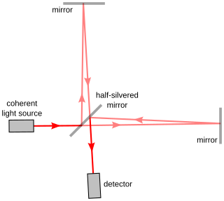
The Michelson interferometer is a common configuration for optical interferometry and was invented by the 19/20th-century American physicist Albert Abraham Michelson. Using a beam splitter, a light source is split into two arms. Each of those light beams is reflected back toward the beamsplitter which then combines their amplitudes using the superposition principle. The resulting interference pattern that is not directed back toward the source is typically directed to some type of photoelectric detector or camera. For different applications of the interferometer, the two light paths can be with different lengths or incorporate optical elements or even materials under test.

Optical coherence tomography (OCT) is an imaging technique that uses interferometry with short-coherence-length light to obtain micrometer-level depth resolution and uses transverse scanning of the light beam to form two- and three-dimensional images from light reflected from within biological tissue or other scattering media. Short-coherence-length light can be obtained using a superluminescent diode (SLD) with a broad spectral bandwidth or a broadly tunable laser with narrow linewidth. The first demonstration of OCT imaging was published by a team from MIT and Harvard Medical School in a 1991 article in the journal Science. The article introduced the term "OCT" to credit its derivation from optical coherence-domain reflectometry, in which the axial resolution is based on temporal coherence. The first demonstrations of in vivo OCT imaging quickly followed.
Surface metrology is the measurement of small-scale features on surfaces, and is a branch of metrology. Surface primary form, surface fractality, and surface finish are the parameters most commonly associated with the field. It is important to many disciplines and is mostly known for the machining of precision parts and assemblies which contain mating surfaces or which must operate with high internal pressures.
Laser-ultrasonics uses lasers to generate and detect ultrasonic waves. It is a non-contact technique used to measure materials thickness, detect flaws and carry out materials characterization. The basic components of a laser-ultrasonic system are a generation laser, a detection laser and a detector.

A profilometer is a measuring instrument used to measure a surface's profile, in order to quantify its roughness. Critical dimensions as step, curvature, flatness are computed from the surface topography.

Bruce J. Tromberg is an American photochemist and a leading researcher in the field of biophotonics. He is the director of the National Institute of Biomedical Imaging and Bioengineering (NIBIB) within the National Institutes of Health (NIH). Before joining NIH, he was Professor of Biomedical Engineering at The Henry Samueli School of Engineering and of Surgery at the School of Medicine, University of California, Irvine. He was the principal investigator of the Laser Microbeam and Medical Program (LAMMP), and the Director of the Beckman Laser Institute and Medical Clinic at Irvine. He was a co-leader of the Onco-imaging and Biotechnology Program of the NCI Chao Family Comprehensive Cancer Center at Irvine.
ISO 25178: Geometrical Product Specifications (GPS) – Surface texture: areal is an International Organization for Standardization collection of international standards relating to the analysis of 3D areal surface texture.
In optical astronomy, interferometry is used to combine signals from two or more telescopes to obtain measurements with higher resolution than could be obtained with either telescopes individually. This technique is the basis for astronomical interferometer arrays, which can make measurements of very small astronomical objects if the telescopes are spread out over a wide area. If a large number of telescopes are used a picture can be produced which has resolution similar to a single telescope with the diameter of the combined spread of telescopes. These include radio telescope arrays such as VLA, VLBI, SMA, astronomical optical interferometer arrays such as COAST, NPOI and IOTA, resulting in the highest resolution optical images ever achieved in astronomy. The VLT Interferometer is expected to produce its first images using aperture synthesis soon, followed by other interferometers such as the CHARA array and the Magdalena Ridge Observatory Interferometer which may consist of up to 10 optical telescopes. If outrigger telescopes are built at the Keck Interferometer, it will also become capable of interferometric imaging.
Angle-resolved low-coherence interferometry (a/LCI) is an emerging biomedical imaging technology which uses the properties of scattered light to measure the average size of cell structures, including cell nuclei. The technology shows promise as a clinical tool for in situ detection of dysplastic, or precancerous tissue.

A white light scanner (WLS) is a device for performing surface height measurements of an object using coherence scanning interferometry (CSI) with spectrally-broadband, "white light" illumination. Different configurations of scanning interferometer may be used to measure macroscopic objects with surface profiles measuring in the centimeter range, to microscopic objects with surface profiles measuring in the micrometer range. For large-scale non-interferometric measurement systems, see structured-light 3D scanner.
Digital holographic microscopy (DHM) is digital holography applied to microscopy. Digital holographic microscopy distinguishes itself from other microscopy methods by not recording the projected image of the object. Instead, the light wave front information originating from the object is digitally recorded as a hologram, from which a computer calculates the object image by using a numerical reconstruction algorithm. The image forming lens in traditional microscopy is thus replaced by a computer algorithm. Other closely related microscopy methods to digital holographic microscopy are interferometric microscopy, optical coherence tomography and diffraction phase microscopy. Common to all methods is the use of a reference wave front to obtain amplitude (intensity) and phase information. The information is recorded on a digital image sensor or by a photodetector from which an image of the object is created (reconstructed) by a computer. In traditional microscopy, which do not use a reference wave front, only intensity information is recorded and essential information about the object is lost.

Mountains is an image analysis and surface metrology software platform published by the company Digital Surf. Its core is micro-topography, the science of studying surface texture and form in 3D at the microscopic scale. The software is dedicated to profilometers, 3D light microscopes ("MountainsMap"), scanning electron microscopes ("MountainsSEM") and scanning probe microscopes ("MountainsSPIP").
A common-path interferometer is a class of interferometers in which the reference beam and sample beams travel along the same path. Examples include the Sagnac interferometer, Zernike phase-contrast interferometer, and the point diffraction interferometer. A common-path interferometer is generally more robust to environmental vibrations than a "double-path interferometer" such as the Michelson interferometer or the Mach–Zehnder interferometer. Although travelling along the same path, the reference and sample beams may travel along opposite directions, or they may travel along the same direction but with the same or different polarization.

Coherence scanning interferometry (CSI) is any of a class of optical surface measurement methods wherein the localization of interference fringes during a scan of optical path length provides a means to determine surface characteristics such as topography, transparent film structure, and optical properties. CSI is currently the most common interference microscopy technique for areal surface topography measurement. The term "CSI" was adopted by the International Organization for Standardization (ISO).

Intracoronary optical coherence tomography (OCT) is a catheter-based imaging application of optical coherence tomography. Currently prospective trials demonstrate OCT alters morbidity and/or mortality in coronary stenting as discussed below.
Jannick Rolland is the Brian J. Thompson Professor of Optical Engineering at the Institute of Optics at the University of Rochester. She is also the co-founder and CTO of LighTopTech, a women-owner business founded in 2013 to create medical imaging technologies with biomimetic noninvasive imaging technology. At the University of Rochester, she is the Director of the NSF I/UCRC Center for Freeform Optics (CeFO). She is also the Director of the R.E. Hopkins Center for Optical Design and Engineering that engages undergraduates in optical design, fabrication, and metrology.
Speckle variance optical coherence tomography (SV-OCT) is an imaging algorithm for functional optical imaging. Optical coherence tomography is an imaging modality that uses low-coherence interferometry to obtain high resolution, depth-resolved volumetric images. OCT can be used to capture functional images of blood flow, a technique known as optical coherence tomography angiography (OCT-A). SV-OCT is one method for OCT-A that uses the variance of consecutively acquired images to detect flow at the micron scale. SV-OCT can be used to measure the microvasculature of tissue. In particular, it is useful in ophthalmology for visualizing blood flow in retinal and choroidal regions of the eye, which can provide information on the pathophysiology of diseases.

Christine P. Hendon is an electrical engineer and computer scientist and an associate professor in the Department of Electrical Engineering at Columbia University in New York City. Hendon is a pioneer in medical imaging. She develops biomedical optics technologies, using optical coherence tomography and near infrared spectroscopy systems, that enable physicians to perform guided interventional procedures and allow for structure-function dissection of human tissues and organs. Her advances in imaging technologies have led to improved diagnostic abilities and treatments for cardiac arrhythmias as well as breast cancer and preterm birth. She has been recognized for her development of optical imaging catheters for cardiac wall imaging by Forbes 30 under 30, the MIT Technology Review’s 35 Innovators Under 35, and by President Obama with the Presidential Early Career Awards in 2017.
Dual-axis optical coherence tomography (DA-OCT) is an imaging modality that is based on the principles of optical coherence tomography (OCT). These techniques are largely used for medical imaging. OCT is non-invasive and non-contact. It allows for real-time, in situ imaging and provides high image resolution. OCT is analogous to ultrasound but relies on light waves, which makes it faster than ultrasound. In general, OCT has proven to be compact and portable. It is compatible with arterial catheters and endoscopes, which helps diagnose diseases within long internal cavities, including the esophagus and coronary arteries.
References
- ↑ Dufour, Marc L.; Gauthier, Bruno (2003). "Precise surface profilometry based on low-coherence interferometry". In Lessard, Roger A; Lampropoulos, George A (eds.). Applications of Photonic Technology 6. Vol. 5260. p. 173. Bibcode:2003SPIE.5260..173D. doi:10.1117/12.543395. S2CID 135946276.
- ↑ Dufour, M. L.; Lamouche, G.; Vergnole, S.; Gauthier, B.; Padioleau, C.; Hewko, M.; Levesque, S.; Bartulovic, V. (June 2006). "Precise surface profilometry based on low-coherence interferometry". Proceedings of SPIE. Vol. 6343. Quebec City, Quebec, Canada: SPIE. pp. 63431Z.1–7. Retrieved December 14, 2010.
- ↑ Dufour, Marc; Lamouche, G.; Gauthier, B.; Padioleau, C.; Monchalin, J.P. (2006). "Inspection of hard-to-reach industrial parts using small diameter probes" (PDF). SPIE Newsroom . doi:10.1117/2.1200610.0467. S2CID 120476700 . Retrieved December 15, 2010.
- 1 2 Bartulovic, Vuk. "Novacam Technologies Inc". Biofinance - Funding Lifescience companies. Retrieved January 6, 2011.
- ↑ Losert, R. (March 31, 2009). "Solution for NDT Inspection". NDT Magazine. Archived from the original on 2011-01-12. Retrieved November 13, 2020.
- ↑ Sprovieri, John (July 28, 2008). "Quality in Assembly: Fiber-Optic Profilometer Measures Surface Quality". Assembly Magazine. Retrieved December 14, 2010.
- ↑ Guss, G.; Bass, I.; Hackel, R.; Mailhiot, C.; Demos, S.G. (November 6, 2007). "High-resolution 3-D imaging of surface damage sites in fused silica with Optical Coherence Tomography" (PDF). Lawrence Livermore National Laboratory UCRL-PROC-236270. Archived from the original (PDF) on February 11, 2017. Retrieved December 14, 2010.
- ↑ Losert, R. (November 2010), "So Far, Yet so Close: Optical Profilometer Systems Inspect Hard-to-Reach Surfaces", INSPECT Magazine, 11: 42–43, retrieved December 15, 2010
- ↑ Wilson, Andrew (April 1, 2007). "Fiber-based profilometers inspect hard-to-reach surfaces". VisionSystems Design Magazine. Retrieved December 14, 2010.
- ↑ Lamouche, Guy; Dufour, Marc; Hewko, Mark; Gauthier, Bruno; Vergnole, Sébastien; Bisaillon, Charles-Étienne; Monchalin, Jean-Pierre; Sowa, Michael (August 9, 2010), "Intravascular Optical Coherence Tomography on a Beating Heart Model", Journal of Biomedical Optics, 15 (4): 046023–046023–7, Bibcode:2010JBO....15d6023L, doi: 10.1117/1.3475960 , PMID 20799825, S2CID 206429235 , retrieved December 15, 2010