Pain and pleasure

Last updated

Some philosophers, such as Jeremy Bentham, Baruch Spinoza, and Descartes, have hypothesized that the feelings of pain (or suffering) and pleasure are part of a continuum.

Contents

There is strong evidence of biological connections between the neurochemical pathways used for the perception of both pain and pleasure, as well as other psychological rewards.

Perception of pain

Sensory input system

From a stimulus-response perspective, the perception of physical pain starts with the nociceptors, a type of physiological receptor that transmits neural signals to the brain when activated. These receptors are commonly found in the skin, membranes, deep fascias, mucosa, connective tissues of visceral organs, ligaments and articular capsules, muscles, tendons, periosteum, and arterial vessels. [1] Once stimuli are received, the various afferent action potentials are triggered and pass along various fibers and axons of these nociceptive nerve cells into the dorsal horn of the spinal cord through the dorsal roots. A neuroanatomical review of the pain pathway, "Afferent pain pathways" by Almeida, describes various specific nociceptive pathways of the spinal cord: spinothalamic tract, spinoreticular tract, spinomesencephalic tract, spinoparabrachial tract, spinohypothalamic tract, spinocervical tract, postsynaptic pathway of the spinal column. [1]

Neural coding and modulation

Activity in many parts of the brain is associated with pain perception. Some of the known parts for the ascending pathway include the thalamus, hypothalamus, midbrain, lentiform nucleus, somatosensory cortices, insular, prefrontal, anterior and parietal cingulum. [2] Then, there are also the descending pathways for the modulation of pain sensation. One of the brainstem regions responsible for this is the periaqueductal gray of the midbrain, which both relieves pain by behavior as well as inhibits the activity of the nociceptive neurons in the dorsal horn of the spinal cord. Other brainstem sites, such as the parabrachial nucleus, the dorsal raphe, locus coeruleus, and the medullary reticular formation also mediate pain relief and use many different neurotransmitters to either facilitate or inhibit activity of the neurons in the dorsal horn. These neurotransmitters include noradrenaline, serotonin, dopamine, histamine, and acetylcholine.

Perception of pleasure

Pleasure can be considered from many different perspectives, from physiological (such as the hedonic hotspots that are activated during the experience) to psychological (such as the study of behavioral responses towards reward). Pleasure has also often been compared to, or even defined by many neuroscientists as, a form of alleviation of pain. [3]

Neural coding and modulation

Pleasure has been studied in the systems of taste, olfaction, auditory (musical), visual (art), and sexual activity. Neural hotspots involved in the processing of pleasure include the nucleus accumbens, posterior ventral pallidum, amygdala, other cortical and subcortical regions. [4] The prefrontal and limbic regions of the neocortex, particularly the orbitofrontal region of the prefrontal cortex, anterior cingulate cortex, and the insular cortex have all been suggested to be pleasure causing substrates in the brain. [3]

Psychology of pain and pleasure (reward-punishment system)

One approach to evaluating the relationship between pain and pleasure is to consider these two systems as a reward-punishment based system. When pleasure is perceived, one associates it with reward. When pain is perceived, one associates with punishment. Evolutionarily, this makes sense, because often, actions that result in pleasure or chemicals that induce pleasure work towards restoring homeostasis in the body. For example, when the body is hungry, the pleasure of rewarding food to one-self restores the body back to a balanced state of replenished energy. Like so, this can also be applied to pain, because the ability to perceive pain enhances both avoidance and defensive mechanisms that were, and still are, necessary for survival. [5]

Opioid and dopamine systems in pain and pleasure

The neural systems to be explored when trying to look for a neurochemical relationship between pain and pleasure are the opioid and dopamine systems. The opioid system is responsible for the actual experience of the sensation, whereas the dopamine system is responsible for the anticipation or expectation of the experience. Opioids work in the modulation of pleasure or pain relief by either blocking neurotransmitter release or by hyperpolarizing neurons by opening up a potassium channel which effectively temporarily blocks the neuron. [6]

Pain and pleasure on a continuum

Arguments for pain and pleasure on a continuum

It has been suggested as early as 4th century BC that pain and pleasure occurs on a continuum. Aristotle claims this antagonistic relationship in his Rhetoric :

"We may lay it down that Pleasure is a movement, a movement by which the soul as a whole is consciously brought into its normal state of being; and that Pain is the opposite." [7]

He describes pain and pleasure very much like a push-pull concept; human beings will move towards something that causes pleasure and will move away from something that causes pain.

Common neuroanatomy

On an anatomical level, it can be shown the source for the modulation of both pain and pleasure originates from neurons in the same locations, including the amygdala, the pallidum, and the nucleus accumbens. Not only have Siri Leknes and Irene Tracey, two neuroscientists who study pain and pleasure, concluded that pain and reward processing involve many of the same regions of the brain, but also that the functional relationship lies in that pain decreases pleasure and rewards increase analgesia, which is the relief from pain. [8]

Arguments against pain and pleasure on a continuum

Asymmetry between pain and pleasure

Thomas Szasz, the late Professor of Psychiatry Emeritus at the State University of New York Health Science Center in Syracuse, New York, explored how pain and pleasure are not opposites ends of a spectrum in his 1957 book, "Pain and Pleasure -a study of bodily feelings".

Szasz notes that although we often refer to pain and pleasure as opposites in such a way, that this is incorrect; we have receptors for pain, but none in the same way for pleasure; and so it makes sense to ask "where is the pain?" but not "where is the pleasure?". With this vantage point established, the author delves into the topics of metaphorical pain and of legitimacy, of power relations, and of communications, and of myriad others. [9]

Evolutionary hypotheses for the relationship between pain and pleasure

Whether or not pain and pleasure are indeed on a continuum, it still remains scientifically supported that parts of the neural pathways for the two perceptions overlap. There is also scientific evidence that one may have opposing effects on the other. So why would it be evolutionarily advantageous to human beings to develop a relationship between the two perceptions at all?

South African neuroscientists presented evidence that there was a physiological link on a continuum between pain and pleasure in 1980. First, the neuroscientists, Mark Gillman and Fred Lichtigfeld demonstrated that there were two endogenous endorphin systems, one pain producing and the other pain relieving. [10] [11] [12] A short time later they showed that these two systems might also be involved in addiction, which is initially pursued, presumably for the pleasure generating or pain relieving actions of the addictive substance. [13] [14] Soon after they provided evidence that the endorphins system was involved in sexual pleasure. [15]

Morten Kringelbach suggests that this relationship between pain and pleasure would be evolutionarily efficient, because it was necessary to know whether or not to avoid or approach something for survival. According to Norman Doidge, the brain is limited in the sense that it tends to focus on the most used pathways. Therefore, having a common pathway for pain and pleasure could have simplified the way in which human beings have interacted with the environment.[ citation needed ]

Leknes and Tracey offer two theoretical perspectives to why a relationship could be evolutionarily advantageous.

Opponent process theory

The opponent-process theory is a model that views two components as being pairs that are opposite to each other, such that if one component is experienced, the other component will be repressed. Therefore, an increase in pain should bring about a decrease in pleasure, and a decrease in pain should bring about an increase in pleasure or pain relief. This simple model serves the purpose of explaining the evolutionarily significant role of homeostasis in this relationship. This is evident since both seeking pleasure and avoiding pain are important for survival. Leknes and Tracey provide an example: [16]

"In the face of a large food reward, which can only be obtained at the cost of a small amount of pain, for instance, it would be beneficial if the pleasurable food reduced pain unpleasantness." [16]

They then suggest that perhaps a common currency for which human beings determine the importance of the motivation for each perception can allow them to be weighed against each other in order to make a decision best for survival. [16]

Motivation-decision model

The Motivation-Decision Model, suggested by Howard L. Fields, [17] is centered around the concept that decision processes are driven by motivations of highest priority. The model predicts that in the case that there is anything more important than pain for survival will cause the human body to mediate pain by activating the descending pain modulation system described earlier. [8] Thus, it is suggested that human beings have developed the unconscious ability to endure pain or sometimes, even relieve pain if it can be more important for survival to gain a larger reward. It may have been more advantageous to link the pain and pleasure perceptions together to be able to reduce pain to gain a reward necessary for fitness, such as childbirth. Like the opponent-process theory, if the body can induce pleasure or pain relief to decrease the effect of pain, it would allow human beings to be able to make the best evolutionary decisions for survival.

Clinical applications

The following neurological and/or mental diseases have been linked to forms of pain or anhedonia: schizophrenia, depression, addiction, cluster headache, chronic pain. [18]

Animal trials

A great deal of what is known about pain and pleasure today primarily comes from studies conducted with rats and primates. [19]

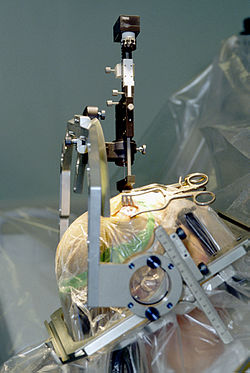
Insertion of electrode during deep brain stimulation surgery using a stereotactic frame Parkinson surgery.jpg
Insertion of electrode during deep brain stimulation surgery using a stereotactic frame

Deep brain stimulation

Deep brain stimulation involves the electrical stimulation of deep brain structures by electrodes implanted into the brain. The effects of this neurosurgery has been studied in patients with Parkinson's disease, tremors, dystonia, epilepsy, depression, obsessive-compulsive disorder, Tourette's syndrome, cluster headache and chronic pain. [18] A fine electrode is inserted into the targeted area of the brain and secured to the skull. This is attached to a pulse generator which is implanted elsewhere on the body under the skin. The surgeon then turns the frequency of the electrode to the voltage and frequency desired. Deep brain stimulation has been shown in several studies to both induce pleasure or even addiction as well as ameliorate pain. For chronic pain, lower frequencies (about 5–50 Hz) have produced analgesic effects, whereas higher frequencies (about 120–180 Hz) have alleviated or stopped pyramidal tremors in Parkinson's patients. [18]

There is still further research necessary into how and why exactly DBS works. However, by understanding the relationship between pleasure and pain, procedures like these can be used to treat patients suffering from a high intensity or longevity of pain. So far, DBS has been recognized as a treatment for Parkinson's disease, tremors, and dystonia by the Food and Drug Administration (FDA). [20] [21]

See also

Related Research Articles

Pleasure is experience that feels good, that involves the enjoyment of something. It contrasts with pain or suffering, which are forms of feeling bad. It is closely related to value, desire and action: humans and other conscious animals find pleasure enjoyable, positive or worthy of seeking. A great variety of activities may be experienced as pleasurable, like eating, having sex, listening to music or playing games. Pleasure is part of various other mental states such as ecstasy, euphoria and flow. Happiness and well-being are closely related to pleasure but not identical with it. There is no general agreement as to whether pleasure should be understood as a sensation, a quality of experiences, an attitude to experiences or otherwise. Pleasure plays a central role in the family of philosophical theories known as hedonism.

The mesolimbic pathway, sometimes referred to as the reward pathway, is a dopaminergic pathway in the brain. The pathway connects the ventral tegmental area in the midbrain to the ventral striatum of the basal ganglia in the forebrain. The ventral striatum includes the nucleus accumbens and the olfactory tubercle.

<span class="mw-page-title-main">Nucleus accumbens</span> Region of the basal forebrain

The nucleus accumbens is a region in the basal forebrain rostral to the preoptic area of the hypothalamus. The nucleus accumbens and the olfactory tubercle collectively form the ventral striatum. The ventral striatum and dorsal striatum collectively form the striatum, which is the main component of the basal ganglia. The dopaminergic neurons of the mesolimbic pathway project onto the GABAergic medium spiny neurons of the nucleus accumbens and olfactory tubercle. Each cerebral hemisphere has its own nucleus accumbens, which can be divided into two structures: the nucleus accumbens core and the nucleus accumbens shell. These substructures have different morphology and functions.

<span class="mw-page-title-main">Dopaminergic pathways</span> Projection neurons in the brain that synthesize and release dopamine

Dopaminergic pathways in the human brain are involved in both physiological and behavioral processes including movement, cognition, executive functions, reward, motivation, and neuroendocrine control. Each pathway is a set of projection neurons, consisting of individual dopaminergic neurons.

Dynorphins (Dyn) are a class of opioid peptides that arise from the precursor protein prodynorphin. When prodynorphin is cleaved during processing by proprotein convertase 2 (PC2), multiple active peptides are released: dynorphin A, dynorphin B, and α/β-neoendorphin. Depolarization of a neuron containing prodynorphin stimulates PC2 processing, which occurs within synaptic vesicles in the presynaptic terminal. Occasionally, prodynorphin is not fully processed, leading to the release of “big dynorphin.” “Big Dynorphin” is a 32-amino acid molecule consisting of both dynorphin A and dynorphin B.

Motivational salience is a cognitive process and a form of attention that motivates or propels an individual's behavior towards or away from a particular object, perceived event or outcome. Motivational salience regulates the intensity of behaviors that facilitate the attainment of a particular goal, the amount of time and energy that an individual is willing to expend to attain a particular goal, and the amount of risk that an individual is willing to accept while working to attain a particular goal.

<span class="mw-page-title-main">Periaqueductal gray</span> Nucleus surrounding the cerebral aqueduct

The periaqueductal gray is a brain region that plays a critical role in autonomic function, motivated behavior and behavioural responses to threatening stimuli. PAG is also the primary control center for descending pain modulation. It has enkephalin-producing cells that suppress pain.

<span class="mw-page-title-main">Nociceptin</span> Chemical compound

Nociceptin/orphanin FQ (N/OFQ), a 17-amino acid neuropeptide, is the endogenous ligand for the nociceptin receptor. Nociceptin acts as a potent anti-analgesic, effectively counteracting the effect of pain-relievers; its activation is associated with brain functions such as pain sensation and fear learning.

<span class="mw-page-title-main">Reward system</span> Group of neural structures responsible for motivation and desire

The reward system is a group of neural structures responsible for incentive salience, associative learning, and positively-valenced emotions, particularly ones involving pleasure as a core component. Reward is the attractive and motivational property of a stimulus that induces appetitive behavior, also known as approach behavior, and consummatory behavior. A rewarding stimulus has been described as "any stimulus, object, event, activity, or situation that has the potential to make us approach and consume it is by definition a reward". In operant conditioning, rewarding stimuli function as positive reinforcers; however, the converse statement also holds true: positive reinforcers are rewarding.

<span class="mw-page-title-main">Nociceptin receptor</span> Protein-coding gene in the species Homo sapiens

The nociceptin opioid peptide receptor (NOP), also known as the nociceptin/orphanin FQ (N/OFQ) receptor or kappa-type 3 opioid receptor, is a protein that in humans is encoded by the OPRL1 gene. The nociceptin receptor is a member of the opioid subfamily of G protein-coupled receptors whose natural ligand is the 17 amino acid neuropeptide known as nociceptin (N/OFQ). This receptor is involved in the regulation of numerous brain activities, particularly instinctive and emotional behaviors. Antagonists targeting NOP are under investigation for their role as treatments for depression and Parkinson's disease, whereas NOP agonists have been shown to act as powerful, non-addictive painkillers in non-human primates.

<span class="mw-page-title-main">Euphoria</span> Intense feelings of well-being

Euphoria is the experience of pleasure or excitement and intense feelings of well-being and happiness. Certain natural rewards and social activities, such as aerobic exercise, laughter, listening to or making music and dancing, can induce a state of euphoria. Euphoria is also a symptom of certain neurological or neuropsychiatric disorders, such as mania. Romantic love and components of the human sexual response cycle are also associated with the induction of euphoria. Certain drugs, many of which are addictive, can cause euphoria, which at least partially motivates their recreational use.

<span class="mw-page-title-main">Frisson</span> Psychophysiological response to rewarding auditory or visual stimuli

Frisson, also known as aesthetic chills or psychogenic shivers, is a psychophysiological response to rewarding stimuli that often induces a pleasurable or otherwise positively-valenced affective state and transient paresthesia, sometimes along with piloerection and mydriasis . The sensation commonly occurs as a mildly to moderately pleasurable emotional response to music with skin tingling; piloerection and pupil dilation not necessarily occurring in all cases.

The ventral pallidum (VP) is a structure within the basal ganglia of the brain. It is an output nucleus whose fibres project to thalamic nuclei, such as the ventral anterior nucleus, the ventral lateral nucleus, and the medial dorsal nucleus. The VP is a core component of the reward system which forms part of the limbic loop of the basal ganglia, a pathway involved in the regulation of motivational salience, behavior, and emotions. It is involved in addiction.

The parabrachial nuclei, also known as the parabrachial complex, are a group of nuclei in the dorsolateral pons that surrounds the superior cerebellar peduncle as it enters the brainstem from the cerebellum. They are named from the Latin term for the superior cerebellar peduncle, the brachium conjunctivum. In the human brain, the expansion of the superior cerebellar peduncle expands the parabrachial nuclei, which form a thin strip of grey matter over most of the peduncle. The parabrachial nuclei are typically divided along the lines suggested by Baxter and Olszewski in humans, into a medial parabrachial nucleus and lateral parabrachial nucleus. These have in turn been subdivided into a dozen subnuclei: the superior, dorsal, ventral, internal, external and extreme lateral subnuclei; the lateral crescent and subparabrachial nucleus along the ventrolateral margin of the lateral parabrachial complex; and the medial and external medial subnuclei

Gaseous signaling molecules are gaseous molecules that are either synthesized internally (endogenously) in the organism, tissue or cell or are received by the organism, tissue or cell from outside and that are used to transmit chemical signals which induce certain physiological or biochemical changes in the organism, tissue or cell. The term is applied to, for example, oxygen, carbon dioxide, sulfur dioxide, nitrous oxide, hydrogen cyanide, ammonia, methane, hydrogen, ethylene, etc.

<span class="mw-page-title-main">Pain in amphibians</span> Ethical issue

Pain is an aversive sensation and feeling associated with actual, or potential, tissue damage. It is widely accepted by a broad spectrum of scientists and philosophers that non-human animals can perceive pain, including pain in amphibians.

<span class="mw-page-title-main">Morten Kringelbach</span> Danish neuroscientist

Morten L Kringelbach is a professor of neuroscience at University of Oxford, UK and Aarhus University, Denmark. He is the director of the 'Centre for Eudaimonia and Human Flourishing', fellow of Linacre College, Oxford and board member of the Empathy Museum.

<span class="mw-page-title-main">Irene Tracey</span> British neuroscientist (born 1966)

Irene Mary Carmel Tracey is Vice-Chancellor of the University of Oxford and former Warden of Merton College, Oxford. She is also Professor of Anaesthetic Neuroscience in the Nuffield Department of Clinical Neurosciences and formerly Pro-Vice-Chancellor at the University of Oxford. She is a co-founder of the Oxford Centre for Functional Magnetic Resonance Imaging of the Brain (FMRIB), now the Wellcome Centre for Integrative Neuroimaging. Her team’s research is focused on the neuroscience of pain, specifically pain perception and analgesia as well as how anaesthetics produce altered states of consciousness. Her team uses multidisciplinary approaches including neuroimaging.

Mark A. Gillman is a South African scholar, neuroscientist, medical consultant and author. He is Emeritus CEO of the S.A. Brain Research Institute and an adviser on substance abuse for Governments in South Africa, the USA, China, and Israel.
References

  1. 1 2 Almeida TF, Roizenblatt S, Tufik S (2004). "Afferent pain pathways: a neuroanatomical review". Brain Res. 1000 (1–2): 40–56. doi:10.1016/j.brainres.2003.10.073. PMID   15053950. S2CID   14772296.{{cite journal}}: CS1 maint: multiple names: authors list (link)
  2. Apkarian, A. V.; Bushnell, M. C.; Treede, R. D.; Zubieta, J. K. (2005). "Human brain mechanisms of pain perception and regulation in health and disease" (PDF). European Journal of Pain . 9 (4): 463–484. doi:10.1016/j.ejpain.2004.11.001. hdl: 2027.42/90300 . PMID   15979027. S2CID   15144693. Archived from the original (PDF) on 2012-04-26. Retrieved 2011-12-02.
  3. 1 2 Kringelbach, M. L.; Berridge, K. C. (2009). "Towards a functional neuroanatomy of pleasure and happiness" (PDF). Trends in Cognitive Sciences. 13 (11): 479–487. doi:10.1016/j.tics.2009.08.006. PMC   2767390 . PMID   19782634. Archived from the original (PDF) on 2018-06-19. Retrieved 2020-04-29.
  4. Berridge, K. C.; Kringelbach, M. L. (2008). "Affective neuroscience of pleasure: reward in humans and animals". Psychopharmacology . 199 (3): 457–480. doi:10.1007/s00213-008-1099-6. PMC   3004012 . PMID   18311558.
  5. Esch T., Stefano G. B. (2004). "The neurobiology of pleasure, reward processes, addiction and their health implications" (PDF). Neuroendocrinology Letters. 25 (4): 235–251. PMID   15361811.
  6. Fields H. L. (2007). "Understanding how opioids contribute to reward and analgesia" (PDF). Regional Anesthesia and Pain Medicine. 32 (3): 242–246. doi:10.1016/j.rapm.2007.01.001. PMID   17543821. S2CID   15293275.
  7. "Aristotle. Rhetoric, 11". Archived from the original on 2012-04-15. Retrieved 2011-11-21.
  8. 1 2 Leknes, S.; Tracey, I. (2008). "A common neurobiology for pain and pleasure" (PDF). Nature Reviews Neuroscience . 9 (4): 314–320. doi:10.1038/nrn2333. PMID   18354400. S2CID   6498126. Archived from the original (PDF) on 2012-04-05. Retrieved 2011-12-02.
  9. Szasz, T.S. (1957) Pain and Pleasure – a study of bodily feelings
  10. Gillman M A, M. A.; Kok, L; Lichtigfeld, F. J. (1980). "Paradoxical effect of naloxone on nitrous oxide analgesia in man". Eur J Pharmacol. 61 (2): 175–177. doi:10.1016/0014-2999(80)90160-0. PMID   6243567.
  11. Gillman, MA; Lichtigfeld, FJ (1981). "A comparison of the effect of morphine sulphate and nitrous oxide analgesia on chronic pain states in man". J Neurol Sci. 45 (1): 41–45. doi:10.1016/0022-510X(81)90186-6. PMID   7205318. S2CID   32640794.
  12. Gillman MA; Kimmel, I; Lichtigfeld FJ, FJ (1981). "The dual system hypothesis of pain perception". Neurological Research. 3 (4): 317–327. doi:10.1080/01616412.1981.11739607. PMID   6175917.
  13. Lichtigfeld, FJ; Gillman, MA. (1982). "The treatment of alcoholic withdrawal states with oxygen and nitrous oxide". S Afr Med J. 61 (10): 349–351. PMID   7064002.
  14. Gillman, MA; Lichtigfeld, FJ (1984). ". The opioid and anti-opioid system in addiction". S Afr Med J. 66 (16): 592. PMID   6495096.
  15. Gillman, MA; Lichtigfeld, FJ (1983). "The effect of nitrous oxide and naloxone on orgasm in human females. A preliminary report". J Sex Res. 19: 49–57. doi:10.1080/00224498309551168.
  16. 1 2 3 Leknes S.; Brooks J. C. W.; Wiech K.; Tracey I. (2008). "Pain relief as an opponent process: a psychophysical investigation". European Journal of Neuroscience. 28 (4): 794–801. doi:10.1111/j.1460-9568.2008.06380.x. PMID   18671736. S2CID   205513577.
  17. Fields, Howard L. (March 2007). "The analgesia-addiction interface: Clinical and neurobiological issues" (PDF). National Institute on Drug Abuse (NIDA). National Institute of Health. Retrieved 14 March 2023.
  18. 1 2 3 Green, A.L., Pereira, E.A., Aziz, T.Z. "Deep brain stimulation and pleasure". Pleasures of the brain. Eds. M. L. Kringelbach, K. C. Berridge. (2010). New York, NY: Oxford University Press, 302-319.
  19. Kringelbach, M. L., & Berridge, K. C. (2010). Pleasures of the Brain. New York, NY: Oxford University Press, Inc.
  20. Ondo, W.; Jankovic, J.; Schwartz, K.; Almaguer, M.; Simpson, R. K. (1998-10-01). "Unilateral thalamic deep brain stimulation for refractory essential tremor and Parkinson's disease tremor". Neurology. 51 (4): 1063–1069. doi:10.1212/WNL.51.4.1063. ISSN   0028-3878. PMID   9781530. S2CID   20986752.
  21. Vercueil, Laurent; Pollak, Pierre; Fraix, Valérie; Caputo, Elena; Moro, Elena; Benazzouz, Abdelhamid; Xie, Jing; Koudsie, Adnan; Benabid, Alim-Louis (2001-08-01). "Deep brain stimulation in the treatment of severe dystonia". Journal of Neurology. 248 (8): 695–700. doi:10.1007/s004150170116. ISSN   1432-1459. PMID   11569899. S2CID   8244088.