Paleoradiology

Last updated
A Peruvian child mummy being placed onto a CT machine for imaging US Navy 110427-N-2531L-135 Tori Randall, Ph.D. prepares a 550-year old Peruvian child mummy for a CT scan.jpg
A Peruvian child mummy being placed onto a CT machine for imaging

Paleoradiology (ancient radiology) is the study of archaeological remains through the use of radiographic techniques, such as X-ray, CT (computer tomography) and micro-CT scans. [1] It is predominately used by archaeologists and anthropologists to examine mummified remains due to its non-invasive nature. [2] Paleoradiologists can discover post-mortem damage to the body, or any artefacts buried with them, while still keeping the remains intact. Radiological images can also contribute evidence about the person's life, such as their age and cause of death. The first recorded use of paleoradiology (although not by that name) was in 1896, just a year after the Rōntgen radiograph was first produced. [3] Although this method of viewing ancient remains is advantageous due to its non-invasive manner, many radiologists lack expertise in archeology and very few radiologists can identify ancient diseases which may be present. [4]

Contents

Techniques

CT

CT scans are most commonly used in paleoradiological studies because they can create images of soft tissue, organs and body cavities of mummified remains without performing an invasive and damaging autopsy. [5] This enables archaeologists and anthropologists to digitally unwrap the remains and reveal what they contain. CT scanners create these images by taking multiple radiographic planes (or cuts) of the body at different angles which records the layers of different structures in the remains. This differs from typical radiographic scans (X-rays) where all the structural layers are documented in one image, which can create shadows and therefore limit their accuracy. [6] [7]

There are several main viewing techniques used in CT imaging. [8]

A 3D image of the skull of an Incan mummy The Wormian Inca bone.png
A 3D image of the skull of an Incan mummy

After these different views of the remains have been obtained, it is possible to create a three-dimensional reconstruction of the body. This brings into focus details which may have been missed on the axial imagining. Algorithm manipulation is used to create the rotational 3D images. [6] [5] In paleoradiology, the 3D images provide a greater understanding of the remains themselves. For example, in 2002, a study of nine Egyptian mummies found that by using the 3D reconstructions they could see the preservation of soft tissues, such as the penis on one male body and braided hair on female remains. The 3D modelling also illustrated discrepancies between the remains, as some had their internal organs removed while others had not. [5]

Due to their ability to take multiple planes of the remains, CT scans are able to virtually 'fly through' the body to assess internal composition and cavities. [2] These techniques are commonly used for diagnostic scans such as colonoscopy and bronchoscopy, and the same method is applied to ancient remains. [5] This enables researchers to digitally look through the remains from the top down, as though they were watching a short video of footage from the interior of the body. The technique presents observable data from the hollow cavities in the chest and abdomen regions. It can demonstrate whether there are internal organs present or if, in the case of Egyptian mummies, linen has been packed to maintain the body shape of the remains. [5]

Micro-CT

Micro-CT is a specialised form of CT scan used to create images with pixels in micrometres. These images are frequently used for bone density examinations and produce greater detail for images of bony structures. This radiology technique is often used to examine the teeth from mummified remains. [6] [9]

Early use (18951970)

Two mummified Egyptian cats with a radiograph of the cat on the left Mummified cats.jpg
Two mummified Egyptian cats with a radiograph of the cat on the left

Radiographs, or X-rays, have been used to study and observe ancient remains since their invention by Wilhelm Röntgen in 1895. This early form of X-ray, sometimes known as Röntgenograms, was immediately used by physicists, anthropologists, anatomists and archaeologists as shown below. [3] [6]

An X-ray of Tutankhamun's skull, the arrow pointing to a possible cause of death Tutankhamunxray.jpg
An X-ray of Tutankhamun's skull, the arrow pointing to a possible cause of death

Current use in archaeology

CT scans are the most common radiological technique used in modern archaeology due to their ability to provide more detailed information about ancient remains (such as soft tissue and blood vessels) and to produce 3D images by taking layers of different angled pictures. Archaeologists are able to collect data such as age, sex, the cause of death, socioeconomic status, mummification and burial practices by analysing the CT images. The images can also reveal whether the remains were subject to ancient diseases or post-mortem damage. [9] Although paleoradiology practices are used on preserved remains such as European bog bodies and frozen bodies from the High Andes, they are more frequently documented in the analysis of Egyptian mummified remains. [4]

Egyptology

Statue of Paramesses (later known as Ramesses I), pharaoh of Egypt during the 19th Dynasty, New Kingdom Egypt Statue Paramesses Munich.JPG
Statue of Paramesses (later known as Ramesses I), pharaoh of Egypt during the 19th Dynasty, New Kingdom Egypt

CT scans are used in Egyptology to gain insight into mummified bodies without risking damage to the integrity of the remains. Hoffman's recent study of nine Egyptian remains discovered new information regarding the mummification practices of Egyptians. The typical process of mummification, as written by Herodotus, involves the removal of the four major internal organs (liver, intestines, lungs and stomach) and placement of them in four canopic jars. The heart is removed, embalmed and placed back into the body as it is an important feature in the journey to the Egyptian afterlife. However, Hoffman discovered that this was not the case in all mummies. Through analysis of CT-produced 3D images, the "fly-through" technique and a combination of axial and coronal images, it was discovered that four of the remains had not had their internal organs removed and in another four the heart could not be identified. [5] Hoffman suggests this may be due to socioeconomic differences between the mummies during the time they were alive. CT scans were further used in Hoffman's study to potentially identify one of the remains as Ramesses I, a Pharaoh during New Kingdom Egypt. It was found that the mummification practices of this particular body were in conjunction with those typically used during the New Kingdom period, as images showed rolled linen placed inside the body in order to preserve its shape. The mummy's arms were also found to be placed across the chest as a symbol of nobility. [5]

Imaging done by Rethy Chhem, in 2004, was able to correct a diagnosis of Ramesses II from X-rays done in 1976. The incorrect diagnosis had been of ankylosing spondylitis, a form of arthritis. However, Chhem perceived that the pharaoh actually had diffuse idiopathic skeletal hyperostosis, a calcification of the joints causing ligaments to attach to the spine. [6] The CT images provided a clearer and more detailed image of the spine when compared with the early X-rays. This enabled the researchers to provide a greater insight into diseases found in ancient remains and achieve a more accurate diagnosis.

A recent CT scan of Tutankhamun in 2006 was able to provide evidence against the 'homicide theory'. [2] [6] A depression fracture noted on the skull from X-rays taken 30 years previously was found to be a post-mortem injury rather than a cause of death. [6] The hole in the head had been created in order to continue the embalming process of mummification. [10] This CT investigation was also able to confirm Tutankhamun's age of death as nineteen and disprove the idea that the young pharaoh had suffered from scoliosis; rather the bend in his spine was from additional post-mortem damage to the body. [10]

Disadvantages

Although the information and evidence gathered by radiological imaging of ancient remains have been largely beneficial to the fields of archaeology and anthropology, not all of the information can be considered accurate due to the lack of radiologists who specialise in paleopathology. Instead, to obtain the most information from CT or X-ray images a team must meet to discuss the findings (e.g. for a skeletal study of the remains, an orthopaedic surgeon, bone pathologist and musculoskeletal radiologist would meet). [2] [4] Another disadvantage to this technique is the low contrast resolution. The researcher may be unable to determine a difference between soft tissue and artefacts left from the embalming process. Due to the decomposed state of some of the mummified remains, it can also be difficult to distinguish internal organs due to shrinkage and a lack of preservation. Post-mortem injuries and damage to the body can also hinder the ability of radiological scans to provide accurate information for researchers. For example, in frozen remains, there can difficulty when differentiating whether a CT suggests the body has air-inflated lung tissue or if there is frozen fluid in the lungs. [4]

Paleoradiology is informative in its ability to assist in determining the age of death of the remains, however, this is not always completely accurate or available information. In a sample of bog bodies, 35% were not able to be identified with any age group and 30% could not be sex determined. A radiological study of an iceman was only able to produce an estimate of 4050 years at age of death. In order to achieve a more accurate time frame, the body would have to be subject to an autopsy or similar physical assessment which would cause irreversible damage to the remains. [4]

Another disadvantage of paleoradiology is the difficulty in transporting the equipment or the artefact/remains to spaces where the images can be taken. [1] [11] For example, in 2005 the mummy of Tutankhamun was imaged using a CT-scanner which had to be brought from the Cairo Museum to tomb KV62 in the Valley of the Kings. [10] This was due to the fragile state of the remains which were unable to be removed from the climate-controlled tomb. In this study, funding came from a five-year grant from the Supreme Council of Antiquities, which was aided by the donation of a Siemens CT-scanner by the National Geographic Society, [10] however typically funding for research can be problematic as equipment is costly and there may not be sufficient interest to prompt donations.

Related Research Articles

<span class="mw-page-title-main">Mummy</span> Human or animal whose skin and organs have been preserved

A mummy is a dead human or an animal whose soft tissues and organs have been preserved by either intentional or accidental exposure to chemicals, extreme cold, very low humidity, or lack of air, so that the recovered body does not decay further if kept in cool and dry conditions. Some authorities restrict the use of the term to bodies deliberately embalmed with chemicals, but the use of the word to cover accidentally desiccated bodies goes back to at least the early 17th century.

<span class="mw-page-title-main">CT scan</span> Medical imaging procedure using X-rays to produce cross-sectional images

A computed tomography scan is a medical imaging technique used to obtain detailed internal images of the body. The personnel that perform CT scans are called radiographers or radiology technologists.

<span class="mw-page-title-main">Radiography</span> Imaging technique using ionizing and non-ionizing radiation

Radiography is an imaging technique using X-rays, gamma rays, or similar ionizing radiation and non-ionizing radiation to view the internal form of an object. Applications of radiography include medical and industrial radiography. Similar techniques are used in airport security,. To create an image in conventional radiography, a beam of X-rays is produced by an X-ray generator and it is projected towards the object. A certain amount of the X-rays or other radiation are absorbed by the object, dependent on the object's density and structural composition. The X-rays that pass through the object are captured behind the object by a detector. The generation of flat two-dimensional images by this technique is called projectional radiography. In computed tomography, an X-ray source and its associated detectors rotate around the subject, which itself moves through the conical X-ray beam produced. Any given point within the subject is crossed from many directions by many different beams at different times. Information regarding the attenuation of these beams is collated and subjected to computation to generate two-dimensional images on three planes which can be further processed to produce a three-dimensional image.

<span class="mw-page-title-main">Ramesses III</span> Egyptian pharaoh 1186–1155 BCE, New Kingdom, 20th Dynasty

Usermaatre Meryamun Ramesses III was the second Pharaoh of the Twentieth Dynasty in Ancient Egypt. He is thought to have reigned from 26 March 1186 to 15 April 1155 BC and is considered to be the last great monarch of the New Kingdom to wield any substantial authority over Egypt.

<span class="mw-page-title-main">Radiology</span> Branch of Medicine

Radiology is the medical discipline that uses medical imaging to diagnose diseases and guide their treatment, within the bodies of humans and other animals. It began with radiography, but today it includes all imaging modalities, including those that use no electromagnetic radiation, as well as others that do, such as computed tomography (CT), fluoroscopy, and nuclear medicine including positron emission tomography (PET). Interventional radiology is the performance of usually minimally invasive medical procedures with the guidance of imaging technologies such as those mentioned above.

<span class="mw-page-title-main">Medical imaging</span> Technique and process of creating visual representations of the interior of a body

Medical imaging is the technique and process of imaging the interior of a body for clinical analysis and medical intervention, as well as visual representation of the function of some organs or tissues (physiology). Medical imaging seeks to reveal internal structures hidden by the skin and bones, as well as to diagnose and treat disease. Medical imaging also establishes a database of normal anatomy and physiology to make it possible to identify abnormalities. Although imaging of removed organs and tissues can be performed for medical reasons, such procedures are usually considered part of pathology instead of medical imaging.

<span class="mw-page-title-main">Angiography</span> Medical imaging technique

Angiography or arteriography is a medical imaging technique used to visualize the inside, or lumen, of blood vessels and organs of the body, with particular interest in the arteries, veins, and the heart chambers. Modern angiography is performed by injecting a radio-opaque contrast agent into the blood vessel and imaging using X-ray based techniques such as fluoroscopy.

<span class="mw-page-title-main">Interventional radiology</span> Medical subspecialty

Interventional radiology (IR) is a medical specialty that performs various minimally-invasive procedures using medical imaging guidance, such as x-ray fluoroscopy, computed tomography, magnetic resonance imaging, or ultrasound. IR performs both diagnostic and therapeutic procedures through very small incisions or body orifices. Diagnostic IR procedures are those intended to help make a diagnosis or guide further medical treatment, and include image-guided biopsy of a tumor or injection of an imaging contrast agent into a hollow structure, such as a blood vessel or a duct. By contrast, therapeutic IR procedures provide direct treatment—they include catheter-based medicine delivery, medical device placement, and angioplasty of narrowed structures.

<span class="mw-page-title-main">Scintigraphy</span> Diagnostic imaging test in nuclear medicine

Scintigraphy, also known as a gamma scan, is a diagnostic test in nuclear medicine, where radioisotopes attached to drugs that travel to a specific organ or tissue (radiopharmaceuticals) are taken internally and the emitted gamma radiation is captured by gamma cameras, which are external detectors that form two-dimensional images in a process similar to the capture of x-ray images. In contrast, SPECT and positron emission tomography (PET) form 3-dimensional images and are therefore classified as separate techniques from scintigraphy, although they also use gamma cameras to detect internal radiation. Scintigraphy is unlike a diagnostic X-ray where external radiation is passed through the body to form an image.

<span class="mw-page-title-main">Radiographer</span> Healthcare professional

Radiographers, also known as radiologic technologists, diagnostic radiographers and medical radiation technologists are healthcare professionals who specialise in the imaging of human anatomy for the diagnosis and treatment of pathology. Radiographers are infrequently, and almost always erroneously, known as x-ray technicians. In countries that use the title radiologic technologist they are often informally referred to as techs in the clinical environment; this phrase has emerged in popular culture such as television programmes. The term radiographer can also refer to a therapeutic radiographer, also known as a radiation therapist.

<span class="mw-page-title-main">Projectional radiography</span> Formation of 2D images using X-rays

Projectional radiography, also known as conventional radiography, is a form of radiography and medical imaging that produces two-dimensional images by X-ray radiation. The image acquisition is generally performed by radiographers, and the images are often examined by radiologists. Both the procedure and any resultant images are often simply called 'X-ray'. Plain radiography or roentgenography generally refers to projectional radiography. Plain radiography can also refer to radiography without a radiocontrast agent or radiography that generates single static images, as contrasted to fluoroscopy, which are technically also projectional.

<span class="mw-page-title-main">High-resolution computed tomography</span> Diagnostic imaging test

High-resolution computed tomography (HRCT) is a type of computed tomography (CT) with specific techniques to enhance image resolution. It is used in the diagnosis of various health problems, though most commonly for lung disease, by assessing the lung parenchyma. On the other hand, HRCT of the temporal bone is used to diagnose various middle ear diseases such as otitis media, cholesteatoma, and evaluations after ear operations.

<span class="mw-page-title-main">The Younger Lady</span> Informal name for a mummy found in tomb KV35

The Younger Lady is the informal name given to an ancient Egyptian mummy discovered within tomb KV35 in the Valley of the Kings by archaeologist Victor Loret in 1898. The mummy also has been given the designation KV35YL and 61072, and currently resides in the Egyptian Museum in Cairo. Through recent DNA tests, this mummy has been identified as the mother of the pharaoh Tutankhamun and a daughter of pharaoh Amenhotep III and his Great Royal Wife Tiye. Early speculation that this mummy was the remains of Nefertiti was argued to be incorrect, as nowhere is Nefertiti accorded the title "King's daughter."

Tutankhamun's mummy was discovered by English Egyptologist Howard Carter and his team on 28 October 1925 in tomb KV62 of Egypt's Valley of the Kings. Tutankhamun was the 13th pharaoh of the 18th Dynasty of the New Kingdom of Egypt, making his mummy over 3,300 years old. Tutankhamun's mummy is the only royal mummy to have been found entirely undisturbed.

A digital autopsy is a non-invasive autopsy in which digital imaging technology, such as with computerized tomography (CT) or magnetic resonance imaging (MRI) scans, is used to develop three-dimensional images for a virtual exploration of a human body.

Cultural property imaging is a necessary part of long term preservation of cultural heritage. While the physical conditions of objects will change over time, imaging serves as a way to document and represent heritage in a moment in time of the life of the item. Different methods of imaging produce results that are applicable in various circumstances. Not every method is appropriate for every object, and not every object needs to be imaged by multiple methods. In addition to preservation and conservation-related concerns, imaging can also serve to enhance research and study of cultural heritage.

<span class="mw-page-title-main">Focal plane tomography</span> Imaging technique using moving X-ray machines

In radiography, focal plane tomography is tomography by simultaneously moving the X-ray generator and X-ray detector so as to keep a consistent exposure of only the plane of interest during image acquisition. This was the main method of obtaining tomographs in medical imaging until the late-1970s. It has since been largely replaced by more advanced imaging techniques such as CT and MRI. It remains in use today in a few specialized applications, such as for acquiring orthopantomographs of the jaw in dental radiography.

<span class="mw-page-title-main">History of computed tomography</span> History of CT scanning technology

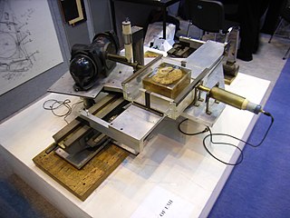
The history of X-ray computed tomography dates back to at least 1917 with the mathematical theory of the Radon transform In the early 1900s an Italian radiologist named Alessandro Vallebona invented tomography which used radiographic film to see a single slice of the body. In October 1963, William H. Oldendorf received a U.S. patent for a "radiant energy apparatus for investigating selected areas of interior objects obscured by dense material". The first clinical CT scan was performed in 1971 using a scanner invented by Sir Godfrey Hounsfield.

<span class="mw-page-title-main">317a and 317b mummies</span> Daughters of Egyptian pharaoh Tutankhamun

Mummies 317a and 317b were the infant daughters of the ancient Egyptian pharaoh Tutankhamun of the Eighteenth Dynasty. Their mother is presumed to be Ankhesenamun, his only known wife, who has been tentatively identified through DNA testing as the mummy KV21A. 317a was born prematurely at 5–6 months' gestation, and 317b was born at or near full term. They are assumed to have been stillborn or died shortly after birth.

Sahar Saleem is a professor of radiology at Cairo University where she specialises in paleoradiology, the use of radiology to study mummies. She discovered the knife wound in the throat of Ramesses III, which was most likely the cause of his death.

References

  1. 1 2 Chhem, Rethy (2004). "Paleoradiology: current status and future challenges". Canadian Association of Radiologists Journal. 55 (4): 198–199. PMID   15362341. ProQuest   235996145.
  2. 1 2 3 4 5 Cosmacini, P; Piacentini, P (2008). "Notes on the history of the radiological study of Egyptian mummies: from X-rays to new imaging techniques". La Radiologia Medica. 113 (5): 615–626. doi:10.1007/s11547-008-0280-7. PMID   18523844. S2CID   371894.
  3. 1 2 3 4 5 6 7 8 9 Boni, Thomas; Chhem, Rethy (2004). "History of paleoradiology: early published literature, 1896-1921". Canadian Association of Radiologists Journal. 55 (4): 203–210. PMID   15362342. ProQuest   235982587.
  4. 1 2 3 4 5 Boni, Thomas; Chhem, Rethy (2004). "Diagnostic paleoradiology of mummified tissue: interpretation and pitfalls". Canadian Association of Radiologists Journal. 55 (4): 218–227. PMID   15362344. ProQuest   236112998.
  5. 1 2 3 4 5 6 7 8 Hoffman, Heidi; Torres, William E.; Ernst, Randy D. (2002). "Paleoradiology: Advanced CT in the Evaluation of Nine Egyptian Mummies". RadioGraphics. 22 (2): 377–385. doi:10.1148/radiographics.22.2.g02mr13377. ISSN   0271-5333. PMID   11896227.
  6. 1 2 3 4 5 6 7 8 9 10 11 Brothwell, D; Chhem, R (2008). Paleoradiology Imaging Mummies and Fossils. Springer. doi:10.1007/978-3-540-48833-0. ISBN   978-3-540-48832-3.
  7. 1 2 Beckett, Ronald G. (2014-02-28). "Paleoimaging: a review of applications and challenges". Forensic Science, Medicine, and Pathology. 10 (3): 423–436. doi:10.1007/s12024-014-9541-z. ISSN   1547-769X. PMID   24682794. S2CID   5448680.
  8. "The Basics - Neuroradiology". sites.google.com. University of Wisconsin. 2018. Retrieved 2018-09-30.
  9. 1 2 Chhem, Rethy K. (2006-06-08). "Paleoradiology: imaging disease in mummies and ancient skeletons". Skeletal Radiology. 35 (11): 803–804. doi:10.1007/s00256-006-0144-y. ISSN   0364-2348. PMID   16847646. S2CID   1692339.
  10. 1 2 3 4 Hawass, Zahi (2013). "The Death of Tutankhamun". International Journal on Humanistic Ideology. 6 (1): 13–27. ProQuest   1469896500.
  11. Eppenberger, Patrick E.; Cavka, Mislav; Habicht, Michael E.; Galassi, Francesco M.; Rühli, Frank (2018-06-20). "Radiological findings in ancient Egyptian canopic jars: comparing three standard clinical imaging modalities (x-rays, CT and MRI)". European Radiology Experimental. 2 (1): 12. doi: 10.1186/s41747-018-0048-3 . ISSN   2509-9280. PMC   6008346 . PMID   29951641.