Related Research Articles

Skin is the layer of usually soft, flexible outer tissue covering the body of a vertebrate animal, with three main functions: protection, regulation, and sensation.
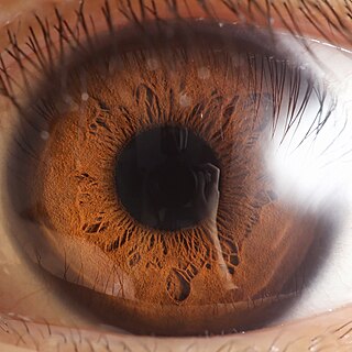
In most mammals and birds, the iris is a thin, annular structure in the eye, responsible for controlling the diameter and size of the pupil, and thus the amount of light reaching the retina. Eye color is defined by the iris. In optical terms, the pupil is the eye's aperture, while the iris is the diaphragm.

The esophagus or oesophagus, colloquially known also as the food pipe, food tube, or gullet, is an organ in vertebrates through which food passes, aided by peristaltic contractions, from the pharynx to the stomach. The esophagus is a fibromuscular tube, about 25 cm (10 in) long in adults, that travels behind the trachea and heart, passes through the diaphragm, and empties into the uppermost region of the stomach. During swallowing, the epiglottis tilts backwards to prevent food from going down the larynx and lungs. The word oesophagus is from Ancient Greek οἰσοφάγος (oisophágos), from οἴσω (oísō), future form of φέρω + ἔφαγον.

A fascia is a generic term for macroscopic membranous bodily structures. Fasciae are classified as superficial, deep, visceral, and parietal, and further designated according to their anatomical location.

A skin condition, also known as cutaneous condition, is any medical condition that affects the integumentary system—the organ system that encloses the body and includes skin, nails, and related muscle and glands. The major function of this system is as a barrier against the external environment.

The ciliary body is a part of the eye that includes the ciliary muscle, which controls the shape of the lens, and the ciliary epithelium, which produces the aqueous humor. The aqueous humor is produced in the non-pigmented portion of the ciliary body. The ciliary body is part of the uvea, the layer of tissue that delivers oxygen and nutrients to the eye tissues. The ciliary body joins the ora serrata of the choroid to the root of the iris.

The subcutaneous tissue, also called the hypodermis, hypoderm, subcutis, superficial fascia, is the lowermost layer of the integumentary system in vertebrates. The types of cells found in the layer are fibroblasts, adipose cells, and macrophages. The subcutaneous tissue is derived from the mesoderm, but unlike the dermis, it is not derived from the mesoderm's dermatome region. It consists primarily of loose connective tissue, and contains larger blood vessels and nerves than those found in the dermis. It is a major site of fat storage in the body.

The myometrium is the middle layer of the uterine wall, consisting mainly of uterine smooth muscle cells but also of supporting stromal and vascular tissue. Its main function is to induce uterine contractions.

The lips are a horizontal pair of soft appendages attached to the jaws and are the most visible part of the mouth of many animals, including humans. Vertebrate lips are soft, movable and serve to facilitate the ingestion of food and the articulation of sound and speech. Human lips are also a somatosensory organ, and can be an erogenous zone when used in kissing and other acts of intimacy.
The panniculus is a dense layer of fatty tissue consisting of excess subcutaneous fat within the lower abdominal region. Panniculi can form after rapid weight loss, as seen with strict exercise plans—in this case, the abdominal fat is successfully reduced, but excess skin is left behind which hangs loosely over the area. It can be a result of obesity and can be mistaken for a tumor or hernia. Abdominal panniculus can be removed during abdominal panniculectomy, a type of abdominoplasty. A panniculus can also be the result of loose tissues after pregnancy or massive weight loss.

The abdomen is the part of the body between the thorax (chest) and pelvis, in humans and in other vertebrates. The abdomen is the front part of the abdominal segment of the torso. The area occupied by the abdomen is called the abdominal cavity. In arthropods it is the posterior tagma of the body; it follows the thorax or cephalothorax.

The gluteal muscles, often called glutes, are a group of three muscles which make up the gluteal region commonly known as the buttocks: the gluteus maximus, gluteus medius and gluteus minimus. The three muscles originate from the ilium and sacrum and insert on the femur. The functions of the muscles include extension, abduction, external rotation, and internal rotation of the hip joint.

Morphea is a form of scleroderma that mainly involves isolated patches of hardened skin on the face, hands, and feet, or anywhere else on the body, usually with no internal organ involvement. However, in Deep Morphea inflammation and sclerosis can be found in the deep dermis, panniculus, fascia, superficial muscle and bone.
The panniculus adiposus is the fatty layer of the subcutaneous tissues, superficial to a deeper vestigial layer of muscle, the panniculus carnosus.

The thoracic wall or chest wall is the boundary of the thoracic cavity.

Erinaceus is a genus of hedgehog from the family of Erinaceidae. There are four main species of Erinaceus. The range is all across Europe, throughout the Middle East, parts of Russia, and extending to northern China and Korea. The European hedgehog has been introduced to New Zealand.

In the context of human evolution, human vestigiality involves those traits occurring in humans that have lost all or most of their original function through evolution. Although structures called vestigial often appear functionless, a vestigial structure may retain lesser functions or develop minor new ones. In some cases, structures once identified as vestigial simply had an unrecognized function. Vestigial organs are sometimes called rudimentary organs. Many human characteristics are also vestigial in other primates and related animals.

The buttocks are two rounded portions of the exterior anatomy of most mammals, located on the posterior of the pelvic region. In humans, the buttocks are located between the lower back and the perineum. They are composed of a layer of exterior skin and underlying subcutaneous fat superimposed on a left and right gluteus maximus and gluteus medius muscles. The two gluteus maximus muscles are the largest muscles in the human body. They are responsible for movements such as straightening the body into the upright (standing) posture when it is bent at the waist; maintaining the body in the upright posture by keeping the hip joints extended; and propelling the body forward via further leg (hip) extension when walking or running.

Anatomical terminology is a form of scientific terminology used by anatomists, zoologists, and health professionals such as doctors, physicians, and pharmacists.

The axillary arch is a variant of the latissimus dorsi muscle in humans. It is found as a slip of muscle or fascia extending between the latissimus dorsi muscle and the pectoralis major. There is considerable variation in the exact position of its origin and insertions as well as its blood and nerve supply. The arch may occur on one or both sides of the body. A meta-analysis revealed that the axillary arch had an overall prevalence of 5.3% of limbs.
References
- ↑ McGrath, J.A.; Eady, R.A.; Pope, F.M. (2004). Rook's Textbook of Dermatology (Seventh Edition). Blackwell Publishing. Page 3.1. ISBN 978-0-632-06429-8.