
The lymphatic system is part of the vascular system and an important part of the immune system, comprising a large network of lymphatic vessels that carry a clear fluid called lymph directionally towards the heart. The lymphatic system was first described in the seventeenth century independently by Olaus Rudbeck and Thomas Bartholin. Unlike the circulatory system, the lymphatic system is not a closed system. The human circulatory system processes an average of 20 litres of blood per day through capillary filtration, which removes plasma while leaving the blood cells. Roughly 17 litres of the filtered plasma is reabsorbed directly into the blood vessels, while the remaining three litres remain in the interstitial fluid. One of the main functions of the lymph system is to provide an accessory return route to the blood for the surplus three litres.

A lymph node or lymph gland is an ovoid or kidney-shaped organ of the lymphatic system, and of the adaptive immune system, that is widely present throughout the body. They are linked by the lymphatic vessels as a part of the circulatory system. Lymph nodes are major sites of B and T lymphocytes, and other white blood cells. Lymph nodes are important for the proper functioning of the immune system, acting as filters for foreign particles and cancer cells. Lymph nodes do not have a detoxification function, which is primarily dealt with by the liver and kidneys.

A lymphocyte is one of the subtypes of a white blood cell in a vertebrate's immune system. Lymphocytes include natural killer cells, T cells, and B cells. They are the main type of cell found in lymph, which prompted the name "lymphocyte".
There are at least two anatomical structures called a Malpighian corpuscle. They are also known as:
The Cords of Billroth are found in the red pulp of the spleen between the sinusoids, consisting of fibrils and connective tissue cells with a large population of monocytes and macrophages. These cords contain half of the mouse body's monocytes as a reserve so that after tissue injury these monocytes can move in and aid locally sourced monocytes in wound healing.
Reticular connective tissue is a type of connective tissue with a network of reticular fibers, made of type III collagen. Reticular fibers are not unique to reticular connective tissue, but only in this type are they dominant.
Gut-associated lymphoid tissue (GALT) is a component of the mucosa-associated lymphoid tissue (MALT) which works in the immune system to protect the body from invasion in the gut.

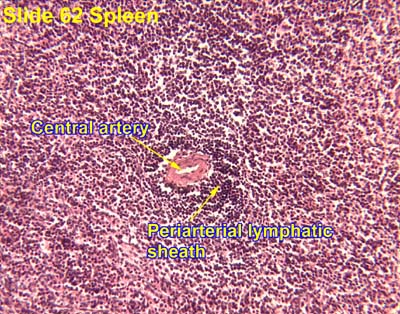
The marginal zone is the region at the interface between the non-lymphoid red pulp and the lymphoid white-pulp of the spleen.

Myeloid tissue, in the bone marrow sense of the word myeloid, is tissue of bone marrow, of bone marrow cell lineage, or resembling bone marrow, and myelogenous tissue is any tissue of, or arising from, bone marrow; in these senses the terms are usually used synonymously, as for example with chronic myeloid/myelogenous leukemia.

Germinal centers or germinal centres (GCs) are sites within secondary lymphoid organs – lymph nodes and the spleen where mature B cells proliferate, differentiate, and mutate their antibody genes, and switch the class of their antibodies during a normal immune response to an infection. These develop dynamically after the activation of follicular B cells by T-dependent antigen.
The red pulp of the spleen is composed of connective tissue known also as the cords of Billroth and many splenic sinusoids that are engorged with blood, giving it a red color. Its primary function is to filter the blood of antigens, microorganisms, and defective or worn-out red blood cells.
Splenic marginal zone lymphoma (SMZL) is a type of cancer made up of B-cells that replace the normal architecture of the white pulp of the spleen. The neoplastic cells are both small lymphocytes and larger, transformed lymphoblasts, and they invade the mantle zone of splenic follicles and erode the marginal zone, ultimately invading the red pulp of the spleen. Frequently, the bone marrow and splenic hilar lymph nodes are involved along with the peripheral blood. The neoplastic cells circulating in the peripheral blood are termed villous lymphocytes due to their characteristic appearance.
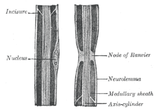
Myelin incisures, are small pockets of cytoplasm left behind during the Schwann cell myelination process. They are histological evidence of the small amount of cytoplasm that remains in the inner layer of the myelin sheath created by Schwann cells wrapping tightly around an axon.

In the peripheral nervous system, the myelin sheath of each axon in a nerve is wrapped in a delicate protective sheath known as the endoneurium. Within the nerve, axons targeting the same anatomical location are bundled together into groups known as fascicles, each surrounded by another protective sheath known as the perineurium. Several fascicles may be in turn bundled together with a blood supply and fatty tissue within yet another sheath, the epineurium. This grouping structure is analogous to the muscular organization system of epimysium, perimysium and endomysium.
The endoneurium is a layer of delicate connective tissue around the myelin sheath of each myelinated nerve fiber in the peripheral nervous system. Its component cells are called endoneurial cells. The endoneuria with their enclosed nerve fibers are bundled into groups called nerve fascicles, each fascicle within its own protective sheath called a perineurium. In sufficiently large nerves multiple fascicles, each with its blood supply and fatty tissue, may be bundled within yet another sheath, the epineurium.

The trabecular arteries are the name of the branches of the splenic artery after it passes into the trabeculae of the spleen, where it branches.
When these arteries then reach the white pulp, and become covered with periarteriolar lymphoid sheaths, the name changes again to central arteries. Branches of the central arteries are given to the red pulp, and these are called penicillar arteries).

The trabecular veins are the largest veins inside the spleen. It drains the blood collected in the sinuses of the pulp.
Follicular B cells are a type of B cell that reside in primary and secondary lymphoid follicles of secondary and tertiary lymphoid organs, including spleen and lymph nodes. Antibody responses against proteins are believed to involve follicular B cell pathways in secondary lymphoid organs.


