Related Research Articles

Haematopoiesis is the formation of blood cellular components. All cellular blood components are derived from haematopoietic stem cells. In a healthy adult human, roughly ten billion to a hundred billion new blood cells are produced per day, in order to maintain steady state levels in the peripheral circulation.
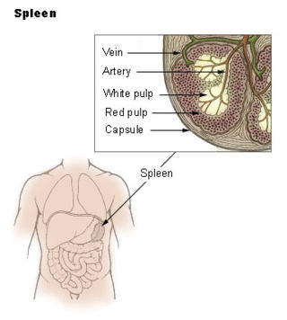
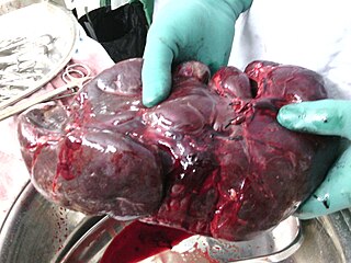
The spleen is an organ found in almost all vertebrates. Similar in structure to a large lymph node, it acts primarily as a blood filter. The word spleen comes from Ancient Greek σπλήν (splḗn).

The lymphatic system, or lymphoid system, is an organ system in vertebrates that is part of the immune system, and complementary to the circulatory system. It consists of a large network of lymphatic vessels, lymph nodes, lymphoid organs, lymphoid tissues and lymph. Lymph is a clear fluid carried by the lymphatic vessels back to the heart for re-circulation. The Latin word for lymph, lympha, refers to the deity of fresh water, "Lympha".
A splenectomy is the surgical procedure that partially or completely removes the spleen. The spleen is an important organ in regard to immunological function due to its ability to efficiently destroy encapsulated bacteria. Therefore, removal of the spleen runs the risk of overwhelming post-splenectomy infection, a medical emergency and rapidly fatal disease caused by the inability of the body's immune system to properly fight infection following splenectomy or asplenia.

Hereditary spherocytosis (HS) is a congenital hemolytic disorder, wherein a genetic mutation coding for a structural membrane protein phenotype leads to a spherical shaping of erythrocytic cellular morphology. As erythrocytes are sphere-shaped (spherocytosis), rather than the normal biconcave disk-shaped, their morphology interferes with these cells' abilities to be flexible during circulation throughout the entirety of the body – arteries, arterioles, capillaries, venules, veins, and organs. This difference in shape also makes the red blood cells more prone to rupture under osmotic stress, mechanical stress, or both. Cells with these dysfunctional proteins are degraded in the spleen, which leads to a shortage of erythrocytes resulting in hemolytic anemia.
In immunology, the mononuclear phagocyte system or mononuclear phagocytic system (MPS) also known as the reticuloendothelial system or macrophage system is a part of the immune system that consists of the phagocytic cells located in reticular connective tissue. The cells are primarily monocytes and macrophages, and they accumulate in lymph nodes and the spleen. The Kupffer cells of the liver and tissue histiocytes are also part of the MPS. The mononuclear phagocyte system and the monocyte macrophage system refer to two different entities, often mistakenly understood as one.
There are at least two anatomical structures called a Malpighian corpuscle. They are also known as:

Reticular fibers, reticular fibres or reticulin is a type of fiber in connective tissue composed of type III collagen secreted by reticular cells. They are mainly composed reticulin protein and form a network or mesh. Reticular fibers crosslink to form a fine meshwork (reticulin). This network acts as a supporting mesh in soft tissues such as liver, bone marrow, and the tissues and organs of the lymphatic system.
Reticular connective tissue is a type of connective tissue with a network of reticular fibers, made of type III collagen. Reticular fibers are not unique to reticular connective tissue, but only in this type they are dominant.
The marginal zone is the region at the interface between the non-lymphoid red pulp and the lymphoid white-pulp of the spleen.

Extramedullary hematopoiesis refers to hematopoiesis occurring outside of the medulla of the bone. It can be physiologic or pathologic.

In human anatomy, the hepatic portal system or portal venous system is the system of veins comprising the portal vein and its tributaries. The other portal venous systems in the body are the renal portal system, and the hypophyseal portal system.

Kurloff cells were described as mononuclear cells in the peripheral blood and organs of the guinea pig, capybara, paca, agouti and cavie. The Kurloff cell contains a characteristic proteoglycan-containing inclusion body. In the guinea pig, Kurloff cells are more numerous in the adult female than the adult male. A marked increase in the number of circulating Kurloff cells is present in the peripheral blood during pregnancy and after estrogen treatment in male and female animals. A relatively smaller number of cells take place in immature, non-pregnant, and non-estrogen-treated animals. The exact function of Kurloff cells remains unknown, but it has some of the characteristics of both monocytes and lymphocytes. In guinea-pigs, it has been proposed that Kurloff cells mainly involve in the function of the immune system, such as acting as a natural killer cell and preventing damage to the trophoblast by maternal defensive cells. Also, Kurloff cells present antibody-dependent cytotoxic activity in vitro.
White pulp is a histological designation for regions of the spleen, that encompasses approximately 25% of splenic tissue. White pulp consists entirely of lymphoid tissue.
The red pulp of the spleen is composed of connective tissue known also as the cords of Billroth and many splenic sinusoids that are engorged with blood, giving it a red color. Its primary function is to filter the blood of antigens, microorganisms, and defective or worn-out red blood cells.
Splenic marginal zone lymphoma (SMZL) is a type of marginal zone lymphoma, a cancer made up of B-cells that replace the normal architecture of the white pulp of the spleen. The neoplastic cells are both small lymphocytes and larger, transformed lymphoblasts, and they invade the mantle zone of splenic follicles and erode the marginal zone, ultimately invading the red pulp of the spleen. Frequently, the bone marrow and splenic hilar lymph nodes are involved along with the peripheral blood. The neoplastic cells circulating in the peripheral blood are termed villous lymphocytes due to their characteristic appearance.

An accessory spleen is a small nodule of splenic tissue found apart from the main body of the spleen. Accessory spleens are found in approximately 10 percent of the population and are typically around 1 centimetre in diameter. They may resemble a lymph node or a small spleen. They form either by the result of developmental anomalies or trauma. They are medically significant in that they may result in interpretation errors in diagnostic imaging or continued symptoms after therapeutic splenectomy. Polysplenia is the presence of multiple accessory spleens rather than one normal spleen.

The trabecular arteries are the name of the branches of the splenic artery after it passes into the trabeculae of the spleen, where it branches. When these arteries then reach the white pulp, and become covered with periarteriolar lymphoid sheaths, the name changes again to central arteries. Branches of the central arteries are given to the red pulp, and these are called penicillar arteries).

The trabecular veins are the largest veins inside the spleen. They drain the blood collected in the sinuses of the pulp.
Splenosis is the result of spleen tissue breaking off the main organ and implanting at another site inside the body. This is called heterotopic autotransplantation of the spleen. It most commonly occurs as a result of traumatic splenic rupture or abdominal surgery. Depending on the location of the spleen, the new piece usually implants in another part of the abdominal cavity. Single case reports also describe splenosis in the thoracic cavity, in subcutaneous tissue, in the liver or in the cranial cavity. Splenosis must be distinguished from the presence of additional spleens, which are innate and are the result of differences in embryological development. Additionally, splenosis must be differentiated from malignant tumors which may look similar when imaged.
References
- ↑ Swirski, FK; Nahrendorf, M; Etzrodt, M; Wildgruber, M; Cortez-Retamozo, V; Panizzi, P; Figueiredo, J-L; Kohler, RH; Chudnovskiy, A; Waterman, P; Aikawa, E; Mempel, TR; Libby, P; Weissleder, R; Pittet, MJ (2009). "Identification of Splenic Reservoir Monocytes and Their Deployment to Inflammatory Sites". Science. 325: 612–616. doi:10.1126/science.1175202. PMC 2803111 . PMID 19644120.
- ↑ Chapter 12, page 425 in: Mitchell, Richard Sheppard; Kumar, Vinay; Abbas, Abul K.; Fausto, Nelson. Robbins Basic Pathology. Philadelphia: Saunders. ISBN 1-4160-2973-7. 8th edition.
- ↑ synd/2727 at Who Named It?