Placentation

Last updated
Placentation
Placentation.svg
Placentation in the human resulting from cleavage at various gestational ages
Details
Identifiers
Latin placentatio
MeSH D010929
Anatomical terminology

Placentation refers to the formation, type and structure, or arrangement of the placenta. The function of placentation is to transfer nutrients, respiratory gases, and water from maternal tissue to a growing embryo, and in some instances to remove waste from the embryo. Placentation is best known in live-bearing mammals (theria), but also occurs in some fish, reptiles, amphibians, a diversity of invertebrates, and flowering plants. In vertebrates, placentas have evolved more than 100 times independently, with the majority of these instances occurring in squamate reptiles.

Contents

The placenta can be defined as an organ formed by the sustained apposition or fusion of fetal membranes and parental tissue for physiological exchange. [1] This definition is modified from the original Mossman (1937) [2] definition, which constrained placentation in animals to only those instances where it occurred in the uterus.

In mammals

In live bearing mammals, the placenta forms after the embryo implants into the wall of the uterus. The developing fetus is connected to the placenta via an umbilical cord. Mammalian placentas can be classified based on the number of tissues separating the maternal from the fetal blood. These include:

endotheliochorial placentation
In this type of placentation, the chorionic villi are in contact with the endothelium of maternal blood vessels. (e.g. in most carnivores like cats and dogs)
epitheliochorial placentation
Chorionic villi, growing into the apertures of uterine glands ( epithelium). (e.g. in ruminants, horses, whales, lower primates, dugongs)
hemochorial placentation
In hemochorial placentation maternal blood comes in direct contact with the fetal chorion, which it does not in the other two types. [3] It may avail for more efficient transfer of nutrients etc., but is also more challenging for the systems of gestational immune tolerance to avoid rejection of the fetus. [4] (e.g. in higher order primates, including humans, and also in rabbits, guinea pigs, mice, and rats) [5]

During pregnancy, placentation is the formation and growth of the placenta inside the uterus. It occurs after the implantation of the embryo into the uterine wall and involves the remodeling of blood vessels in order to supply the needed amount of blood. In humans, placentation takes place 7–8 days after fertilization.

In humans, the placenta develops in the following manner. Chorionic villi (from the embryo) on the embryonic pole grow, forming chorion frondosum. Villi on the opposite side (abembryonic pole) degenerate and form the chorion laeve (or chorionic laevae), a smooth surface. The endometrium (from the mother) over the chorion frondosum (this part of the endometrium is called the decidua basalis) forms the decidual plate. The decidual plate is tightly attached to the chorion frondosum and goes on to form the actual placenta. Endometrium on the opposite side to the decidua basalis is the decidua parietalis. This fuses with the chorion laevae, thus filling up the uterine cavity. [6]

In the case of twins, dichorionic placentation refers to the presence of two placentas (in all dizygotic and some monozygotic twins). Monochorionic placentation occurs when monozygotic twins develop with only one placenta and bears a higher risk of complications during pregnancy. Abnormal placentation can lead to an early termination of pregnancy, for example in pre-eclampsia.

In lizards and snakes

As placentation often results during the evolution of live birth, the more than 100 origins of live birth in lizards and snakes (Squamata) have seen close to an equal number of independent origins of placentation. This means that the occurrence of placentation in squamata is more frequent than in all other vertebrates combined, [7] making them ideal for research on the evolution of placentation and viviparity itself. In most squamates two separate placentae form, utilising separate embryonic tissue (the chorioallantoic and yolk-sac placentae). In species with more complex placentation, we see regional specialisation for gas, [8] amino acid, [9] and lipid transport. [10] Placentae form following implantation into uterine tissue (as seen in mammals) and formation is likely facilitated by a plasma membrane transformation. [11]

Most reptiles exhibit strict epitheliochorial placentation (e.g. Pseudemoia entrecasteauxii) however at least two examples of endotheliochorial placentation have been identified (Mabuya sp. and Trachylepis ivensi ). [12] Unlike eutherian mammals, epitheliochorial placentation is not maintained by maternal tissue as embryos do not readily invade tissues outside of the uterus. [13]

Research

The placenta is an organ that has evolved multiple times independently, [14] evolved relatively recently in some lineages, and exists in intermediate forms in living species; for these reasons it is an outstanding model to study the evolution of complex organs in animals. [1] [15] Research into the genetic mechanisms that underpin the evolution of the placenta have been conducted in a diversity of animals including reptiles, [16] [17] seahorses, [18] and mammals. [19]

The genetic processes that support the evolution of the placenta can be best understood by separating those that result in the evolution of new structures within the animal and those that result in the evolution of new functions within the placenta. [1]

Evolution of placental structures

In all placental animals, placentas have evolved through the utilisation of existing tissues. [1] In viviparous mammals and reptiles placentas form from the intimate interaction of the uterus and a series of embryonic membranes including the chorioallantoic and yolk sac membranes. In guppies placental tissues form between the ovarian tissue and the egg membrane. In pipefish placentas form following the interaction with the egg and the skin.

Despite the placenta forming from pre-existing tissues, in many instances new structures can evolve within these pre-existing tissues. For example, in male seahorses the underbelly skin has become highly modified to form a pouch in which embryos can develop. In mammals and some reptiles, including the viviparous southern grass skink, the uterus becomes regionally specialised to support placental functions, within each of these regions being a new specialised uterine structure. In the southern grass skink three distinct regions of the placenta form which likely perform different functions; the placentome supports nutrient transfer via membrane bound transport proteins, the paraplacentome supports the exchange of respiratory gasses, and the yolk sac placenta supports lipid transport via apocrine secretion. [16] [20]

Evolution of placental functions

Placental functions include nutrient transport, gas exchange, maternal-fetal communication, and waste removal from the embryo. [1] These functions have evolved by a series of general processes such as re-purposing processes found in the ancestral tissues from which a placenta is derived, recruiting the expression of genes expressed elsewhere in the organism to perform new functions in placental tissues, and the evolution of new molecular processes following the formation of new placenta specific genes.

In mammals, maternal-fetal communication occurs via the production of a range of signalling molecules and their receptors in the chorioallantoic membrane of the embryo and the endometrium of the mother. Examination of these tissues in egg-laying and other independently evolved live bearing vertebrates has shown us that many of these signalling molecules are expressed widely in vertebrate species and were probably expressed in the ancestral amniote vertebrate. [17] This suggests that maternal fetal communication has evolved by utilising the existing signalling molecules and their receptors, from which placental tissues are derived.

In plants

In flowering plants, placentation is the attachment of ovules inside the ovary. [21] The ovules inside a flower's ovary (which later become the seeds inside a fruit) are attached via funiculi, the plant part equivalent to an umbilical cord. The part of the ovary where the funiculus attaches is referred to as the placenta.

In botany, the term placentation most commonly refers to the arrangement of ovules inside an ovary. Placentation types include:

See also

Related Research Articles

<span class="mw-page-title-main">Gestation</span> Period during the carrying of an embryo

Gestation is the period of development during the carrying of an embryo, and later fetus, inside viviparous animals. It is typical for mammals, but also occurs for some non-mammals. Mammals during pregnancy can have one or more gestations at the same time, for example in a multiple birth.

<span class="mw-page-title-main">Uterus</span> Female sex organ in mammals

The uterus or womb is the organ in the reproductive system of most female mammals, including humans, that accommodates the embryonic and fetal development of one or more embryos until birth. The uterus is a hormone-responsive sex organ that contains glands in its lining that secrete uterine milk for embryonic nourishment.

<span class="mw-page-title-main">Placenta</span> Organ that connects the fetus to the uterine wall

The placenta is a temporary embryonic and later fetal organ that begins developing from the blastocyst shortly after implantation. It plays critical roles in facilitating nutrient, gas and waste exchange between the physically separate maternal and fetal circulations, and is an important endocrine organ, producing hormones that regulate both maternal and fetal physiology during pregnancy. The placenta connects to the fetus via the umbilical cord, and on the opposite aspect to the maternal uterus in a species-dependent manner. In humans, a thin layer of maternal decidual (endometrial) tissue comes away with the placenta when it is expelled from the uterus following birth. Placentas are a defining characteristic of placental mammals, but are also found in marsupials and some non-mammals with varying levels of development.

The oviduct in mammals is the passageway from an ovary. In human females, this is more usually known as the fallopian tube or uterine tube. The eggs travel along the oviduct. These eggs will either be fertilized by spermatozoa to become a zygote, or will degenerate in the body. Normally, these are paired structures, but in birds and some cartilaginous fishes, one or the other side fails to develop, and only one functional oviduct can be found.

<span class="mw-page-title-main">Amnion</span> Innermost membranous sac that surrounds and protects the developing embryo

The amnion is a membrane that closely covers the human and various other embryos when first formed. It fills with amniotic fluid, which causes the amnion to expand and become the amniotic sac that provides a protective environment for the developing embryo. The amnion, along with the chorion, the yolk sac and the allantois protect the embryo. In birds, reptiles and monotremes, the protective sac is enclosed in a shell. In marsupials and placental mammals, it is enclosed in a uterus.

<span class="mw-page-title-main">Viviparity</span> Development of the embryo inside mother

In animals, viviparity is development of the embryo inside the body of the mother, with the maternal circulation providing for the metabolic needs of the embryo's development, until the mother gives birth to a fully or partially developed juvenile that is at least metabolically independent. This is opposed to oviparity, where the embryos develop independently outside the mother in eggs until they are developed enough to break out as hatchlings; and ovoviviparity, where the embryos are developed in eggs that remain carried inside the mother's body until the hatchlings emerge from the mother as juveniles, similar to a live birth.

<span class="mw-page-title-main">Gestational hypertension</span> Medical condition

Gestational hypertension or pregnancy-induced hypertension (PIH) is the development of new hypertension in a pregnant woman after 20 weeks' gestation without the presence of protein in the urine or other signs of pre-eclampsia. Gestational hypertension is defined as having a blood pressure greater than 140/90 on two occasions at least 6 hours apart.

<span class="mw-page-title-main">Trophoblast</span> Early embryonic structure that gives rise to the placenta

The trophoblast is the outer layer of cells of the blastocyst. Trophoblasts are present four days after fertilization in humans. They provide nutrients to the embryo and develop into a large part of the placenta. They form during the first stage of pregnancy and are the first cells to differentiate from the fertilized egg to become extraembryonic structures that do not directly contribute to the embryo. After blastulation, the trophoblast is contiguous with the ectoderm of the embryo and is referred to as the trophectoderm. After the first differentiation, the cells in the human embryo lose their totipotency because they can no longer form a trophoblast. They become pluripotent stem cells.

Antepartum bleeding, also known as antepartum haemorrhage (APH) or prepartum hemorrhage, is genital bleeding during pregnancy after the 28th week of pregnancy up to delivery.

<span class="mw-page-title-main">Decidua</span> Part of uterus modified in pregnancy

The decidua is the modified mucosal lining of the uterus that forms every month, in preparation for pregnancy. It is shed off each month when there is no fertilised egg to support. The decidua is under the influence of progesterone. Endometrial cells become highly characteristic. The decidua forms the maternal part of the placenta and remains for the duration of the pregnancy. After birth the decidua is shed together with the placenta.

<span class="mw-page-title-main">Vasa praevia</span> Condition in which fetal blood vessels cross or run near the internal opening of the uterus

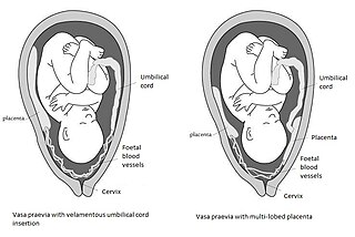
Vasa praevia is a condition in which fetal blood vessels cross or run near the internal opening of the uterus. These vessels are at risk of rupture when the supporting membranes rupture, as they are unsupported by the umbilical cord or placental tissue.

<span class="mw-page-title-main">Implantation (embryology)</span> First stage of pregnancy

Implantation, also known as nidation, is the stage in the embryonic development of mammals in which the blastocyst hatches, attaches, adheres, and invades into the wall of the female's uterus. Implantation is the first stage of gestation, and, when successful, the female is considered to be pregnant. An implanted embryo is detected by the presence of increased levels of human chorionic gonadotropin (hCG) in a pregnancy test. The implanted embryo will receive oxygen and nutrients in order to grow.

<span class="mw-page-title-main">Decidualization</span>

Decidualization is a process that results in significant changes to cells of the endometrium in preparation for, and during, pregnancy. This includes morphological and functional changes to endometrial stromal cells (ESCs), the presence of decidual white blood cells (leukocytes), and vascular changes to maternal arteries. The sum of these changes results in the endometrium changing into a structure called the decidua. In humans, the decidua is shed during childbirth.

<span class="mw-page-title-main">Uterine gland</span>

Uterine glands or endometrial glands are tubular glands, lined by a simple columnar epithelium, found in the functional layer of the endometrium that lines the uterus. Their appearance varies during the menstrual cycle. During the proliferative phase, uterine glands appear long due to estrogen secretion by the ovaries. During the secretory phase, the uterine glands become very coiled with wide lumens and produce a glycogen-rich secretion known as histotroph or uterine milk. This change corresponds with an increase in blood flow to spiral arteries due to increased progesterone secretion from the corpus luteum. During the pre-menstrual phase, progesterone secretion decreases as the corpus luteum degenerates, which results in decreased blood flow to the spiral arteries. The functional layer of the uterus containing the glands becomes necrotic, and eventually sloughs off during the menstrual phase of the cycle.

<i>Pseudemoia entrecasteauxii</i> Species of lizard

Pseudemoia entrecasteauxii, also known commonly as Entrecasteaux's skink, the southern grass skink, the tussock cool-skink, and the tussock skink, is a species of lizard in the family Scincidae. The species is endemic to Australia.

Immune tolerance in pregnancy or maternal immune tolerance is the immune tolerance shown towards the fetus and placenta during pregnancy. This tolerance counters the immune response that would normally result in the rejection of something foreign in the body, as can happen in cases of spontaneous abortion. It is studied within the field of reproductive immunology.

<span class="mw-page-title-main">Fetal membranes</span> Amnion and chorion which surround and protect a developing fetus

The fetal membranes are the four extraembryonic membranes, associated with the developing embryo, and fetus in humans and other mammals. They are the amnion, chorion, allantois, and yolk sac. The amnion and the chorion are the chorioamniotic membranes that make up the amniotic sac which surrounds and protects the embryo. The fetal membranes are four of six accessory organs developed by the conceptus that are not part of the embryo itself, the other two are the placenta, and the umbilical cord.

<span class="mw-page-title-main">Placental expulsion</span>

Placental expulsion occurs when the placenta comes out of the birth canal after childbirth. The period from just after the baby is expelled until just after the placenta is expelled is called the third stage of labor.

<span class="mw-page-title-main">Mammalian reproduction</span> Most mammals are viviparous, giving birth to live young

Most mammals are viviparous, giving birth to live young. However, the five species of monotreme, the platypuses and the echidnas, lay eggs. The monotremes have a sex determination system different from that of most other mammals. In particular, the sex chromosomes of a platypus are more like those of a chicken than those of a therian mammal.

<span class="mw-page-title-main">Pregnancy in fish</span>

Pregnancy has been traditionally defined as the period of time eggs are incubated in the body after the egg-sperm union. Although the term often refers to placental mammals, it has also been used in the titles of many international, peer-reviewed, scientific articles on fish, e.g. Consistent with this definition, there are several modes of reproduction in fish, providing different amounts of parental care. In ovoviviparity, there is internal fertilization and the young are born live but there is no placental connection or significant trophic (feeding) interaction; the mother's body maintains gas exchange but the unborn young are nourished by egg yolk. There are two types of viviparity in fish. In histotrophic viviparity, the zygotes develop in the female's oviducts, but she provides no direct nutrition; the embryos survive by eating her eggs or their unborn siblings. In hemotrophic viviparity, the zygotes are retained within the female and are provided with nutrients by her, often through some form of placenta.

References

  1. 1 2 3 4 5 Griffith, OW; Wagner, GP (2017). "The placenta as a model for understanding the origin and evolution of vertebrate organs". Nature Ecology and Evolution. 1 (4): 0072. Bibcode:2017NatEE...1...72G. doi:10.1038/s41559-017-0072. PMID   28812655. S2CID   32213223.
  2. Mossman, H. Comparative Morphogenesis of the Fetal Membranes and Accessory Uterine Structures Vol. 26 (Carnegie Institution of Washington, 1937).
  3. thefreedictionary.com > hemochorial placenta Citing: Dorland's Medical Dictionary for Health Consumers. Copyright 2007 by Saunders
  4. Elliot, M.; Crespi, B. (2006). "Placental invasiveness mediates the evolution of hybrid inviability in mammals". The American Naturalist. 168 (1): 114–120. doi:10.1086/505162. PMID   16874618. S2CID   16661549.
  5. Claim for guinea pigs, rabbits, mice, and rats taken from: Thornburg KL, Faber JJ (October 1976). "The steady state concentration gradients of an electron-dense marker (ferritin) in the three-layered hemochorial placenta of the rabbit". J. Clin. Invest. 58 (4): 912–25. doi:10.1172/JCI108544. PMC   333254 . PMID   965495.
  6. T.W. Sadler, Langman's Medical Embryology, 11th edition, Lippincott & Wilkins
  7. Blackburn, DG; Flemming, AF (2009). "Morphology, development, and evolution of fetal membranes and placentation in squamate reptiles". J. Exp. Zool. (Mol. Dev. Evol.). 312B (6): 579–589. Bibcode:2009JEZB..312..579B. doi: 10.1002/jez.b.21234 . PMID   18683170.
  8. Adams, S. M., Biazik, J. M., Thompson, M. B., & Murphy, C. R. (2005). Cyto‐epitheliochorial placenta of the viviparous lizard Pseudemoia entrecasteauxii: A new placental morphotype. Journal of morphology, 264(3), 264-276.Chicago
  9. Itonaga, K.; Wapstra, E.; Jones, S. M. (2012). "A novel pattern of placental leucine transfer during mid to late gestation in a highly placentotrophic viviparous lizard". Journal of Experimental Zoology Part B: Molecular and Developmental Evolution. 318 (4): 308–315. Bibcode:2012JEZB..318..308I. doi:10.1002/jez.b.22446. PMID   22821866.
  10. Griffith, O. W., Ujvari, B., Belov, K., & Thompson, M. B. (2013). Placental lipoprotein lipase (LPL) gene expression in a placentotrophic lizard, Pseudemoia entrecasteauxii. Journal of Experimental Zoology Part B: Molecular and Developmental Evolution.
  11. Murphy, C. R.; Hosie, M. J.; Thompson, M. B. (2000). "The plasma membrane transformation facilitates pregnancy in both reptiles and mammals". Comparative Biochemistry and Physiology A. 127 (4): 433–439. doi:10.1016/s1095-6433(00)00274-9. PMID   11154940.
  12. Blackburn, D. G., & Flemming, A. F. (2010). Reproductive specializations in a viviparous African skink: Implications for evolution and biological conservation.
  13. Griffith, O. W.; Van Dyke, J. U.; Thompson, M. B. (2013). "No implantation in an extra-uterine pregnancy of a placentotrophic reptile". Placenta. 34 (6): 510–511. doi:10.1016/j.placenta.2013.03.002. PMID   23522396.
  14. Griffith, OW; Blackburn, DG; Brandley, MC; Van Dyke, JU; Whittington, CW; Thompson, MB (2015). "Ancestral state reconstructions require biological evidence to test evolutionary hypotheses: A case study examining the evolution of reproductive mode in squamate reptiles". J Exp Zool B. 493 (6): 493–503. Bibcode:2015JEZB..324..493G. doi:10.1002/jez.b.22614. PMID   25732809.
  15. Griffith, Oliver (23 February 2017). "Using the placenta to understand how complex organs evolve". The Conversation. Retrieved 24 February 2017.
  16. 1 2 Griffith, OW; Brandley, MC; Belov, K; Thompson, MB (2016). "Reptile pregnancy is underpinned by complex changes in uterine gene expression: a comparative analysis of the uterine transcriptome in viviparous and oviparous lizards". Genome Biol Evol. 8 (10): 3226–3239. doi:10.1093/gbe/evw229. PMC   5174741 . PMID   27635053.
  17. 1 2 Griffith, OW; Brandley, MC; Whittington, CM; Belov, K; Thompson, MB (2016). "Comparative genomics of hormonal signaling in the chorioallantoic membrane of oviparous and viviparous amniotes". Gen Comp Endocrinol. 244: 19–29. doi:10.1016/j.ygcen.2016.04.017. PMID   27102939.
  18. Whittington, CW; Griffith, OW; Qi, W; Thompson, MB; Wilson, AB (2015). "Seahorse brood pouch transcriptome reveals common genes associated with vertebrate pregnancy". Mol Biol Evol. 32 (12): 3114–3131. doi: 10.1093/molbev/msv177 . hdl: 20.500.11850/110832 . PMID   26330546.
  19. Lynch, V. J.; Nnamani, M. C.; Kapusta, A.; Brayer, K.; Plaza, S. L.; Mazur, E. C.; Graf, A. (2015). "Ancient transposable elements transformed the uterine regulatory landscape and transcriptome during the evolution of mammalian pregnancy". Cell Reports. 10 (4): 551–561. doi:10.1016/j.celrep.2014.12.052. PMC   4447085 . PMID   25640180.
  20. Griffith, OW; Ujvari, B; Belov, K; Thompson, MB (2013). "Placental lipoprotein lipase (LPL) gene expression in a placentotrophic lizard, Pseudemoia entrecasteauxii". J Exp Zool B. 320 (7): 465–470. doi:10.1002/jez.b.22526. PMID   23939756.
  21. "Flowers" At: Botany Online At: University of Hamburg Department of Biology. (see External links below).