Related Research Articles

Magnetic resonance imaging (MRI) is a medical imaging technique used in radiology to form pictures of the anatomy and the physiological processes of the body. MRI scanners use strong magnetic fields, magnetic field gradients, and radio waves to generate images of the organs in the body. MRI does not involve X-rays or the use of ionizing radiation, which distinguishes it from computed tomography (CT) and positron emission tomography (PET) scans. MRI is a medical application of nuclear magnetic resonance (NMR) which can also be used for imaging in other NMR applications, such as NMR spectroscopy.
Technological applications of superconductivity include:
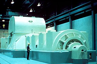
In electricity generation, a generator is a device that converts motion-based power or fuel-based power into electric power for use in an external circuit. Sources of mechanical energy include steam turbines, gas turbines, water turbines, internal combustion engines, wind turbines and even hand cranks. The first electromagnetic generator, the Faraday disk, was invented in 1831 by British scientist Michael Faraday. Generators provide nearly all the power for electrical grids.

Oxford Instruments plc is a United Kingdom manufacturing and research company that designs and manufactures tools and systems for industry and research. The company is headquartered in Abingdon, Oxfordshire, England, with sites in the United Kingdom, United States, Europe, and Asia. It is listed on the London Stock Exchange and is a constituent of the FTSE 250 Index.
The tesla is the unit of magnetic flux density in the International System of Units (SI).
Siemens Healthineers is a German company which provides healthcare solutions and services. It was spun off from its parent company Siemens in 2017, which retains a 75% stake. Siemens Healthineers is the parent company for several medical technology companies and is headquartered in Erlangen, Germany.

Interventional magnetic resonance imaging, also interventional MRI or IMRI, is the use of magnetic resonance imaging (MRI) to do interventional radiology procedures.

A dynamo is an electrical generator that creates direct current using a commutator. Dynamos were the first electrical generators capable of delivering power for industry, and the foundation upon which many other later electric-power conversion devices were based, including the electric motor, the alternating-current alternator, and the rotary converter.
Nuclear magnetic resonance (NMR) in the geomagnetic field is conventionally referred to as Earth's field NMR (EFNMR). EFNMR is a special case of low field NMR.
The Wolfson Brain Imaging Centre (WBIC) is a UK Biomedical Imaging Centre, located at Addenbrooke's Hospital, Cambridge, England, on the Cambridge Bio-Medical Campus at the southwestern end of Hills Road. It is a division of the Department of Clinical Neurosciences of the University of Cambridge.

Nuclear magnetic resonance (NMR) is a physical phenomenon in which nuclei in a strong constant magnetic field are perturbed by a weak oscillating magnetic field and respond by producing an electromagnetic signal with a frequency characteristic of the magnetic field at the nucleus. This process occurs near resonance, when the oscillation frequency matches the intrinsic frequency of the nuclei, which depends on the strength of the static magnetic field, the chemical environment, and the magnetic properties of the isotope involved; in practical applications with static magnetic fields up to ca. 20 tesla, the frequency is similar to VHF and UHF television broadcasts (60–1000 MHz). NMR results from specific magnetic properties of certain atomic nuclei. Nuclear magnetic resonance spectroscopy is widely used to determine the structure of organic molecules in solution and study molecular physics and crystals as well as non-crystalline materials. NMR is also routinely used in advanced medical imaging techniques, such as in magnetic resonance imaging (MRI).
Radiofrequency coils are the receivers, and sometimes also the transmitters, of radiofrequency (RF) signals in equipment used in magnetic resonance imaging (MRI).

The physics of magnetic resonance imaging (MRI) concerns fundamental physical considerations of MRI techniques and technological aspects of MRI devices. MRI is a medical imaging technique mostly used in radiology and nuclear medicine in order to investigate the anatomy and physiology of the body, and to detect pathologies including tumors, inflammation, neurological conditions such as stroke, disorders of muscles and joints, and abnormalities in the heart and blood vessels among others. Contrast agents may be injected intravenously or into a joint to enhance the image and facilitate diagnosis. Unlike CT and X-ray, MRI uses no ionizing radiation and is, therefore, a safe procedure suitable for diagnosis in children and repeated runs. Patients with specific non-ferromagnetic metal implants, cochlear implants, and cardiac pacemakers nowadays may also have an MRI in spite of effects of the strong magnetic fields. This does not apply on older devices, and details for medical professionals are provided by the device's manufacturer.
A shim is a device used to adjust the homogeneity of a magnetic field. Shims received their name from the purely mechanical shims used to adjust position and parallelity of the pole faces of an electromagnet. Coils used to adjust the homogeneity of a magnetic field by changing the current flowing through it were called "electrical current shims" because of their similar function.
Intraoperative magnetic resonance imaging (iMRI) refers to an operating room configuration that enables surgeons to image the patient via an MRI scanner while the patient is undergoing surgery, particularly brain surgery. iMRI reduces the risk of damaging critical parts of the brain and helps confirm that the surgery was successful or if additional resection is needed before the patient's head is closed and the surgery completed.
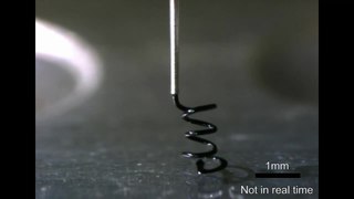
A microcoil is a tiny electrical conductor such as a wire in the shape of a spiral or helix which could be a solenoid or a planar structure.
Synthetic MRI is a simulation method in Magnetic Resonance Imaging (MRI), for generating contrast weighted images based on measurement of tissue properties. The synthetic (simulated) images are generated after an MR study, from parametric maps of tissue properties. It is thereby possible to generate several contrast weightings from the same acquisition. This is different from conventional MRI, where the signal acquired from the tissue is used to generate an image directly, often generating only one contrast weighting per acquisition. The synthetic images are similar in appearance to those normally acquired with an MRI scanner.

In a medical facility, such as a hospital or clinic, a gantry holds radiation detectors and/or a radiation source used to diagnose or treat a patient's illness. Radiation sources may produce gamma radiation, x-rays, electromagnetic radiation, or magnetic fields depending on the purpose of the device.

Magnetic resonance imaging (MRI) is in general a safe technique, although injuries may occur as a result of failed safety procedures or human error. During the last 150 years, thousands of papers focusing on the effects or side effects of magnetic or radiofrequency fields have been published. They can be categorized as incidental and physiological. Contraindications to MRI include most cochlear implants and cardiac pacemakers, shrapnel and metallic foreign bodies in the eyes. The safety of MRI during the first trimester of pregnancy is uncertain, but it may be preferable to other options. Since MRI does not use any ionizing radiation, its use generally is favored in preference to CT when either modality could yield the same information.
References
- ↑ Mitra, Esha (2018-06-08). "Tata Trusts develops portable MRI scanner". The Hindu . India. Retrieved 2019-12-12.
- ↑ "Hyperfine and Yale School of Medicine collaborate on world's first portable MRI technology". EurekAlert!. Washington, DC, U.S.: AAAS news release . Retrieved 2019-12-12.
- ↑ Ren, Zhi Hua; Obruchkov, Sergei; Lu, Dong; Dykstra, Robin; Huang, Shao Ying (2017-11-01). "A low-field portable magnetic resonance imaging system for head imaging". 2017 Progress in Electromagnetics Research Symposium - Fall (PIERS - FALL). pp. 3042–3044. doi:10.1109/PIERS-FALL.2017.8293655. ISBN 978-1-5386-1211-8. S2CID 3400077 . Retrieved 2019-12-12– via Researchgate.
- ↑ "Mobile MRI Scanner". www.siemens-healthineers.com. Archived from the original on 2019-12-12.
- ↑ Sarracanie M, LaPierre CD, Salameh N, Waddington DEJ, Witzel T & Rosen MS, Low-Cost High-Performance MRI. Sci Rep 5, 15177 (2015). https://doi.org/10.1038/srep15177
- ↑ Tsai LL, Mair RW, Rosen MS, Patz S, Walsworth RL. An open-access, very-low-field MRI system for posture-dependent 3He human lung imaging. J Magn Reson. 2008 Aug;193(2):274-85. https://doi.org/10.1016/j.jmr.2008.05.016. Epub 2008 May 24. PMID 18550402; PMCID: PMC2572034
- ↑ "Terranova | Magritek". Archived from the original on 2014-07-20.
- ↑ S. Y. Huang, Z. H. REN, S. OBRUCHKOV, J. GONG, R. DYKSTRA, W. YU, " Portable Low-cost MRI System based on Permanent Magnets/Magnet Arrays", Investigative Magnetic Resonance Imaging(2019), 23 (3):179, http://dx.doi.org/10.13104/imri.2019.23.3.179
- ↑ "Magnetic Resonance Imaging". www.siemens-healthineers.com. Munich , Germany: Siemens Healthineers . Retrieved 2019-12-12.
- ↑ O-scan, Esaote
- ↑ Terada, Yasuhiko, et al. "Improved reliability in skeletal age assessment using a pediatric hand MR scanner with a 0.3 T permanent magnet." Magnetic Resonance in Medical Sciences 13.3 (2014): 215–219
- ↑ J. McGinley, et al, ISMRM 2018 0944
- ↑ E. Esparza-Coss and D. Cole, “A Low Cost MR/Permanent Magnet Prototype,” Second Mexican Symp on Med Physics. American Institute of Physics Conf. Proc., 1998.
- ↑ J. Gong, S. Y. Huang, Z. H. Ren, and Wenwei Yu, “Effects of Encoding Fields of Permanent Magnet Arrays on Image Quality in Low-field Portable MRI Systems”, IEEE Access, vol. 7, pp. 80310-80327, 2019, doi: 10.1109/ACCESS.2019.2923118
- ↑ K. Halbach, Nuclear instruments and methods, vol. 169, no. 1, pp. 1–10, 1980.
- ↑ Cooley, C. Z.et al. Magn. Resonance Medicine73, 872–883 (2015)
- ↑ Z. H. Ren, W. C. Mu, and S.Y.Huang, “Design and Optimization of a Ring-Pair Permanent Magnet Array for Head Imaging in a Low-field Portable MRI System”, IEEE Transactions on Magnetics, Volume 55, Issue 1, Jan. 2019
- ↑ Z. H. Ren, J Gong, and S.Y.Huang, “An Irregular-shaped Ring-Pair Magnet Array with a Linear Field Gradient for 2D Head Imaging in Low-field Portable MRI”, IEEE Access 7, 48715-48724, 2019
- 1 2 T-O LIANG, Y. H. KOH, T. QIU, E. LI, W. YU, and S. Y. HUANG, “High-Performance Permanent Magnet Array Design by a Fast Genetic Algorithm (GA)-based Optimization for Low-Field Portable MRI,” Journal of Magnetic Resonance (JMR), Vol. 345, Dec. 2022, 107309, https://doi.org/10.1016/j.jmr.2022.107309
- ↑ T-O LIANG, Y. H. KOH, T. QIU, E. LI, W. YU, and S. Y. HUANG, “MagTetris: A Simulator for Fast Magnetic Field and Force Calculation for Permanent Magnet Array Designs,” Volume 352, July 2023, 107463, https://doi.org/10.1016/j.jmr.2023.107463