Related Research Articles

Skin is the layer of usually soft, flexible outer tissue covering the body of a vertebrate animal, with three main functions: protection, regulation, and sensation.

In male human anatomy, the glans penis or penile glans, commonly referred to as the glans, is the bulbous structure at the distal end of the human penis that is the human male's most sensitive erogenous zone and primary anatomical source of sexual pleasure. The glans penis is present in the male reproductive organs of humans and most other mammals where it may appear smooth, spiny, elongated or divided. It is externally lined with mucosal tissue, which creates a smooth texture and glossy appearance. In humans, the glans is located over the distal ends of the corpora cavernosa and is a continuation of the corpus spongiosum of the penis. At the summit appears the urinary meatus and at the base forms the corona glandis. An elastic band of tissue, known as the frenulum, runs on its ventral surface. In men who are not circumcised, it is completely or partially covered by a fold of skin called the foreskin. In adults, the foreskin can generally be retracted over and past the glans manually or sometimes automatically during an erection.

Pre-ejaculate is a clear, colorless, viscous fluid that is emitted from the urethra of the penis during sexual arousal. It is similar in composition to semen but has distinct chemical differences. The presence of sperm in the fluid is variable from low to absent. Pre-ejaculate functions as a lubricant and an acid neutralizer.
Smegma is a combination of shed skin cells, skin oils, and moisture. It occurs in both male and female mammalian genitalia. In females, it collects around the clitoris and in the folds of the labia minora; in males, smegma collects under the foreskin.

The bulbourethral glands or Cowper's glands are two small exocrine and accessory glands in the reproductive system of many male mammals. They are homologous to Bartholin's glands in females. The bulbourethral glands are responsible for producing a pre-ejaculate fluid called Cowper's fluid, which is secreted during sexual arousal, neutralizing the acidity of the urethra in preparation for the passage of sperm cells. The paired glands are found adjacent to the urethra just below the prostate, seen best by screening (medicine) MRI as a tool in preventative healthcare in males. Screening MRI may be performed when there is a positive prostate-specific antigen on basic laboratory tests. Prostate cancer is the second-most common cause of cancer-related mortality in males in the USA.

Phimosis is a condition in which the foreskin of the penis cannot stretch to allow it to be pulled back past the glans. A balloon-like swelling under the foreskin may occur with urination. In teenagers and adults, it may result in pain during an erection, but is otherwise not painful. Those affected are at greater risk of inflammation of the glans, known as balanitis, and other complications.
The preputial mucosa of the penis is the epithelium of the inside of the prepuce, or foreskin. To differentiate it from the cutaneous skin of the outside of the prepuce, it is sometimes referred to as the inner mucosa. It starts at the ridged band of the prepuce and continues to the coronal sulcus, where it meets the epithelium of the glans and penile shaft. The preputial mucosa is devoid of hair, as is the cutaneous surface.

A sebaceous gland or oil gland is a microscopic exocrine gland in the skin that opens into a hair follicle to secrete an oily or waxy matter, called sebum, which lubricates the hair and skin of mammals. In humans, sebaceous glands occur in the greatest number on the face and scalp, but also on all parts of the skin except the palms of the hands and soles of the feet. In the eyelids, meibomian glands, also called tarsal glands, are a type of sebaceous gland that secrete a special type of sebum into tears. Surrounding the female nipple, areolar glands are specialized sebaceous glands for lubricating the nipple. Fordyce spots are benign, visible, sebaceous glands found usually on the lips, gums and inner cheeks, and genitals.

Sweat glands, also known as sudoriferous or sudoriparous glands, from Latin sudor 'sweat', are small tubular structures of the skin that produce sweat. Sweat glands are a type of exocrine gland, which are glands that produce and secrete substances onto an epithelial surface by way of a duct. There are two main types of sweat glands that differ in their structure, function, secretory product, mechanism of excretion, anatomic distribution, and distribution across species:
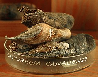
Castoreum is a yellowish exudate from the castor sacs of mature beavers. Beavers use castoreum in combination with urine to scent mark their territory. Both beaver sexes have a pair of castor sacs and a pair of anal glands, located in two cavities under the skin between the pelvis and the base of the tail. The castor sacs are not true glands on a cellular level, hence references to these structures as preputial glands, castor glands, or scent glands are misnomers.

Scent gland are exocrine glands found in most mammals. They produce semi-viscous secretions which contain pheromones and other semiochemical compounds. These odor-messengers indicate information such as status, territorial marking, mood, and sexual behaviour. The odor may be subliminal—not consciously detectable. Though it is not their primary function, the salivary glands may also function as scent glands in some animals.

Pearly penile papules are benign, small bumps or spots on the human penis. They vary in size from 1–4 mm, are pearly or flesh-colored, smooth and dome-topped or filiform, and appear in one or, several rows around the corona, the ridge of the head of the penis and sometimes on the penile shaft. They are painless, non-cancerous and not harmful. The medical condition of having such papules is called hirsutoid papillomatosis or hirsuties papillaris coronae glandis.

The frenulum of the penis, often known simply as the frenulum or frenum, is a thin elastic strip of tissue on the underside of the glans and the neck of the human penis. In men who are not circumcised, it also connects the foreskin to the glans and the ventral mucosa. In adults, the frenulum is typically supple enough to allow manual movement of the foreskin over the glans and help retract the foreskin during erection. In flaccid state, it tightens to narrow the foreskin opening.

Melanocortin 5 receptor (MC5R) is a protein that in humans is encoded by the MC5R gene. It is located on the chromosome 18 in the human genome. When the MC5R was disrupted in transgenic mice, it induced disruption of their exocrine glands and resulted in decreased production of sebum.

The corona of glans penis or penis crown refers to the rounded projecting border or flare that forms at the base of the glans in human males. The corona overhangs a mucosal surface, known as the neck of the penis, which separates the shaft and the glans. The deep retro-glandular coronal sulcus forms between the corona and the neck of the penis. The two sides of the corona merge on the ventral midline forming the septum glandis. The circumference of the corona is richly innervated and is described as a highly erogenous area of the glans.

In male human anatomy, the foreskin, also known as the prepuce, is the double-layered fold of skin, mucosal and muscular tissue at the distal end of the human penis that covers the glans and the urinary meatus. The foreskin is attached to the glans by an elastic band of tissue, known as the frenulum. The outer skin of the foreskin meets with the inner preputial mucosa at the area of the mucocutaneous junction. The foreskin is mobile, fairly stretchable and sustains the glans in a moist environment. Except for humans, a similar structure known as a penile sheath appears in the male sexual organs of all primates and the vast majority of mammals.

The preorbital gland is a paired exocrine gland found in many species of artiodactyls, which is homologous to the lacrimal gland found in humans. These glands are trenchlike slits of dark blue to black, nearly bare skin extending from the medial canthus of each eye. They are lined by a combination of sebaceous and sudoriferous glands, and they produce secretions which contain pheromones and other semiochemical compounds. Ungulates frequently deposit these secretions on twigs and grass as a means of communication with other animals.
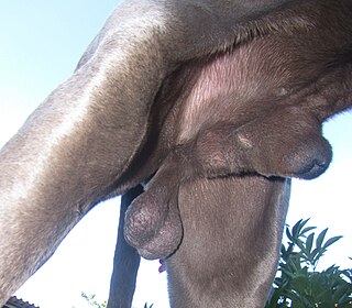
Almost all mammal penises have foreskins or prepuces, although in non-human cases, the foreskin is usually a sheath into which the whole penis is retracted. In koalas, the foreskin contains naturally occurring bacteria that play an important role in fertilization. In some bat species, the prepuce contains an erectile tissue structure called the accessory corpus cavernosus.

The septum glandis, also septum of the glans, refers to the fibrous partition of the ventral aspect of the glans penis that separates the two glans wings in the ventral midline. The septum extends from the urethral meatus through the glanular urethra and ends in the tunica albuginea of the human penis. Externally it is attached to the frenulum which extends lower on the neck of the penis.

Wolves communicate using vocalizations, body postures, scent, touch, and taste. The lunar phases have no effect on wolf vocalisation. Despite popular belief, wolves do not howl at the Moon. Gray wolves howl to assemble the pack, usually before and after hunts, to pass on an alarm particularly at a den site, to locate each other during a storm or while crossing unfamiliar territory, and to communicate across great distances. Other vocalisations include growls, barks and whines. Wolves do not bark as loudly or continuously as dogs do but they bark a few times and then retreat from a perceived danger. Aggressive or self-assertive wolves are characterized by their slow and deliberate movements, high body posture and raised hackles, while submissive ones carry their bodies low, sleeken their fur, and lower their ears and tail. Raised leg urination is considered to be one of the most important forms of scent communication in the wolf, making up 60–80% of all scent marks observed.
References
- 1 2 Mech, L. David; Boitani, Luigi, eds. (2003). Wolves: Behaviour, Ecology and Conservation. University of Chicago Press. ISBN 0-226-51696-2.
- ↑ Martin-Alguacil N, Schober J, Kow LM, Pfaff D (December 2008). "Oestrogen receptor expression and neuronal nitric oxide synthase in the clitoris and preputial gland structures of mice". BJU Int. 102 (11): 1719–23. doi: 10.1111/j.1464-410X.2008.07989.x . PMID 18793302.
- ↑ Clapperton, B. Kay; Fordham, R. A.; Sparksman, R. I. (1987). "Preputial glands of the ferret Mustela furo (Carnivora: Mustelidae)". Journal of Zoology. 212 (2): 356–361. doi:10.1111/j.1469-7998.1987.tb05998.x.
- ↑ Cave, A. J. E. "The preputial glands of ceratotherium." Mammalia 30.1 (1966): 153-159.
- ↑ Odend'hal, Stewart; Miller, Karl V.; Demarais, Stephen (1996). "Preputial glands in Artiodactyla" (PDF). Journal of Mammalogy. 77 (2): 417–421. doi: 10.2307/1382818 . JSTOR 1382818.
- ↑ Van Heerden, Joseph. "The role of integumental glands in the social and mating behaviour of the hunting dog Lycaon pictus (Temminck, 1820)." (1981).
- ↑ "Perfumes combine hundreds of ingredients to create 1 fragrance". The Galveston Daily News . Vol. 149, no. 144. AP. September 1, 1991. p. 44. Retrieved 5 December 2015.
- ↑ Kruger L (December 2003). "Edward Tyson's 1680 account of the 'porpess' brain and its place in the history of comparative neurology". Journal of the History of the Neurosciences . 12 (4): 339–49. doi:10.1076/jhin.12.4.339.27915. PMID 15069865. S2CID 23830465.
- ↑ Cowper, W (1724) [1694], Myotomia reformata; or, An anatomical treatise on the muscles of the human body, London
- ↑ Tyson's gland at Who Named It?
- ↑ Batistatou, Anna; Panelos, John; Zioga, Aikaterini; Charalabopoulos, Konstantinos A. (October 2006). "Ectopic Modified Sebaceous Glands in Human Penis". International Journal of Surgical Pathology . 14 (4): 355–6. doi:10.1177/1066896906291779. PMID 17041207. S2CID 24295783.
- ↑ Parkash, Satya; S. Jeyakumar; K. Subramanyan; S. Chaudhuri (August 1973). "Human subpreputial collection: its nature and formation". The Journal of Urology . 110 (2): 211–212. doi:10.1016/s0022-5347(17)60164-2. PMID 4722614.
- ↑ "Surgical Temptation: A chance to cut, a chance to cure? | National Sexuality Resource Center (NSRC)". Archived from the original on April 30, 2009. Retrieved 2009-11-29.
- ↑ Hyman AB, Brownstein MH (January 1969). "Tyson's "glands." Ectopic sebaceous glands and papillomatosis penis". Arch Dermatol. 99 (1): 31–6. doi:10.1001/archderm.99.1.31. PMID 5761803.[ permanent dead link ]
- ↑ Parkash S, Rao R, Venkatesan K, Ramakrishnan S. Sub-preputial wetness: its nature. Annals of National Medical Science (India) 1982; 18: 109-12