
In human anatomy, the arm refers to the upper limb in common usage, although academically the term specifically means the upper arm between the glenohumeral joint and the elbow joint. The distal part of the upper limb between the elbow and the radiocarpal joint is known as the forearm or "lower" arm, and the extremity beyond the wrist is the hand.

The brachial artery is the major blood vessel of the (upper) arm. It is the continuation of the axillary artery beyond the lower margin of teres major muscle. It continues down the ventral surface of the arm until it reaches the cubital fossa at the elbow. It then divides into the radial and ulnar arteries which run down the forearm. In some individuals, the bifurcation occurs much earlier and the ulnar and radial arteries extend through the upper arm. The pulse of the brachial artery is palpable on the anterior aspect of the elbow, medial to the tendon of the biceps, and, with the use of a stethoscope and sphygmomanometer, often used to measure the blood pressure.
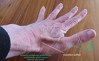
The anatomical snuff box or snuffbox or foveola radialis is a triangular deepening on the radial, dorsal aspect of the hand—at the level of the carpal bones, specifically, the scaphoid and trapezium bones forming the floor. The name originates from the use of this surface for placing and then sniffing powdered tobacco, or "snuff." It is sometimes referred to by its French name tabatière.
The forearm is the region of the upper limb between the elbow and the wrist. The term forearm is used in anatomy to distinguish it from the arm, a word which is used to describe the entire appendage of the upper limb, but which in anatomy, technically, means only the region of the upper arm, whereas the lower "arm" is called the forearm. It is homologous to the region of the leg that lies between the knee and the ankle joints, the crus.

The femoral artery is a large artery in the thigh and the main arterial supply to the thigh and leg. The femoral artery gives off the deep femoral artery and descends along the anteromedial part of the thigh in the femoral triangle. It enters and passes through the adductor canal, and becomes the popliteal artery as it passes through the adductor hiatus in the adductor magnus near the junction of the middle and distal thirds of the thigh.

In human anatomy, the radial artery is the main artery of the lateral aspect of the forearm.
In medicine, Allen's test or the Allen test is a medical sign used in physical examination of arterial blood flow to the hands. It was named for Edgar Van Nuys Allen, who described the original version of the test in 1942.

An arterial line is a thin catheter inserted into an artery.

The cubital fossa, chelidon or inside of elbow is the area on the anterior side of the upper part between the arm and forearm of a human or other hormid animals. It lies anteriorly to the elbow when in standard anatomical position.
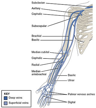
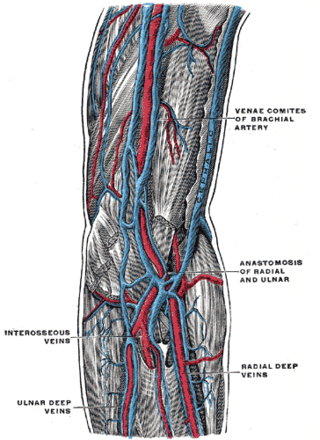
In human anatomy, the brachial veins are venae comitantes of the brachial artery in the arm proper. Because they are deep to muscle, they are considered deep veins. Their course is that of the brachial artery : they begin where radial veins and ulnar veins join. They end at the inferior border of the teres major muscle. At this point, the brachial veins join the basilic vein to form the axillary vein.
The ulnar artery is the main blood vessel, with oxygenated blood, of the medial aspects of the forearm. It arises from the brachial artery and terminates in the superficial palmar arch, which joins with the superficial branch of the radial artery. It is palpable on the anterior and medial aspect of the wrist.

In human anatomy, the internal thoracic artery (ITA), also known as the internal mammary artery, is an artery that supplies the anterior chest wall and the breasts. It is a paired artery, with one running along each side of the sternum, to continue after its bifurcation as the superior epigastric and musculophrenic arteries.
The ulnar veins are venae comitantes of the ulnar artery. They drain the superficial venous palmar arch. They arise in the hand and terminate by uniting with the radial veins to form the brachial veins. They mostly drain the medial aspect of the forearm. They receive the venae comitantes of the anterior and posterior interosseous arteries near the elbow, as well as a large branch from the median cubital vein. The ulnar veins are larger than the radial veins.

In anatomy, the left and right common carotid arteries (carotids) are arteries that supply the head and neck with oxygenated blood; they divide in the neck to form the external and internal carotid arteries.
Vena comitans is Latin for accompanying vein and is also known as a satellite vein. It refers to a vein that is usually paired, with both veins lying on the sides of an artery. Because they are generally found in pairs, they are often referred to by their plural form: venae comitantes.

Cortical radial arteries, formerly known as interlobular arteries, are renal blood vessels given off at right angles from the side of the arcuate arteries looking toward the cortical substance. The interlobular arteries pass directly outward between the medullary rays to reach the fibrous tunic, where they end in the capillary network of this part.

The arcuate vessels of the uterus are a component of the blood supply of the uterus. They are arteries and veins that branch from the uterine arteries and veins, respectively, with additional anastomoses from the ovarian arteries and veins, and penetrate and assume a circumferential course in the myometrium.

Anterior spinal veins are veins that receive blood from the anterior spinal cord.

The following outline is provided as an overview of and topical guide to human anatomy:

