Reflectron

Last updated
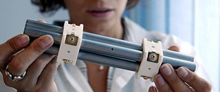
An ion mirror (right) attached to a flight tube (left) of the reflectron. Voltages applied to a stack of metal plates create the electric field reflecting the ions back to the flight tube. Reflectron.jpg
An ion mirror (right) attached to a flight tube (left) of the reflectron. Voltages applied to a stack of metal plates create the electric field reflecting the ions back to the flight tube.

A reflectron (mass reflectron) is a type of time-of-flight mass spectrometer (TOF MS) that comprises a pulsed ion source, field-free region, ion mirror, and ion detector and uses a static or time dependent electric field in the ion mirror to reverse the direction of travel of the ions entering it. Using the reflectron, one can substantially diminish a spread of flight times of the ions with the same mass-to-charge ratio (m/z) caused by spread in kinetic energy of these ions measured at the exit from the ion source.

Contents

Development

In the reflection, the higher energy ion (red) takes a longer path but arrives at the detector at the same time as the lower energy ion (blue) of the same mass. Reflectron schematic.gif
In the reflection, the higher energy ion (red) takes a longer path but arrives at the detector at the same time as the lower energy ion (blue) of the same mass.

The idea of improving mass resolution in TOF MS by implementing the reflection of ions from a region with retarding electric field (the ion mirror) has been first proposed by Russian scientist S. G. Alikhanov. [1] In 1973, the dual-stage reflectron utilizing an ion mirror with two regions of homogeneous field was built in a laboratory of Boris Aleksandrovich Mamyrin. [2] [3] Mass resolution of the reflectron measured over broad mass range is much larger than that in a simpler (so-called linear) time-of-flight mass spectrometer comprising a pulsed ion source, flight tube, and ion detector. The masses of ions analyzed in the reflectron can span from a few daltons to a few million daltons. Sensitivity in the reflectron used for the analysis of ions produced in vacuum by photo or electron ionization, e.g., matrix-assisted laser desorption/ionization source, can be lower than in linear TOF MS due to post-source decay - a dissociation of vibrationally-excited molecular ions (often referred as metastable ions).

Single-stage reflectron

Schematic drawing of a single-stage reflectron. Reflectron ss.gif
Schematic drawing of a single-stage reflectron.

A single-stage reflectron is equipped with an ion mirror that has a single electric field region. The distribution of electric potential along the central axis of the ion mirror can be linear or non-linear. Also, the electric field in the mirror can be constant or time-dependent. In single-stage reflectrons with homogeneous field, a zero field in a field-free region of a flight tube and the homogeneous field inside the ion mirror are separated by highly transparent (~95%) metal grid. The grid position is then referred as the entrance (exit) to the ion mirror and is used to calculate the retarding electric field. The single-stage reflector utilizing homogeneous field can be used to attain high mass resolution in cases where the variation of energies of ions leaving the ion source is small (typically less than a few per cent). Time of flight t of the ions with mass m, charge q, kinetic energy U is

where L is the path length of the ions in a field-free space, Lm is the length of ion mirror, Um is the voltage applied across the mirror. To find a first-order compensation condition for flight time t with respect to spread dU in ion energy U, the following condition should be fulfilled

Assume that the kinetic energy of the ions in the field-free region equals the ion potential energy near the stop point of the ions inside the mirror (we assume that this stop point is very close to the back electrode of the mirror, i.e. Um = U). From here it follows that

In practice, the mirror length should be 10-20% longer to accommodate all ions whose kinetic energy is spread over some interval.

So, the electric field Em in the mirror of a single-stage reflector should be

In case of a wider variation of dU, the relative width of the time-of-flight peaks dt/t in such a reflectron is determined by the uncompensated part of the flight time t(U) proportional to the second derivative

.

where k is a constant depending on the parameters of the single-stage reflector.

Dual-stage reflectron

Schematic drawing of an ion mirror with high- and low-field regions (dual-stage reflectron). Reflectron ds.gif
Schematic drawing of an ion mirror with high- and low-field regions (dual-stage reflectron).

The mirror in a dual-stage reflectron has two regions (stages) with different fields. This makes it possible to zero both the first and second derivatives of t(U) with respect to energy U. That is why dual-stage reflectrons can compensate flight times over larger variations in ion kinetic energy compared to single-stage ones. This type of reflectrons is typically employed in orthogonal acceleration (oa) TOF MS. "Classical" (i.e., Mamyrin's) design includes two highly transparent conductive grids separating regions with homogeneous fields. In general, the first stage (section) of the reflectron has high electric field, in this section the ions decelerate losing 2/3 or more of their kinetic energy depending on the reflectron parameters; [4] the second stage has lower field, in this stage the ions are repelled toward the first region. Mass resolution in dual-stage reflectron is mainly determined by ion scattering on the grids, [5] the spread of kinetic energy of ions leaving the pulsed ion source, and accuracy of mechanical alignment. To diminish effect of scattering, the length of the first deceleration region should be relatively large. Ion scattering makes using triple- and further stage reflectrons impractical.

The effect of ion scattering on mass resolution in single- and dual-stage reflectrons can be diminished by utilizing polarized grid geometry. [6]

Gridless reflectron

The gridless design of the reflectron usually comprises two stages with individually adjustable voltages: a decelerating region, where the ions lose about two-thirds of their kinetic energy, and repelling region, where the ions reverse their direction of motion. The symmetry of gridless reflectron is typically a cylindrical one, though a 2D design comprising two parallel flat electrode systems can be utilized for the same purpose of flight-time compensation of energy spread the ions acquire at the exit from the ion source. [7] The gridless reflectron almost always includes a thick electrostatic Einzel lens placed at its front or some distance. The curved potential distribution in a gridless reflectron geometrically affects the trajectories of reflected ions and therefore the gridless reflectron either focus or defocus the ions, which depends on a chosen field profile. In addition, one needs to take into account that the lensing also affects the time-of-flight of ions traversing different sections of the reflectron. Due to the positive voltages in the reflectron with respect to that applied to the field-free drift region (this region is often kept at the ground potential), the reflectron entrance acts like the first half of a "positive" electrostatic lens (Einzel lens where the central electrode is kept at a positive potential with respect to two outer electrodes) causing the ion beam to diverge when entering the reflectron. A positive (decelerating) lens affects the ion flight times as well as the spread of ion flight times (on-axis vs. off-axis ions) more strongly than a negative (accelerating) lens does under similar focusing conditions because in positive Einzel lens the ions are moving along the extended (i.e., more lengthy) off-axis trajectories at lower ion energies. To minimize the positive lens effect produced by the gridless reflectron, one must add a negative Einzel lens near the reflectron exit, which conducts the geometric focusing, i.e., directs the converging ion beam toward the ion detector and compensate for the flight time spread. The reflectron with the negative Einzel lens placed near its exit is sometimes referred as the Frey mirror. [8] As earlier as 1985, Frey et al. [9] reported on the gridless reflector that demonstrated mass resolution over 10,000 while mass analyzing the laser-ablated plumes that exhibited 3.3% kinetic energy spread at the exit of the ion source. In the 1980s, there were suggested several approaches to a design of gridless reflectrons, mainly aimed at finding the middle ground between higher transmission (i.e., directing a significant per cent of exiting ions toward the ion detector) and target mass resolution. [10] [11]

One implementation of gridless reflectron utilizes a curved field where the electric potential V(x) along the mirror axis depends non-linearly on distance x to the mirror entrance. Time of flight compensation for ions with different kinetic energy can be obtained by adjusting voltage on the elements producing the electric field inside the mirror, which values follow the equation of an arc of a circle: R2 = V(x)2 + kx2, where k and R are some constants. [12] [13]

The electric potential in some other implementation of gridless reflectron (a so-called quadratic-field reflectron) is proportional to a square of a distance x to the mirror entrance: V(x)=kx2 thus exhibiting a case of one-dimensional harmonic field. If both the ion source and the detector are placed at the reflectron entrance and if the ions travel in a close proximity of the ion mirror axis, the flight times of ions in the quadratic-field reflectron are almost independent on ion kinetic energy. [14]

A gridless reflectron with nonlinear field, which comprised only three cylindrical elements was also demonstrated. [15] Bergmann et al. implemented an original numerical approach to finding voltage distribution across the stack of the metal electrodes to create a nonlinear field in different regions of the reflectron to provide conditions for both geometrical focusing and compensation of flight times caused by the spread of kinetic energies of ions entering the reflectron at different angles. [16]

Post-source decay

A post-source decay (PSD) is a process specific to the ion source utilizing matrix-assisted laser desorption/ionization and operating in vacuum. In the post-source decay, parent ions (typically of several keV kinetic energy) fragment in a process of laser-induced fragmentation or high-energy collision-induced dissociation (HE CID). Time interval suitable for observation of the post-source decay in the reflectron starts after the precursors (parent ions) leave the ion source and ends prior to the moment when the precursors enter the ion mirror. [17] The kinetic energy of fragment ions of mass m in the post-source decay significantly differs from that of parent ions of mass M and is proportional to m/M. So, the distribution of kinetic energies for the PSD ions is extremely large. Not surprisingly, it cannot be compensated in "classic" single or double-stage reflectrons. To achieve acceptable mass resolution for PSD ions with masses typically distributed over broad mass range, these ions are accelerated to energies substantially (at least, a factor of 4 [18] ) exceeding the initial energy of precursor ions. Use of gridless curved-field mirror or that with time-dependent field also improves the mass resolution for fragment ions generated in the post-source decay.

Related Research Articles

<span class="mw-page-title-main">Atom probe</span> Field ion microscope coupled with a mass spectrometer

The atom probe was introduced at the 14th Field Emission Symposium in 1967 by Erwin Wilhelm Müller and J. A. Panitz. It combined a field ion microscope with a mass spectrometer having a single particle detection capability and, for the first time, an instrument could “... determine the nature of one single atom seen on a metal surface and selected from neighboring atoms at the discretion of the observer”.

<span class="mw-page-title-main">Mass spectrometry</span> Analytical technique based on determining mass to charge ratio of ions

Mass spectrometry (MS) is an analytical technique that is used to measure the mass-to-charge ratio of ions. The results are presented as a mass spectrum, a plot of intensity as a function of the mass-to-charge ratio. Mass spectrometry is used in many different fields and is applied to pure samples as well as complex mixtures.

<span class="mw-page-title-main">Time of flight</span> Timing of substance within a medium

Time of flight (ToF) is the measurement of the time taken by an object, particle or wave to travel a distance through a medium. This information can then be used to measure velocity or path length, or as a way to learn about the particle or medium's properties. The traveling object may be detected directly or indirectly. Time of flight technology has found valuable applications in the monitoring and characterization of material and biomaterials, hydrogels included.

<span class="mw-page-title-main">Tandem mass spectrometry</span> Type of mass spectrometry

Tandem mass spectrometry, also known as MS/MS or MS2, is a technique in instrumental analysis where two or more stages of analysis using one or more mass analyzer are performed with an additional reaction step in between these analyses to increase their abilities to analyse chemical samples. A common use of tandem MS is the analysis of biomolecules, such as proteins and peptides.
<span class="mw-page-title-main">Quadrupole mass analyzer</span> Type of mass spectrometer

In mass spectrometry, the quadrupole mass analyzer is a type of mass analyzer originally conceived by Nobel laureate Wolfgang Paul and his student Helmut Steinwedel. As the name implies, it consists of four cylindrical rods, set parallel to each other. In a quadrupole mass spectrometer (QMS) the quadrupole is the mass analyzer – the component of the instrument responsible for selecting sample ions based on their mass-to-charge ratio (m/z). Ions are separated in a quadrupole based on the stability of their trajectories in the oscillating electric fields that are applied to the rods.
<span class="mw-page-title-main">Matrix-assisted laser desorption/ionization</span> Ionization technique

In mass spectrometry, matrix-assisted laser desorption/ionization (MALDI) is an ionization technique that uses a laser energy-absorbing matrix to create ions from large molecules with minimal fragmentation. It has been applied to the analysis of biomolecules and various organic molecules, which tend to be fragile and fragment when ionized by more conventional ionization methods. It is similar in character to electrospray ionization (ESI) in that both techniques are relatively soft ways of obtaining ions of large molecules in the gas phase, though MALDI typically produces far fewer multi-charged ions.
<span class="mw-page-title-main">Sector mass spectrometer</span> Class of mass spectrometer

A sector instrument is a general term for a class of mass spectrometer that uses a static electric (E) or magnetic (B) sector or some combination of the two as a mass analyzer. Popular combinations of these sectors have been the EB, BE, three-sector BEB and four-sector EBEB (electric-magnetic-electric-magnetic) instruments. Most modern sector instruments are double-focusing instruments in that they focus the ion beams both in direction and velocity.

<span class="mw-page-title-main">Ion mobility spectrometry</span> Analytical technique used to separate and identify ionized molecules in the gas phase

Ion mobility spectrometry (IMS) It is a method of conducting analytical research that separates and identifies ionized molecules present in the gas phase based on the mobility of the molecules in a carrier buffer gas. Even though it is used extensively for military or security objectives, such as detecting drugs and explosives, the technology also has many applications in laboratory analysis, including studying small and big biomolecules. IMS instruments are extremely sensitive stand-alone devices, but are often coupled with mass spectrometry, gas chromatography or high-performance liquid chromatography in order to achieve a multi-dimensional separation. They come in various sizes, ranging from a few millimeters to several meters depending on the specific application, and are capable of operating under a broad range of conditions. IMS instruments such as microscale high-field asymmetric-waveform ion mobility spectrometry can be palm-portable for use in a range of applications including volatile organic compound (VOC) monitoring, biological sample analysis, medical diagnosis and food quality monitoring. Systems operated at higher pressure are often accompanied by elevated temperature, while lower pressure systems (1–20 hPa) do not require heating.

An einzel lens, or unipotential lens, is a charged particle electrostatic lens that focuses without changing the energy of the beam. It consists of three or more sets of cylindrical or rectangular apertures or tubes in series along an axis. It is used in ion optics to focus ions in flight, which is accomplished through manipulation of the electric field in the path of the ions.

<span class="mw-page-title-main">Time-of-flight mass spectrometry</span> Method of mass spectrometry

Time-of-flight mass spectrometry (TOFMS) is a method of mass spectrometry in which an ion's mass-to-charge ratio is determined by a time of flight measurement. Ions are accelerated by an electric field of known strength. This acceleration results in an ion having the same kinetic energy as any other ion that has the same charge. The velocity of the ion depends on the mass-to-charge ratio. The time that it subsequently takes for the ion to reach a detector at a known distance is measured. This time will depend on the velocity of the ion, and therefore is a measure of its mass-to-charge ratio. From this ratio and known experimental parameters, one can identify the ion.

Static secondary-ion mass spectrometry, or static SIMS is a secondary ion mass spectrometry technique for chemical analysis including elemental composition and chemical structure of the uppermost atomic or molecular layer of a solid which may be a metal, semiconductor or plastic with insignificant disturbance to its composition and structure. It is one of the two principal modes of operation of SIMS, which is the mass spectrometry of ionized particles emitted by a solid surface upon bombardment by energetic primary particles.

<span class="mw-page-title-main">Boris Mamyrin</span>

Boris Aleksandrovich Mamyrin was a Soviet and Russian physicist, best known for his invention of the electrostatic ion mirror mass spectrometer known as the reflectron.

<span class="mw-page-title-main">Mass-analyzed ion-kinetic-energy spectrometry</span>

Mass-analyzed ion kinetic-energy spectrometry (MIKES) is a mass spectrometry technique by which mass spectra are obtained from a sector instrument that incorporates at least one magnetic sector plus one electric sector in reverse geometry. The accelerating voltage V, and the magnetic field B, are set to select the precursor ions of a particular m/z. The precursor ions then dissociate or react in an electric field-free region between the two sectors. The ratio of the kinetic energy to charge of the product ions are analyzed by scanning the electric sector field E. The width of the product ion spectrum peaks is related to the kinetic energy release distribution for the dissociation process.

<span class="mw-page-title-main">Ion-mobility spectrometry–mass spectrometry</span>

Ion mobility spectrometry–mass spectrometry (IMS-MS) is an analytical chemistry method that separates gas phase ions based on their interaction with a collision gas and their masses. In the first step, the ions are separated according to their mobility through a buffer gas on a millisecond timescale using an ion mobility spectrometer. The separated ions are then introduced into a mass analyzer in a second step where their mass-to-charge ratios can be determined on a microsecond timescale. The effective separation of analytes achieved with this method makes it widely applicable in the analysis of complex samples such as in proteomics and metabolomics.

A hybrid mass spectrometer is a device for tandem mass spectrometry that consists of a combination of two or more m/z separation devices of different types.

<span class="mw-page-title-main">Collision-induced dissociation</span> Mass spectrometry technique to induce fragmentation of selected ions in the gas phase

Collision-induced dissociation (CID), also known as collisionally activated dissociation (CAD), is a mass spectrometry technique to induce fragmentation of selected ions in the gas phase. The selected ions are usually accelerated by applying an electrical potential to increase the ion kinetic energy and then allowed to collide with neutral molecules. In the collision, some of the kinetic energy is converted into internal energy which results in bond breakage and the fragmentation of the molecular ion into smaller fragments. These fragment ions can then be analyzed by tandem mass spectrometry.

Photoelectron photoion coincidence spectroscopy (PEPICO) is a combination of photoionization mass spectrometry and photoelectron spectroscopy. It is largely based on the photoelectric effect. Free molecules from a gas-phase sample are ionized by incident vacuum ultraviolet (VUV) radiation. In the ensuing photoionization, a cation and a photoelectron are formed for each sample molecule. The mass of the photoion is determined by time-of-flight mass spectrometry, whereas, in current setups, photoelectrons are typically detected by velocity map imaging. Electron times-of-flight are three orders of magnitude smaller than those of ions, which allows electron detection to be used as a time stamp for the ionization event, starting the clock for the ion time-of-flight analysis. In contrast with pulsed experiments, such as REMPI, in which the light pulse must act as the time stamp, this allows to use continuous light sources, e.g. a discharge lamp or a synchrotron light source. No more than several ion–electron pairs are present simultaneously in the instrument, and the electron–ion pairs belonging to a single photoionization event can be identified and detected in delayed coincidence.

<span class="mw-page-title-main">Robert J. Cotter</span>

Robert J. Cotter was an American chemist and mass spectrometrist. His research contributed to many early advances in the field of time-of-flight mass spectrometry. From 1998 to 2000 he was president of the American Society for Mass Spectrometry. Cotter was also a co-investigator on the Mars Organic Molecule Analyzer (MOMA) project, developing a miniaturized, low power consumption ion trap/time-of-flight mass spectrometer that was to be deployed with the ExoMars rover, now the Rosalind Franklin rover.
<span class="mw-page-title-main">Miniature mass spectrometer</span>

A miniature mass spectrometer (MMS) is a type of mass spectrometer (MS) which has small size and weight and can be understood as a portable or handheld device. Current lab-scale mass spectrometers however, usually weigh hundreds of pounds and can cost on the range from thousands to millions of dollars. One purpose of producing MMS is for in situ analysis. This in situ analysis can lead to much simpler mass spectrometer operation such that non-technical personnel like physicians at the bedside, firefighters in a burning factory, food safety inspectors in a warehouse, or airport security at airport checkpoints, etc. can analyze samples themselves saving the time, effort, and cost of having the sample run by a trained MS technician offsite. Although, reducing the size of MS can lead to a poorer performance of the instrument versus current analytical laboratory standards, MMS is designed to maintain sufficient resolutions, detection limits, accuracy, and especially the capability of automatic operation. These features are necessary for the specific in-situ applications of MMS mentioned above.

References

  1. Alikhanov, S. G. (1957). "A new impulse technique for ion mass measurement". Sov. Phys. JETP. 4: 452.
  2. Mamyrin, B. A.; Karataev, V. I.; Shmikk, D. V.; Zagulin, V. A. (1973). "The mass-reflectron, a new nonmagnetic time-of-flight mass spectrometer with high resolution". Sov. Phys. JETP. 37: 45. Bibcode:1973JETP...37...45M.
  3. Mamyrin, Boris (2001-03-22), "Time-of-flight mass spectrometry (concepts, achievements, and prospects)", International Journal of Mass Spectrometry, 206 (3): 251–266, Bibcode:2001IJMSp.206..251M, doi:10.1016/S1387-3806(00)00392-4.
  4. Moskovets, E. (1991). "Optimization of the reflecting system parameters in the mass-reflectron". Applied Physics B. 53 (4): 253. Bibcode:1991ApPhB..53..253M. doi:10.1007/BF00357146. S2CID   123303425.
  5. Bergmann, T.; Martin, T. P.; Schaber, H. (1989). "High‐resolution time‐of‐flight mass spectrometers: Part I. Effects of field distortions in the vicinity of wire meshes". Rev. Sci. Instrum. 60 (3): 347. Bibcode:1989RScI...60..347B. doi:10.1063/1.1140436.
  6. D.S. Selby, V. Mlynski, M. Guilhaus, Demonstrating the effect of the ‘polarised grid geometry’ for orthogonal acceleration time-of-flight mass spectrometers, Rapid Communications in Mass Spectrometry, 14(7), 616 (2000).
  7. Pomozov, T. V.; Yavor, M. I.; A. N. Verentchikov, A.N. (2012). "Reflectrons with ion orthogonal acceleration based on planar gridless mirrors". Technical Physics. 57 (4): 550. Bibcode:2012JTePh..57..550P. doi:10.1134/S106378421204024X. S2CID   255232494.
  8. US 4731532,Frey, Rudiger&Schlag, Edward W.,"Time of flight mass spectrometer using an ion reflector",published 1988-03-15, assigned to Bruker Analytische Mestechnik GmbH
  9. Frey, R.; Weiss, G; Kaminski, H.; Schlag, E.W. (1985). "A High-Resolution Time-of-Flight Mass Spectrometer Using Laser Resonance Ionization". Z. Naturforsch. A. 40a (12): 1349. Bibcode:1985ZNatA..40.1349F. doi: 10.1515/zna-1985-1225 . S2CID   94998602.
  10. Berger, C. (1983). "Compensator role of the electrostatic mirror in time of flight mass spectrometry". Int. J. Mass Spectrom. Ion Phys. 46: 63. Bibcode:1983IJMSI..46...63B. doi:10.1016/0020-7381(83)80053-9.
  11. Grix, R.; Kutscher, R.; Li, J.; Grüner, U.; Wollnik, H.; Matsuda, H. (1988). "A time-of-flight mass analyzer with high resolving power". Rapid Commun. Mass Spectrom. 2 (5): 83. Bibcode:1988RCMS....2...83G. doi:10.1002/rcm.1290020503.
  12. Cornish, Timothy J.; Cotter, RJ (1993), "A curved-field reflectron for improved energy focusing of product ions in time-of-flight mass spectrometry", Rapid Communications in Mass Spectrometry, 7 (11): 1037–1040, Bibcode:1993RCMS....7.1037C, doi:10.1002/rcm.1290071114, PMID   8280914
  13. Cotter, R.; Iltchenko, S; Wang, D (2005), "The curved-field reflectron: PSD and CID without scanning, stepping or lifting", International Journal of Mass Spectrometry, 240 (3): 169, Bibcode:2005IJMSp.240..169C, doi:10.1016/j.ijms.2004.09.022
  14. Flensburg, J.; Haid, D; Blomberg, J; Bielawski, J; Ivansson, D (2004), "Applications and performance of a MALDI-TOF mass spectrometer with quadratic field reflectron technology", Journal of Biochemical and Biophysical Methods, 60 (3): 319–334, doi:10.1016/j.jbbm.2004.01.010, PMID   15345299
  15. Zhang, Jun; Enke, Christie G. (2000). "Simple cylindrical ion mirror with three elements". Journal of the American Society for Mass Spectrometry. 11 (9): 759–764. doi:10.1016/S1044-0305(00)00145-8. ISSN   1044-0305. PMID   10976882. S2CID   9022672.
  16. Bergmann, T.; Martin, T. P.; Schaber, H. (1990). "High resolution time-of-flight mass spectrometers. Part III. Reflector design". Review of Scientific Instruments. 61 (10): 2592. Bibcode:1990RScI...61.2592B. doi:10.1063/1.1141843. ISSN   0034-6748.
  17. Kaufmann, R.; Kirsch, D.; Spengler, B. (1994), "Sequenching of peptides in a time-of-flight mass spectrometer: evaluation of postsource decay following matrix-assisted laser desorption ionisation (MALDI)", International Journal of Mass Spectrometry and Ion Processes, 131: 355–385, Bibcode:1994IJMSI.131..355K, doi:10.1016/0168-1176(93)03876-N
  18. Kurnosenko, Sergey; Moskovets, Eugene (2010). "On the high-resolution mass analysis of the product ions in tandem time-of-flight (TOF/TOF) mass spectrometers using a time-dependent re-acceleration technique". Rapid Communications in Mass Spectrometry. 24 (1): 63–74. doi:10.1002/rcm.4356. ISSN   0951-4198. PMID   19960493.

Further reading

· ·