Robertsonian translocation

Last updated
A Robertsonian translocation. The short arms of the chromosomes (shown on right) are often lost Robertsonian translocation.png
A Robertsonian translocation. The short arms of the chromosomes (shown on right) are often lost

Robertsonian translocation (ROB) is a chromosomal abnormality where the entire long arms of two different chromosomes become fused to each other. It is the most common form of chromosomal translocation in humans, affecting 1 out of every 1,000 babies born. [1] It does not usually cause medical problems, though some people may produce gametes with an incorrect number of chromosomes, resulting in a risk of miscarriage. In rare cases this translocation results in Down syndrome and Patau syndrome. [2] Robertsonian translocations result in a reduction in the number of chromosomes. A Robertsonian evolutionary fusion, which may have occurred in the common ancestor of humans and other great apes, is the reason humans have 46 chromosomes while all other primates have 48. Detailed DNA studies of chimpanzee, orangutan, gorilla and bonobo apes has determined that where human chromosome 2 is present in our DNA in all four great apes this is split into two separate chromosomes typically numbered 2a and 2b. [3] [4] Similarly, the fact that horses have 64 chromosomes and donkeys 62, and that they can still have common, albeit usually infertile, offspring, [5] may be due to a Robertsonian evolutionary fusion at some point in the descent of today's donkeys from their common ancestor. [6]

Contents

Mechanism

Chromosome arms can have different length ratios. Robertsonian translocation occurs in acrocentric chromosome pairs (number II in the image), where the short arms are fairly short but not very short. A: Short arm (p arm)
B: Centromere
C: Long arm (q arm)
D: Sister chromatids Centromere Placement.svg
Chromosome arms can have different length ratios. Robertsonian translocation occurs in acrocentric chromosome pairs (number II in the image), where the short arms are fairly short but not very short. A: Short arm (p arm)
B: Centromere
C: Long arm (q arm)
D: Sister chromatids

All chromosomes in animals have a long arm (known as q) and a short arm (known as p), separated by a region called the centromere. Robertsonian translocations can only occur between chromosomes which have the centromere very close to one end. This means these chromosomes have a long arm which is particularly long, and a short arm which is particularly short. These are known as acrocentric chromosomes. Humans have five of these acrocentric chromosomes: 13, 14, 15, 21 and 22. When these chromosomes break at their centromeres, the two resulting long arms may fuse. The result is a single, large chromosome with a metacentric centromere. This form of rearrangement is a Robertsonian translocation.[ citation needed ]

This type of translocation may involve homologous (paired) or non-homologous chromosomes. Owing to the acrocentric nature of the chromosomes involved, the long arms of these chromosomes contain the majority of genetic material contained on the original chromosomes. The short arms also join to form a smaller reciprocal product, which typically contains only nonessential genes also present elsewhere in the genome, and is usually lost within a few cell divisions. This type of translocation is cytologically visible, and can reduce chromosome number (in humans, from 23 to 22). However, the smaller chromosome carries so few essential genes that its loss is usually clinically insignificant. [7] [8]

Consequences

In humans, when a Robertsonian translocation joins the long arm of chromosome 21 with the long arm of chromosomes 14 or 15, the heterozygous carrier is phenotypically normal because there are two copies of all major chromosome arms and hence two copies of all essential genes. [9] However, the progeny of this carrier may inherit an unbalanced trisomy 21, causing Down syndrome. [10]

A Robertsonian translocation in balanced form results in no excess or deficit of genetic material and causes no health difficulties. In unbalanced forms, Robertsonian translocations cause chromosomal deletions or addition and result in syndromes of multiple malformations, including trisomy 13 (Patau syndrome) and trisomy 21 (Down syndrome). The most frequent forms of Robertsonian translocations are between chromosomes 13 and 14, 14 and 21, and 14 and 15. [2]

A Robertsonian translocation results when the long arms of two acrocentric chromosomes fuse at the centromere and the two short arms are lost. If, for example, the long arms of chromosomes 13 and 14 fuse, no significant genetic material is lost—and the person is completely normal in spite of the translocation. Common Robertsonian translocations are confined to the acrocentric chromosomes 13, 14, 15, 21 and 22, because the short arms of these chromosomes encode for rRNA which is present in multiple copies. [11]

Most people with Robertsonian translocations have only 45 chromosomes in each of their cells, yet all essential genetic material is present, and they appear normal. Their children, however, may either be normal, carry the fusion chromosome (depending which chromosome is represented in the gamete), or they may inherit a missing or extra long arm of an acrocentric chromosome (phenotype affected). Genetic counseling and genetic testing is offered to families that may be carriers of chromosomal translocations. [12]

Rarely, the same translocation may be present homozygously if heterozygous parents with the same Robertsonian translocation have children. The result may be viable offspring with 44 chromosomes. [13] Outside of humans, Przewalski's horse has 66 chromosomes, while both of domesticated horses and the tarpan have 64 chromosomes and donkeys have 62; it is thought that the difference is due to a Robertsonian translocation. [6]

Nomenclature

Human karyotype with annotated bands and sub-bands as used for the nomenclature of chromosome abnormalities. It shows dark and white regions as seen on G banding. Each row is vertically aligned at centromere level. It shows 22 homologous autosomal chromosome pairs, both the female (XX) and male (XY) versions of the two sex chromosomes, as well as the mitochondrial genome (at bottom left).
.mw-parser-output .hatnote{font-style:italic}.mw-parser-output div.hatnote{padding-left:1.6em;margin-bottom:0.5em}.mw-parser-output .hatnote i{font-style:normal}.mw-parser-output .hatnote+link+.hatnote{margin-top:-0.5em}
Further information: Karyotype Human karyotype with bands and sub-bands.png
Human karyotype with annotated bands and sub-bands as used for the nomenclature of chromosome abnormalities. It shows dark and white regions as seen on G banding. Each row is vertically aligned at centromere level. It shows 22 homologous autosomal chromosome pairs, both the female (XX) and male (XY) versions of the two sex chromosomes, as well as the mitochondrial genome (at bottom left).

The International System for Human Cytogenomic Nomenclature (ISCN) is an international standard for human chromosome nomenclature, which includes band names, symbols and abbreviated terms used in the description of human chromosome and chromosome abnormalities. Abbreviations include rob for Robertsonian translocations. [14] For example, rob(21;21)(q10;q10) causes Down syndrome. [15]

Name

Robertsonian translocations are named after the American zoologist and cytogeneticist William Rees Brebner Robertson (1881–1941) who first described a Robertsonian translocation in grasshoppers in 1916. [7] They are also called whole-arm translocations or centric-fusion translocations. [16] [17]

Related Research Articles

<span class="mw-page-title-main">Autosome</span> Any chromosome other than a sex chromosome

An autosome is any chromosome that is not a sex chromosome. The members of an autosome pair in a diploid cell have the same morphology, unlike those in allosomal pairs, which may have different structures. The DNA in autosomes is collectively known as atDNA or auDNA.

<span class="mw-page-title-main">Chromosome</span> DNA molecule containing genetic material of a cell

A chromosome is a package of DNA with part or all of the genetic material of an organism. In most chromosomes, the very long thin DNA fibers are coated with nucleosome-forming packaging proteins; in eukaryotic cells the most important of these proteins are the histones. These proteins, aided by chaperone proteins, bind to and condense the DNA molecule to maintain its integrity. These chromosomes display a complex three-dimensional structure, which plays a significant role in transcriptional regulation.

<span class="mw-page-title-main">Centromere</span> Specialized DNA sequence of a chromosome that links a pair of sister chromatids

The centromere links a pair of sister chromatids together during cell division. This constricted region of chromosome connects the sister chromatids, creating a short arm (p) and a long arm (q) on the chromatids. During mitosis, spindle fibers attach to the centromere via the kinetochore.

<span class="mw-page-title-main">Chromosome 15q partial deletion</span> Medical condition

Chromosome 15q partial deletion is a rare human genetic disorder, caused by a chromosomal aberration in which the long ("q") arm of one copy of chromosome 15 is deleted, or partially deleted. Like other chromosomal disorders, this increases the risk of birth defects, developmental delay and learning difficulties, however, the problems that can develop depend very much on what genetic material is missing. If the mother's copy of the chromosomal region 15q11-13 is deleted, Angelman syndrome (AS) can result. The sister syndrome Prader-Willi syndrome (PWS) can result if the father's copy of the chromosomal region 15q11-13 is deleted. The smallest observed region that can result in these syndromes when deleted is therefore called the PWS/AS critical region. In addition to deletions, uniparental disomy of chromosome 15 also gives rise to the same genetic disorders, indicating that genomic imprinting must occur in this region.

<span class="mw-page-title-main">Karyotype</span> Photographic display of total chromosome complement in a cell

A karyotype is the general appearance of the complete set of chromosomes in the cells of a species or in an individual organism, mainly including their sizes, numbers, and shapes. Karyotyping is the process by which a karyotype is discerned by determining the chromosome complement of an individual, including the number of chromosomes and any abnormalities.

<span class="mw-page-title-main">Aneuploidy</span> Presence of an abnormal number of chromosomes in a cell

Aneuploidy is the presence of an abnormal number of chromosomes in a cell, for example a human cell having 45 or 47 chromosomes instead of the usual 46. It does not include a difference of one or more complete sets of chromosomes. A cell with any number of complete chromosome sets is called a euploid cell.

<span class="mw-page-title-main">Uniparental disomy</span> Medical condition

Uniparental disomy (UPD) occurs when a person receives two copies of a chromosome, or of part of a chromosome, from one parent and no copy from the other. UPD can be the result of heterodisomy, in which a pair of non-identical chromosomes are inherited from one parent or isodisomy, in which a single chromosome from one parent is duplicated. Uniparental disomy may have clinical relevance for several reasons. For example, either isodisomy or heterodisomy can disrupt parent-specific genomic imprinting, resulting in imprinting disorders. Additionally, isodisomy leads to large blocks of homozygosity, which may lead to the uncovering of recessive genes, a similar phenomenon seen in inbred children of consanguineous partners.

<span class="mw-page-title-main">Patau syndrome</span> Chromosomal disorder in which there are three copies of chromosome 13

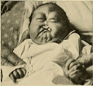
Patau syndrome is a syndrome caused by a chromosomal abnormality, in which some or all of the cells of the body contain extra genetic material from chromosome 13. The extra genetic material disrupts normal development, causing multiple and complex organ defects.

<span class="mw-page-title-main">Trisomy 18</span> Chromosomal disorder in which there are three copies of chromosome 18

Trisomy 18, also known as Edwards syndrome, is a genetic disorder caused by the presence of a third copy of all or part of chromosome 18. Many parts of the body are affected. Babies are often born small and have heart defects. Other features include a small head, small jaw, clenched fists with overlapping fingers, and severe intellectual disability.

<span class="mw-page-title-main">Nondisjunction</span> Failure to separate properly during cell division

Nondisjunction is the failure of homologous chromosomes or sister chromatids to separate properly during cell division (mitosis/meiosis). There are three forms of nondisjunction: failure of a pair of homologous chromosomes to separate in meiosis I, failure of sister chromatids to separate during meiosis II, and failure of sister chromatids to separate during mitosis. Nondisjunction results in daughter cells with abnormal chromosome numbers (aneuploidy).

<span class="mw-page-title-main">Chromosomal translocation</span> Phenomenon that results in unusual rearrangement of chromosomes

In genetics, chromosome translocation is a phenomenon that results in unusual rearrangement of chromosomes. This includes balanced and unbalanced translocation, with two main types: reciprocal, and Robertsonian translocation. Reciprocal translocation is a chromosome abnormality caused by exchange of parts between non-homologous chromosomes. Two detached fragments of two different chromosomes are switched. Robertsonian translocation occurs when two non-homologous chromosomes get attached, meaning that given two healthy pairs of chromosomes, one of each pair "sticks" and blends together homogeneously.

<span class="mw-page-title-main">Small supernumerary marker chromosome</span> Abnormal partial or mixed chromosome

A small supernumerary marker chromosome (sSMC) is an abnormal extra chromosome. It contains copies of parts of one or more normal chromosomes and like normal chromosomes is located in the cell's nucleus, is replicated and distributed into each daughter cell during cell division, and typically has genes which may be expressed. However, it may also be active in causing birth defects and neoplasms. The sSMC's small size makes it virtually undetectable using classical cytogenetic methods: the far larger DNA and gene content of the cell's normal chromosomes obscures those of the sSMC. Newer molecular techniques such as fluorescence in situ hybridization, next generation sequencing, comparative genomic hybridization, and highly specialized cytogenetic G banding analyses are required to study it. Using these methods, the DNA sequences and genes in sSMCs are identified and help define as well as explain any effect(s) it may have on individuals.

<span class="mw-page-title-main">Isochromosome</span>

An isochromosome is an unbalanced structural abnormality in which the arms of the chromosome are mirror images of each other. The chromosome consists of two copies of either the long (q) arm or the short (p) arm because isochromosome formation is equivalent to a simultaneous duplication and deletion of genetic material. Consequently, there is partial trisomy of the genes present in the isochromosome and partial monosomy of the genes in the lost arm.

<span class="mw-page-title-main">Chromosome 13</span> Human chromosome

Chromosome 13 is one of the 23 pairs of chromosomes in humans. People normally have two copies of this chromosome. Chromosome 13 spans about 113 million base pairs and represents between 3.5 and 4% of the total DNA in cells.

<span class="mw-page-title-main">Chromosome 15</span> Human chromosome

Chromosome 15 is one of the 23 pairs of chromosomes in humans. People normally have two copies of this chromosome. Chromosome 15 spans about 99.7 million base pairs and represents between 3% and 3.5% of the total DNA in cells. Chromosome 15 is an acrocentric chromosome, with a very small short arm, which contains few protein coding genes among its 19 million base pairs. It has a larger long arm that is gene rich, spanning about 83 million base pairs.

A dicentric chromosome is an abnormal chromosome with two centromeres. It is formed through the fusion of two chromosome segments, each with a centromere, resulting in the loss of acentric fragments and the formation of dicentric fragments. The formation of dicentric chromosomes has been attributed to genetic processes, such as Robertsonian translocation and paracentric inversion. Dicentric chromosomes have important roles in the mitotic stability of chromosomes and the formation of pseudodicentric chromosomes. Their existence has been linked to certain natural phenomena such as irradiation and have been documented to underlie certain clinical syndromes, notably Kabuki syndrome. The formation of dicentric chromosomes and their implications on centromere function are studied in certain clinical cytogenetics laboratories.

<span class="mw-page-title-main">Genetics of Down syndrome</span>

Down syndrome is a chromosomal abnormality characterized by the presence of an extra copy of genetic material on chromosome 21, either in whole or part. The effects of the extra copy varies greatly from individual to individual, depending on the extent of the extra copy, genetic background, environmental factors, and random chance. Down syndrome can occur in all human populations, and analogous effects have been found in other species, such as chimpanzees and mice. In 2005, researchers have been able to create transgenic mice with most of human chromosome 21.

A chromosomal abnormality, chromosomal anomaly, chromosomal aberration, chromosomal mutation, or chromosomal disorder is a missing, extra, or irregular portion of chromosomal DNA. These can occur in the form of numerical abnormalities, where there is an atypical number of chromosomes, or as structural abnormalities, where one or more individual chromosomes are altered. Chromosome mutation was formerly used in a strict sense to mean a change in a chromosomal segment, involving more than one gene. Chromosome anomalies usually occur when there is an error in cell division following meiosis or mitosis. Chromosome abnormalities may be detected or confirmed by comparing an individual's karyotype, or full set of chromosomes, to a typical karyotype for the species via genetic testing.

Trisomic rescue is a genetic phenomenon in which a fertilized ovum containing three copies of a chromosome loses one of these chromosomes to form a diploid chromosome complement. If both of the retained chromosomes come from the same parent, then uniparental disomy results. If the retained chromosomes come from different parents then there are no phenotypic or genotypic anomalies. The mechanism of trisomic rescue has been well confirmed in vivo, and alternative mechanisms that occur in trisomies are rare in comparison.

William Rees Brebner Robertson was an American zoologist and early cytogeneticist who discovered the chromosomal rearrangement named in his honour, Robertsonian translocation, the most common structural chromosomal abnormalities seen in humans that result in syndromes of multiple malformations, including trisomy 13 Patau syndrome and trisomy 21 Down syndrome.

References

  1. E. Therman, B. Susman and C. Denniston. The nonrandom participation of human acrocentric chromosomes in Robertsonian translocations. Annals of Human Genetics 1989;53:49-65.
  2. 1 2 "Unique: Rare Chromosome Disorder Support Group" (PDF). Archived from the original (PDF) on 2019-02-18. Retrieved 2019-02-17.
  3. Chiatante G, Giannuzzi G, Calabrese FM, Eichler EE, Ventura M (1 July 2017). "Centromere Destiny in Dicentric Chromosomes: New Insights from the Evolution of Human Chromosome 2 Ancestral Centromeric Region". Molecular Biology and Evolution . 34 (7): 1669–1681. doi:10.1093/molbev/msx108. PMC   5722054 . PMID   28333343.
  4. Pratas D, Silva RM, Pinho AJ, Ferreira PJ (18 May 2015). "An alignment-free method to find and visualise rearrangements between pairs of DNA sequences". Scientific Reports . 5 (1): 1203. Bibcode:2015NatSR...510203P. doi:10.1038/srep10203. PMC   4434998 . PMID   25984837.
  5. More details under Mule and Hinny.
  6. 1 2 Chowdhary BP (2013-01-22). Equine Genomics. John Wiley & Sons. ISBN   978-1-118-52212-7.
  7. 1 2 Robertson WRB. Chromosome studies. I. Taxonomic relationships shown in the chromosomes of Tettigidae and Acrididae. V-shaped chromosomes and their significance in Acrididae, Locustidae and Gryllidae: chromosome and variation. J Morph 1916;27:179-331.
  8. Hartwell L, Hood L, Goldberg M, Reynolds A, Silver L (2011). Genetics From Genes to Genomes, 4e. New York: McGraw-Hill. pp. 443, 454. ISBN   978-0-07-352526-6.
  9. Peter J. Russel; Essential Genetics 2003
  10. Plaiasu V (September 2017). "Down Syndrome - Genetics and Cardiogenetics". Maedica. 12 (3): 208–213. ISSN   1841-9038. PMC   5706761 . PMID   29218069.
  11. Guarracino A, Buonaiuto S, Potapova T, Rhie A, Koren S, Rubinstein B, Fischer C, Gerton J, Phillippy A, Colonna V, Garrison E (2022). "Recombination between heterologous human acrocentric chromosomes". bioRxiv. doi:10.1101/2022.08.15.504037. hdl: 2117/393184 . S2CID   251647679.
  12. Sheets KB, Crissman BG, Feist CD, Sell SL, Johnson LR, Donahue KC, Masser-Frye D, Brookshire GS, Carre AM, LaGrave D, Brasington CK (October 2011). "Practice Guidelines for Communicating a Prenatal or Postnatal Diagnosis of Down Syndrome: Recommendations of the National Society of Genetic Counselors". Journal of Genetic Counseling. 20 (5): 432–441. doi: 10.1007/s10897-011-9375-8 . ISSN   1059-7700. PMID   21618060. S2CID   19308113.
  13. Martinez-Castro P, Ramos MC, Rey JA, Benitez J, Sanchez Cascos A (1984). "Homozygosity for a Robertsonian translocation (13q14q) in three offspring of heterozygous parents". Cytogenet Cell Genet. 38 (4): 310–2. doi:10.1159/000132080. PMID   6510025.
  14. "ISCN Symbols and Abbreviated Terms". Coriell Institute for Medical Research. Retrieved 2022-10-27.
  15. Rajasekhar M, Rekharao RM, Shetty H, Gopinath PM, Satyamoorthy K (2010). "Cytogenetic Analysis of 1400 Referral Cases: Manipal Experience". International Journal of Human Genetics. 10 (1–3). Kamla Raj Enterprises: 49–55. doi:10.1080/09723757.2010.11886084. ISSN   0972-3757. S2CID   55971437.
  16. Yip MY (April 2014). "Uniparental disomy in Robertsonian translocations: strategies for uniparental disomy testing". Translational Pediatrics. 3 (2): 9807–9107. doi:10.3978/j.issn.2224-4336.2014.03.03. ISSN   2224-4344. PMC   4729106 . PMID   26835328.
  17. Slijepcevic P (1998-05-01). "Telomeres and mechanisms of Robertsonian fusion". Chromosoma. 107 (2): 136–140. doi:10.1007/s004120050289. ISSN   1432-0886. PMID   9601982. S2CID   11712171.