The clavicle, collarbone, or keybone is a slender, S-shaped long bone approximately 6 inches (15 cm) long that serves as a strut between the shoulder blade and the sternum (breastbone). There are two clavicles, one on the left and one on the right. The clavicle is the only long bone in the body that lies horizontally. Together with the shoulder blade, it makes up the shoulder girdle. It is a palpable bone and, in people who have less fat in this region, the location of the bone is clearly visible. It receives its name from Latin clavicula 'little key' because the bone rotates along its axis like a key when the shoulder is abducted. The clavicle is the most commonly fractured bone. It can easily be fractured by impacts to the shoulder from the force of falling on outstretched arms or by a direct hit.

The trapezius is a large paired trapezoid-shaped surface muscle that extends longitudinally from the occipital bone to the lower thoracic vertebrae of the spine and laterally to the spine of the scapula. It moves the scapula and supports the arm.

The scapula, also known as the shoulder blade, is the bone that connects the humerus with the clavicle. Like their connected bones, the scapulae are paired, with each scapula on either side of the body being roughly a mirror image of the other. The name derives from the Classical Latin word for trowel or small shovel, which it was thought to resemble.

The brachial plexus is a network of nerves formed by the anterior rami of the lower four cervical nerves and first thoracic nerve. This plexus extends from the spinal cord, through the cervicoaxillary canal in the neck, over the first rib, and into the armpit, it supplies afferent and efferent nerve fibers to the chest, shoulder, arm, forearm, and hand.

The upper limbs or upper extremities are the forelimbs of an upright-postured tetrapod vertebrate, extending from the scapulae and clavicles down to and including the digits, including all the musculatures and ligaments involved with the shoulder, elbow, wrist and knuckle joints. In humans, each upper limb is divided into the arm, forearm and hand, and is primarily used for climbing, lifting and manipulating objects.

The levator scapulae is a slender skeletal muscle situated at the back and side of the neck. It originates from the transverse processes of the four uppermost cervical vertebrae; it inserts onto the upper portion of the medial border of the scapula. It is innervated by the cervical nerves C3-C4, and frequently also by the dorsal scapular nerve. As the Latin name suggests, its main function is to lift the scapula.

The rhomboid major is a skeletal muscle of the back that connects the scapula with the vertebrae of the spinal column. It originates from the spinous processes of the thoracic vertebrae T2–T5 and supraspinous ligament; it inserts onto the lower portion of the medial border of the scapula. It acts together with the rhomboid minor to keep the scapula pressed against thoracic wall and to retract the scapula toward the vertebral column.

In human anatomy, the rhomboid minor is a small skeletal muscle of the back that connects the scapula to the vertebrae of the spinal column. It arises from the nuchal ligament, and the 7th cervical and 1st thoracic vertebrae and intervening supraspinous ligaments; it inserts onto the medial border of the scapula. It is innervated by the dorsal scapular nerve.

Pectoralis minor muscle is a thin, triangular muscle, situated at the upper part of the chest, beneath the pectoralis major in the human body. It arises from ribs III-V; it inserts onto the coracoid process of the scapula. It is innervated by the medial pectoral nerve. Its function is to stabilise the scapula by holding it fast in position against the chest wall.

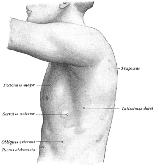
The axilla is the area on the human body directly under the shoulder joint. It includes the axillary space, an anatomical space within the shoulder girdle between the arm and the thoracic cage, bounded superiorly by the imaginary plane between the superior borders of the first rib, clavicle and scapula, medially by the serratus anterior muscle and thoracolumbar fascia, anteriorly by the pectoral muscles and posteriorly by the subscapularis, teres major and latissimus dorsi muscle.

The shoulder joint is structurally classified as a synovial ball-and-socket joint and functionally as a diarthrosis and multiaxial joint. It involves an articulation between the glenoid fossa of the scapula and the head of the humerus. Due to the very loose joint capsule that gives a limited interface of the humerus and scapula, it is the most mobile joint of the human body.
The shoulder girdle or pectoral girdle is the set of bones in the appendicular skeleton which connects to the arm on each side. In humans it consists of the clavicle and scapula; in those species with three bones in the shoulder, it consists of the clavicle, scapula, and coracoid. Some mammalian species have only the scapula.

The posterior triangle is a region of the neck.

The transverse cervical artery is an artery in the neck and a branch of the thyrocervical trunk, running at a higher level than the suprascapular artery.

The supraspinous fossa of the posterior aspect of the scapula is smaller than the infraspinous fossa, concave, smooth, and broader at its vertebral than at its humeral end. Its medial two-thirds give origin to the Supraspinatus.

The triangle of auscultation is a relative thinning of the musculature of the back, situated along the medial border of the scapula which allows for improved listening to the lungs.
A winged scapula is a skeletal medical condition in which the shoulder blade protrudes from a person's back in an abnormal position.

The axillary spaces are anatomic spaces. through which axillary contents leave the axilla. They consist of the quadrangular space, triangular space, and triangular interval. It is bounded by teres major, teres minor, medial border of the humerus, and long head of triceps brachii.

The following outline is provided as an overview of and topical guide to human anatomy:
The Eden–Lange procedure is an orthopedic procedure to alleviate the symptoms of trapezius palsy when more conservative measures, such as spontaneous resolution and surgical nerve repair are not promising. The rhomboid major, rhomboid minor, and levator scapulae muscles are transferred laterally along the scapula to replace the functions of the lower, middle, and upper fibers of the trapezius, respectively. The transferred muscles hold the scapula in a more medial and upwardly rotated position, without winging.
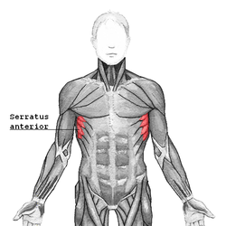
 Position of serratus anterior muscle. Scapulae are shown as semi-transparent.
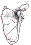 Left scapula, costal surface. Attachment point for the SA is seen on the left border.
 Serratus anterior muscle
 The axillary artery and its branches. The SA is seen alongside the pec minor.
 Nerves of the left upper extremity. The SA is seen to the left of the red line.
 Serratus anterior muscle. Anterior thoracic wall. External abdominal oblique muscle. Deep dissection, anterior view.
 Clearly visible serratus anterior muscle of the artistic gymnast Calvin Currie during preparation of the parallel bars at the Austrian Future Cup in Linz.