
The nephron is the minute or microscopic structural and functional unit of the kidney. It is composed of a renal corpuscle and a renal tubule. The renal corpuscle consists of a tuft of capillaries called a glomerulus and a cup-shaped structure called Bowman's capsule. The renal tubule extends from the capsule. The capsule and tubule are connected and are composed of epithelial cells with a lumen. A healthy adult has 1 to 1.5 million nephrons in each kidney. Blood is filtered as it passes through three layers: the endothelial cells of the capillary wall, its basement membrane, and between the foot processes of the podocytes of the lining of the capsule. The tubule has adjacent peritubular capillaries that run between the descending and ascending portions of the tubule. As the fluid from the capsule flows down into the tubule, it is processed by the epithelial cells lining the tubule: water is reabsorbed and substances are exchanged ; first with the interstitial fluid outside the tubules, and then into the plasma in the adjacent peritubular capillaries through the endothelial cells lining that capillary. This process regulates the volume of body fluid as well as levels of many body substances. At the end of the tubule, the remaining fluid—urine—exits: it is composed of water, metabolic waste, and toxins.

Epithelium or epithelial tissue is a thin, continuous, protective layer of compactly packed cells with a little intercellular matrix. Epithelial tissues line the outer surfaces of organs and blood vessels throughout the body, as well as the inner surfaces of cavities in many internal organs. An example is the epidermis, the outermost layer of the skin. Epithelial tissue is one of the four basic types of animal tissue, along with connective tissue, muscle tissue and nervous tissue. These tissues also lack blood or lymph supply. The tissue is supplied by nerves.
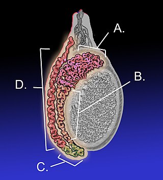
The epididymis is an elongated tubular structure attached to the posterior side of each one of the two male reproductive glands, the testicles. It is a single, narrow, tightly coiled tube in adult humans, 6 to 7 centimetres in length; uncoiled the tube would be approximately 6 m long. It connects the testicle to the vas deferens in the male reproductive system. The epididymis serves as an interconnection between the multiple efferent ducts at the rear of a testicle (proximally), and the vas deferens (distally). Its primary function is the storage, maturation and transport of sperm cells.

The juxtaglomerular apparatus is a structure in the kidney that regulates the function of each nephron, the functional units of the kidney. The juxtaglomerular apparatus is named because it is next to (juxta-) the glomerulus.

The collecting duct system of the kidney consists of a series of tubules and ducts that physically connect nephrons to a minor calyx or directly to the renal pelvis. The collecting duct system is the last part of nephron and participates in electrolyte and fluid balance through reabsorption and excretion, processes regulated by the hormones aldosterone and vasopressin.

Renal physiology is the study of the physiology of the kidney. This encompasses all functions of the kidney, including maintenance of acid-base balance; regulation of fluid balance; regulation of sodium, potassium, and other electrolytes; clearance of toxins; absorption of glucose, amino acids, and other small molecules; regulation of blood pressure; production of various hormones, such as erythropoietin; and activation of vitamin D.
The proximal tubule is the segment of the nephron in kidneys which begins from the renal pole of the Bowman's capsule to the beginning of loop of Henle. At this location, the glomerular parietal epithelial cells (PECs) lining bowman’s capsule abruptly transition to proximal tubule epithelial cells (PTECs). The proximal tubule can be further classified into the proximal convoluted tubule (PCT) and the proximal straight tubule (PST).

Thyroid follicular cells (also called thyroid epithelial cells or thyrocytes) are the major cell type in the thyroid gland, and are responsible for the production and secretion of the thyroid hormones thyroxine (T4) and triiodothyronine (T3). They form the single layer of cuboidal epithelium that makes up the outer structure of the almost spherical thyroid follicle.

The rete testis is an anastomosing network of delicate tubules located in the hilum of the testicle that carries sperm from the seminiferous tubules to the efferent ducts. It is the homologue of the rete ovarii in females. Its function is to provide a site for fluid reabsorption.
In biology, a tubule is a general term referring to small tube or similar type of structure. Specifically, tubule can refer to:
The development of the urinary system begins during prenatal development, and relates to the development of the urogenital system – both the organs of the urinary system and the sex organs of the reproductive system. The development continues as a part of sexual differentiation.

The male reproductive system consists of a number of sex organs that play a role in the process of human reproduction. These organs are located on the outside of the body, and within the pelvis.
An oncocytoma is a tumor made up of oncocytes, epithelial cells characterized by an excessive amount of mitochondria, resulting in an abundant acidophilic, granular cytoplasm. The cells and the tumor that they compose are often benign but sometimes may be premalignant or malignant.

A pseudostratified epithelium is a type of epithelium that, though comprising only a single layer of cells, has its cell nuclei positioned in a manner suggestive of stratified epithelia. As it rarely occurs as squamous or cuboidal epithelia, it is usually considered synonymous with the term pseudostratified columnar epithelium.

Eccrine sweat glands, sometimes called merocrine glands as their type of secretion is merocrine, are the major sweat glands of the human body. Eccrine sweat glands are found in virtually all skin, with the highest density in the palms of the hands, and soles of the feet, and on the head, but much less on the torso and the extremities. In other mammals, they are relatively sparse, being found mainly on hairless areas such as foot pads. They reach their peak of development in humans, where they may number 200–400/cm2 of skin surface. They produce sweat, a clear, odorless substance, consisting primarily of water. These are present from birth. Their secretory part is present deep inside the dermis.
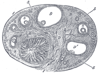
The ovarian surface epithelium, also called the germinal epithelium of Waldeyer, or coelomic epithelium is a layer of simple squamous-to-cuboidal epithelial cells covering the ovary.

The germinal epithelium is the epithelial layer of the seminiferous tubules of the testicles. It is also known as the wall of the seminiferous tubules. The cells in the epithelium are connected via tight junctions.
The development of the reproductive system is the part of embryonic growth that results in the sex organs and contributes to sexual differentiation. Due to its large overlap with development of the urinary system, the two systems are typically described together as the urogenital or genitourinary system.
The development of the gonads is part of the prenatal development of the reproductive system and ultimately forms the testicles in males and the ovaries in females. The gonads initially develop from the mesothelial layer of the peritoneum.
The rock dove, Columbia livia, has a number of special adaptations for regulating water uptake and loss.

