Spatiotemporal gene expression

Last updated
Gene expression patterns are regulated both spatially and temporally in embryos of Drosophila melanogaster. Gene-expression-patterns.png
Gene expression patterns are regulated both spatially and temporally in embryos of Drosophila melanogaster.

Spatiotemporal gene expression is the activation of genes within specific tissues of an organism at specific times during development. Gene activation patterns vary widely in complexity. Some are straightforward and static, such as the pattern of tubulin, which is expressed in all cells at all times in life. Some, on the other hand, are extraordinarily intricate and difficult to predict and model, with expression fluctuating wildly from minute to minute or from cell to cell. Spatiotemporal variation plays a key role in generating the diversity of cell types found in developed organisms; since the identity of a cell is specified by the collection of genes actively expressed within that cell, if gene expression was uniform spatially and temporally, there could be at most one kind of cell.

Contents

Consider the gene wingless, a member of the wnt family of genes. In the early embryonic development of the model organism Drosophila melanogaster , or fruit fly, wingless is expressed across almost the entire embryo in alternating stripes three cells separated. This pattern is lost by the time the organism develops into a larva, but wingless is still expressed in a variety of tissues such as the wing imaginal discs, patches of tissue that will develop into the adult wings. The spatiotemporal pattern of wingless gene expression is determined by a network of regulatory interactions consisting of the effects of many different genes such as even-skipped and Krüppel.

What causes spatial and temporal differences in the expression of a single gene? Because current expression patterns depend strictly on previous expression patterns, there is a regressive problem of explaining what caused the first differences in gene expression. The process by which uniform gene expression becomes spatially and temporally differential is known as symmetry breaking. For example, in the case of embryonic Drosophila development, the genes nanos and bicoid are asymmetrically expressed in the oocyte because maternal cells deposit messenger RNA (mRNA) for these genes in the poles of the egg before it is laid.

The gamma-crystalline promoter drives expression of the green fluorescent protein reporter gene exclusively in the eye of an adult frog. Frog GFP eye.gif
The gamma-crystalline promoter drives expression of the green fluorescent protein reporter gene exclusively in the eye of an adult frog.

Identifying spatiotemporal patterns

One way to identify the expression pattern of a particular gene is to place a reporter gene downstream of its promoter. In this configuration, the promoter gene will cause the reporter gene to be expressed only where and when the gene of interest is expressed. The expression distribution of the reporter gene can be determined by visualizing it. For example, the reporter gene green fluorescent protein can be visualized by stimulating it with blue light and then using a digital camera to record green fluorescent emission.

If the promoter of the gene of interest is unknown, there are several ways to identify its spatiotemporal distribution. Immunohistochemistry involves preparing an antibody with specific affinity for the protein associated with the gene of interest. This distribution of this antibody can then be visualized by a technique such as fluorescent labeling. Immunohistochemistry has the advantages of being methodologically feasible and relatively inexpensive. Its disadvantages include non-specificity of the antibody leading to false positive identification of expression. Poor penetrance of the antibody into the target tissue can lead to false negative results. Furthermore, since immunohistochemistry visualizes the protein generated by the gene, if the protein product diffuses between cells, or has a particularly short or long half-life relative to the mRNA that is used to translate the protein, this can lead to distorted interpretation of which cells are expressing the mRNA.

In situ hybridizations of genes expressed in arteries (top) and veins (bottom) in zebrafish. Blue staining indicates presence of the gene mRNAs. Panels on the left are normal animals, while animals on the right are mutated in the Notch gene. Fish lacking Notch have fewer arteries and more veins at this point in developmental time. Veins and arteries in situ.jpg
In situ hybridizations of genes expressed in arteries (top) and veins (bottom) in zebrafish. Blue staining indicates presence of the gene mRNAs. Panels on the left are normal animals, while animals on the right are mutated in the Notch gene. Fish lacking Notch have fewer arteries and more veins at this point in developmental time.

In situ hybridization is an alternate method in which a "probe," a synthetic nucleic acid with a sequence complementary to the mRNA of the gene, is added to the tissue. This probe is then chemically tagged so that it can be visualized later. This technique enables visualization specifically of mRNA-producing cells without any of the artifacts associated with immunohistochemistry. However, it is notoriously difficult, and requires knowledge of the sequence of DNA corresponding to the gene of interest.

A method called enhancer-trap screening reveals the diversity of spatiotemporal gene expression patterns possible in an organism. In this technique, DNA that encodes a reporter gene is randomly inserted into the genome. Depending on the gene promoters proximal to the insertion point, the reporter gene will be expressed in particular tissues at particular points in development. While enhancer-trap derived expression patterns do not necessarily reflect the actual patterns of expression of specific genes, they reveal the variety of spatiotemporal patterns that are accessible to evolution.

Reporter genes can be visualized in living organisms, but both immunohistochemistry and in situ hybridization must be performed in fixed tissues. Techniques that require fixation of tissue can only generate a single temporal time point per individual organism. However, using live animals instead of fixed tissue can be crucial in dynamically understanding expression patterns over an individual's lifespan. Either way, variation between individuals can confound the interpretation of temporal expression patterns.

Methods to control spatiotemporal gene expression

Several methods are being pursued for controlling gene expression spatially, temporally and in different degrees. One method is by using operon inducer/repressor system which provides temporal control of gene expression. To control gene expression spatially inkjet printers are under development for printing ligands on gel culture. [1] Other popular method involves use of light to control gene expression in spatiotemporal fashion. Since light can also be controlled easily in space, time and degree, several methods of controlling gene expression at DNA and RNA level [2] have been developed and are under study. For example, RNA interference can be controlled using light [3] [4] and also patterning of gene expression has been performed in cell monolayer [5] and in zebrafish embryos using caged morpholino [6] or peptide nucleic acid [7] [8] [9] demonstrating the control of gene expression spatiotemporally. Recently light based control has been shown at DNA level using transgene based system [10] or caged triplex forming oligos [11]

Related Research Articles

Molecular biology is a branch of biology that seeks to understand the molecular basis of biological activity in and between cells, including biomolecular synthesis, modification, mechanisms, and interactions.

<span class="mw-page-title-main">Northern blot</span> Molecular biology technique

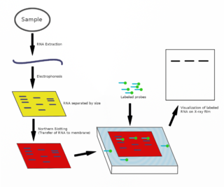
The northern blot, or RNA blot, is a technique used in molecular biology research to study gene expression by detection of RNA in a sample.

<span class="mw-page-title-main">Gene expression</span> Conversion of a genes sequence into a mature gene product or products

Gene expression is the process by which information from a gene is used in the synthesis of a functional gene product that enables it to produce end products, proteins or non-coding RNA, and ultimately affect a phenotype. These products are often proteins, but in non-protein-coding genes such as transfer RNA (tRNA) and small nuclear RNA (snRNA), the product is a functional non-coding RNA. The process of gene expression is used by all known life—eukaryotes, prokaryotes, and utilized by viruses—to generate the macromolecular machinery for life.

<span class="mw-page-title-main">Enhancer (genetics)</span> DNA sequence that binds activators to increase the likelihood of gene transcription

In genetics, an enhancer is a short region of DNA that can be bound by proteins (activators) to increase the likelihood that transcription of a particular gene will occur. These proteins are usually referred to as transcription factors. Enhancers are cis-acting. They can be located up to 1 Mbp away from the gene, upstream or downstream from the start site. There are hundreds of thousands of enhancers in the human genome. They are found in both prokaryotes and eukaryotes.

<span class="mw-page-title-main">Codon usage bias</span> Genetic bias in coding DNA

Codon usage bias refers to differences in the frequency of occurrence of synonymous codons in coding DNA. A codon is a series of three nucleotides that encodes a specific amino acid residue in a polypeptide chain or for the termination of translation.

Gene knockdown is an experimental technique by which the expression of one or more of an organism's genes is reduced. The reduction can occur either through genetic modification or by treatment with a reagent such as a short DNA or RNA oligonucleotide that has a sequence complementary to either gene or an mRNA transcript.

<i>Drosophila</i> embryogenesis Embryogenesis of the fruit fly Drosophila, a popular model system

Drosophila embryogenesis, the process by which Drosophila embryos form, is a favorite model system for genetics and developmental biology. The study of its embryogenesis unlocked the century-long puzzle of how development was controlled, creating the field of evolutionary developmental biology. The small size, short generation time, and large brood size make it ideal for genetic studies. Transparent embryos facilitate developmental studies. Drosophila melanogaster was introduced into the field of genetic experiments by Thomas Hunt Morgan in 1909.

<span class="mw-page-title-main">Functional genomics</span> Field of molecular biology

Functional genomics is a field of molecular biology that attempts to describe gene functions and interactions. Functional genomics make use of the vast data generated by genomic and transcriptomic projects. Functional genomics focuses on the dynamic aspects such as gene transcription, translation, regulation of gene expression and protein–protein interactions, as opposed to the static aspects of the genomic information such as DNA sequence or structures. A key characteristic of functional genomics studies is their genome-wide approach to these questions, generally involving high-throughput methods rather than a more traditional "candidate-gene" approach.

<span class="mw-page-title-main">Morpholino</span> Chemical compound

A Morpholino, also known as a Morpholino oligomer and as a phosphorodiamidate Morpholino oligomer (PMO), is a type of oligomer molecule used in molecular biology to modify gene expression. Its molecular structure contains DNA bases attached to a backbone of methylenemorpholine rings linked through phosphorodiamidate groups. Morpholinos block access of other molecules to small specific sequences of the base-pairing surfaces of ribonucleic acid (RNA). Morpholinos are used as research tools for reverse genetics by knocking down gene function.

<span class="mw-page-title-main">Two-hybrid screening</span> Molecular biology technique

Two-hybrid screening is a molecular biology technique used to discover protein–protein interactions (PPIs) and protein–DNA interactions by testing for physical interactions between two proteins or a single protein and a DNA molecule, respectively.

<span class="mw-page-title-main">Photostimulation</span>

Photostimulation is the use of light to artificially activate biological compounds, cells, tissues, or even whole organisms. Photostimulation can be used to noninvasively probe various relationships between different biological processes, using only light. In the long run, photostimulation has the potential for use in different types of therapy, such as migraine headache. Additionally, photostimulation may be used for the mapping of neuronal connections between different areas of the brain by “uncaging” signaling biomolecules with light. Therapy with photostimulation has been called light therapy, phototherapy, or photobiomodulation.

<i>In situ</i> hybridization Laboratory technique to localize nucleic acids

In situ hybridization (ISH) is a type of hybridization that uses a labeled complementary DNA, RNA or modified nucleic acids strand to localize a specific DNA or RNA sequence in a portion or section of tissue or if the tissue is small enough, in the entire tissue, in cells, and in circulating tumor cells (CTCs). This is distinct from immunohistochemistry, which usually localizes proteins in tissue sections.

<span class="mw-page-title-main">FLP-FRT recombination</span>

In genetics, Flp-FRT recombination is a site-directed recombination technology, increasingly used to manipulate an organism's DNA under controlled conditions in vivo. It is analogous to Cre-lox recombination but involves the recombination of sequences between short flippase recognition target (FRT) sites by the recombinase flippase (Flp) derived from the 2 µ plasmid of baker's yeast Saccharomyces cerevisiae.

<span class="mw-page-title-main">GAL4/UAS system</span> Biochemical method

The GAL4-UAS system is a biochemical method used to study gene expression and function in organisms such as the fruit fly. It is based on the finding by Hitoshi Kakidani and Mark Ptashne, and Nicholas Webster and Pierre Chambon in 1988 that Gal4 binding to UAS sequences activates gene expression. The method was introduced into flies by Andrea Brand and Norbert Perrimon in 1993 and is considered a powerful technique for studying the expression of genes. The system has two parts: the Gal4 gene, encoding the yeast transcription activator protein Gal4, and the UAS, an enhancer to which GAL4 specifically binds to activate gene transcription.

A Riboprobe, abbreviation of RNA probe, is a segment of labelled RNA that can be used to detect a target mRNA or DNA during in situ hybridization. RNA probes can be produced by in vitro transcription of cloned DNA inserted in a suitable plasmid downstream of a viral promoter. Some bacterial viruses code for their own RNA polymerases, which are highly specific for the viral promoters. Using these enzymes, labeled NTPs, and inserts inserted in both forward and reverse orientations, both sense and antisense riboprobes can be generated from a cloned gene.

<span class="mw-page-title-main">Genetic engineering techniques</span> Methods used to change the DNA of organisms

Genetic engineering techniques allow the modification of animal and plant genomes. Techniques have been devised to insert, delete, and modify DNA at multiple levels, ranging from a specific base pair in a specific gene to entire genes. There are a number of steps that are followed before a genetically modified organism (GMO) is created. Genetic engineers must first choose what gene they wish to insert, modify, or delete. The gene must then be isolated and incorporated, along with other genetic elements, into a suitable vector. This vector is then used to insert the gene into the host genome, creating a transgenic or edited organism.

<span class="mw-page-title-main">Photoactivatable probes</span> Cellular players that can be activated by light

Photoactivatable probes, or caged probes, are cellular players that can be triggered by a flash of light. They are used in biological research to study processes in cells. The basic principle is to bring a photoactivatable agent to cells, tissues or even living animals and specifically control its activity by illumination.

The Expression Atlas is a database maintained by the European Bioinformatics Institute that provides information on gene expression patterns from RNA-Seq and Microarray studies, and protein expression from Proteomics studies. The Expression Atlas allows searches by gene, splice variant, protein attribute, disease, treatment or organism part. Individual genes or gene sets can be searched for. All datasets in Expression Atlas have its metadata manually curated and its data analysed through standardised analysis pipelines. There are two components to the Expression Atlas, the Baseline Atlas and the Differential Atlas:

Q-system is a genetic tool that allows to express transgenes in a living organism. Originally the Q-system was developed for use in the vinegar fly Drosophila melanogaster, and was rapidly adapted for use in cultured mammalian cells, zebrafish, worms and mosquitoes. The Q-system utilizes genes from the qa cluster of the bread fungus Neurospora crassa, and consists of four components: the transcriptional activator (QF/QF2/QF2w), the enhancer QUAS, the repressor QS, and the chemical de-repressor quinic acid. Similarly to GAL4/UAS and LexA/LexAop, the Q-system is a binary expression system that allows to express reporters or effectors in a defined subpopulation of cells with the purpose of visualising these cells or altering their function. In addition, GAL4/UAS, LexA/LexAop and the Q-system function independently of each other and can be used simultaneously to achieve a desired pattern of reporter expression, or to express several reporters in different subsets of cells.

This glossary of cellular and molecular biology is a list of definitions of terms and concepts commonly used in the study of cell biology, molecular biology, and related disciplines, including genetics, biochemistry, and microbiology. It is split across two articles:

References

  1. Cohen, DJ; Morfino, RC; Maharbiz, MM (2009). "A Modified Consumer Inkjet for Spatiotemporal Control of Gene Expression". PLOS ONE. 4 (9): e7086. Bibcode:2009PLoSO...4.7086C. doi: 10.1371/journal.pone.0007086 . PMC   2739290 . PMID   19763256.
  2. Ando, Hideki; Furuta, Toshiaki; Tsien, Roger Y.; Okamoto, Hitoshi (2001). "Photo-mediated gene activation using caged RNA/DNA in zebrafish embryos". Nature Genetics. 28 (4): 317–325. doi:10.1038/ng583. PMID   11479592. S2CID   6773535.
  3. Shah, Samit; Rangarajan, Subhashree; Friedman, Simon H. (2005). "Light‐Activated RNA Interference". Angewandte Chemie International Edition. 44 (9): 1328–1332. doi:10.1002/anie.200461458. PMID   15643658.
  4. Mikat, Vera; Heckel, Alexander (2007). "Light-dependent RNA interference with nucleobase-caged siRNAs". RNA. 13 (12): 2341–2347. doi:10.1261/rna.753407. PMC   2080613 . PMID   17951332.
  5. Jain, Piyush K.; Shah, Samit; Friedman, Simon H. (2011). "Patterning of Gene Expression Using New Photolabile Groups Applied to Light Activated RNAi". Journal of the American Chemical Society. 133 (3): 440–446. doi:10.1021/ja107226e. PMID   21162570. S2CID   207058522.
  6. Shestopalov, Ilya A; Chen, James K (2011). "Spatiotemporal Control of Embryonic Gene Expression Using Caged Morpholinos". The Zebrafish: Genetics, Genomics and Informatics. Methods in Cell Biology. Vol. 104. pp. 151–72. doi:10.1016/B978-0-12-374814-0.00009-4. ISBN   9780123748140. PMC   4408312 . PMID   21924162.
  7. Tang, XinJing; Maegawa, Shingo; Weinberg, Eric S.; Dmochowski, Ivan J. (2007). "Regulating Gene Expression in Zebrafish Embryos Using Light-Activated, Negatively Charged Peptide Nucleic Acids". Journal of the American Chemical Society. 129 (36): 11000–11001. doi:10.1021/ja073723s. PMID   17711280.
  8. Alexander Heckel, Günter Mayer, "The Chemical Biology of Nucleic Acids", Chapter 13. Light-Responsive Nucleic Acids for the Spatiotemporal Control of Biological Processes. http://onlinelibrary.wiley.com/doi/10.1002/9780470664001.ch13/summary
  9. Govan, J. M; Deiters (2012) "Activation and Deactivation of Antisense and RNA Interference Function with Light A. From Nucleic Acids Sequences to Molecular Medicine (eds V. A. Erdmann and J. Barciszewski)", Springer, Heidelberg, https://doi.org/10.1007%2F978-3-642-27426-8_11
  10. Wang, X; Chen, X; Yang, Y (2012). "Spatiotemporal control of gene expression by a light-switchable transgene system". Nat Methods. 9 (3): 266–9. doi: 10.1038/nmeth.1892 . PMID   22327833. S2CID   26529717.
  11. Govan, Jeane M.; Uprety, Rajendra; Hemphill, James; Lively, Mark O.; Deiters, Alexander (2012). "Regulation of Transcription through Light-Activation and Light-Deactivation of Triplex-Forming Oligonucleotides in Mammalian Cells". ACS Chem. Biol. 7 (7): 1247–1256. doi:10.1021/cb300161r. PMC   3401312 . PMID   22540192.