Magnetoencephalography (MEG) is a functional neuroimaging technique for mapping brain activity by recording magnetic fields produced by electrical currents occurring naturally in the brain, using very sensitive magnetometers. Arrays of SQUIDs are currently the most common magnetometer, while the SERF magnetometer is being investigated for future machines. Applications of MEG include basic research into perceptual and cognitive brain processes, localizing regions affected by pathology before surgical removal, determining the function of various parts of the brain, and neurofeedback. This can be applied in a clinical setting to find locations of abnormalities as well as in an experimental setting to simply measure brain activity.

Functional neuroimaging is the use of neuroimaging technology to measure an aspect of brain function, often with a view to understanding the relationship between activity in certain brain areas and specific mental functions. It is primarily used as a research tool in cognitive neuroscience, cognitive psychology, neuropsychology, and social neuroscience.
The first neuroimaging technique ever is the so-called 'human circulation balance' invented by Angelo Mosso in the 1880s and able to non-invasively measure the redistribution of blood during emotional and intellectual activity. Then, in the early 1900s, a technique called pneumoencephalography was set. This process involved draining the cerebrospinal fluid from around the brain and replacing it with air, altering the relative density of the brain and its surroundings, to cause it to show up better on an x-ray, and it was considered to be incredibly unsafe for patients. A form of magnetic resonance imaging (MRI) and computed tomography (CT) were developed in the 1970s and 1980s. The new MRI and CT technologies were considerably less harmful and are explained in greater detail below. Next came SPECT and PET scans, which allowed scientists to map brain function because, unlike MRI and CT, these scans could create more than just static images of the brain's structure. Learning from MRI, PET and SPECT scanning, scientists were able to develop functional MRI (fMRI) with abilities that opened the door to direct observation of cognitive activities.
Neurotechnology encompasses any method or electronic device which interfaces with the nervous system to monitor or modulate neural activity.
Vision science is the scientific study of visual perception. Researchers in vision science can be called vision scientists, especially if their research spans some of the science's many disciplines.

The Psychonomic Society is an international scientific society of over 4,500 scientists in the field of experimental psychology. The mission of the Psychonomic Society is to foster the science of cognition through the advancement and communication of basic research in experimental psychology and allied sciences. It is open to international researchers, and almost 40% of members are based outside of North America. Although open to all areas of experimental and cognitive psychology, its members typically study areas such as learning, memory, attention, motivation, perception, categorization, decision making, and psycholinguistics. Its name is taken from the word psychonomics, meaning "the science of the laws of the mind".

Neuroimaging is the use of quantitative (computational) techniques to study the structure and function of the central nervous system, developed as an objective way of scientifically studying the healthy human brain in a non-invasive manner. Increasingly it is also being used for quantitative studies of brain disease and psychiatric illness. Neuroimaging is a highly multidisciplinary research field and is not a medical specialty.

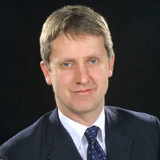
Jonathon Stevens "Jon Driver" was a psychologist and neuroscientist. He was a leading figure in the study of perception, selective attention and multisensory integration in the normal and damaged human brain.

Electroencephalography (EEG) is a method to record an electrogram of the spontaneous electrical activity of the brain. The biosignals detected by EEG have been shown to represent the postsynaptic potentials of pyramidal neurons in the neocortex and allocortex. It is typically non-invasive, with the EEG electrodes placed along the scalp using the International 10-20 system, or variations of it. Electrocorticography, involving surgical placement of electrodes, is sometimes called "intracranial EEG". Clinical interpretation of EEG recordings is most often performed by visual inspection of the tracing or quantitative EEG analysis.
The Human Connectome Project (HCP) is a five-year project sponsored by sixteen components of the National Institutes of Health, split between two consortia of research institutions. The project was launched in July 2009 as the first of three Grand Challenges of the NIH's Blueprint for Neuroscience Research. On September 15, 2010, the NIH announced that it would award two grants: $30 million over five years to a consortium led by Washington University in St. Louis and the University of Minnesota, with strong contributions from Oxford University (FMRIB) and $8.5 million over three years to a consortium led by Harvard University, Massachusetts General Hospital and the University of California Los Angeles.
Anders Martin Dale is a prominent neuroscientist and professor of radiology, neurosciences, psychiatry, and cognitive science at the University of California, San Diego (UCSD), and is one of the world's leading developers of sophisticated computational neuroimaging techniques. He is the founding Director of the Center for Multimodal Imaging Genetics (CMIG) at UCSD.
Jocelyn Faubert is a psychophysicist best known for his work in the fields of visual perception, vision of the elderly, and neuropsychology. Professor Faubert holds the NSERC-Essilor Industrial Research Chair in Visual Perception and Presbyopia. He is the director of the Laboratory of Psychophysics and Visual Perception at the University of Montreal. Professor Faubert has also been involved in the award-winning transfer of research and developments from the laboratory into the commercial domain. He is a co-founder and member of the Board of Directors of CogniSens Inc.
Scholar, educator, and philanthropist Herschel Leibowitz is widely recognized for his research in visual perception and for his symbiotic approach to conducting research that both advanced theory and helped in the understanding and relief of societal problems. His research on transportation safety included studies of nearsightedness during night driving, vision during civil twilight, an illusion that underlies the behavior of motorists involved in auto-train collisions, susceptibility of pilots to illusions caused by visual-vestibular interactions, and the design of aircraft instrument panels.
Mark Steven Cohen is an American neuroscientist and early pioneer of functional brain imaging using magnetic resonance imaging. He currently is a Professor of Psychiatry, Neurology, Radiology, Psychology, Biomedical Physics and Biomedical Engineering at the Semel Institute for Neuroscience and Human Behavior and the Staglin Center for Cognitive Neuroscience. He is also a performing musician.
The sensory enhancement theory assumes that attentional resources will spread until they reach the boundaries of a cued object, including regions that may be obstructed or are overlapping other objects. It has been suggested that sensory enhancement is an essential mechanism that underlies object-based attention. The sensory enhancement theory of object-based attention proposes that when attention is directed to a cued object, the quality of the object’s physical representations improve because the spread of attention facilitates the efficiency of processing the features of the object as a whole. The qualities of the cued object, such as spatial resolution and contrast sensitivity, are therefore more strongly represented in one's memory than the qualities of other objects or locations that received little or no attentional resource. Information processing of these objects also tends to be significantly faster and more accurate as the representations have become more salient.
The White House BRAIN Initiative is a collaborative, public-private research initiative announced by the Obama administration on April 2, 2013, with the goal of supporting the development and application of innovative technologies that can create a dynamic understanding of brain function.
The following outline is provided as an overview of and topical guide to brain mapping:

Russell L. De Valois was an American scientist recognized for his pioneering research on spatial and color vision.

Doris Ying Tsao is an American systems neuroscientist and professor of biology at the University of California, Berkeley. She was formerly on the faculty at the California Institute of Technology. She is recognized for pioneering the use of fMRI with single-unit electrophysiological recordings and for discovering the macaque face patch system for face perception. She is a Howard Hughes Medical Institute Investigator and the director of the T&C Chen Center for Systems Neuroscience. She won a MacArthur "Genius" fellowship in 2018. Tsao was elected a member of the National Academy of Sciences in 2020.
The Werner Reichardt Centre for Integrative Neuroscience (CIN) is the common platform for systems neuroscience at the University of Tübingen in Germany. It was installed as a cluster of excellence within the framework of the Excellence Initiative in 2007/2008. About 90 scientists with their research groups – 21 of which are currently supported with excellence initiative funds – form the CIN's membership. The focus of their work is on basic research in systems neurobiology. Based on an interdisciplinary and integrative approach, it encompasses projects rooted in biology, medicine, physics, computer science and engineering as well as cognition and neurophilosophy.


