
A corrective lens is a transmissive optical device that is worn on the eye to improve visual perception. The most common use is to treat refractive errors: myopia, hypermetropia, astigmatism, and presbyopia. Glasses or "spectacles" are worn on the face a short distance in front of the eye. Contact lenses are worn directly on the surface of the eye. Intraocular lenses are surgically implanted most commonly after cataract removal but can be used for purely refractive purposes.

Far-sightedness, also known as long-sightedness, hypermetropia, and hyperopia, is a condition of the eye where distant objects are seen clearly but near objects appear blurred. This blur is due to incoming light being focused behind, instead of on, the retina due to insufficient accommodation by the lens. Minor hypermetropia in young patients is usually corrected by their accommodation, without any defects in vision. But, due to this accommodative effort for distant vision, people may complain of eye strain during prolonged reading. If the hypermetropia is high, there will be defective vision for both distance and near. People may also experience accommodative dysfunction, binocular dysfunction, amblyopia, and strabismus. Newborns are almost invariably hypermetropic, but it gradually decreases as the newborn gets older.

LASIK or Lasik, commonly referred to as laser eye surgery or laser vision correction, is a type of refractive surgery for the correction of myopia, hyperopia, and an actual cure for astigmatism, since it is in the cornea. LASIK surgery is performed by an ophthalmologist who uses a laser or microkeratome to reshape the eye's cornea in order to improve visual acuity.

An eyeglass prescription is an order written by an eyewear prescriber, such as an optometrist, that specifies the value of all parameters the prescriber has deemed necessary to construct and/or dispense corrective lenses appropriate for a patient. If an eye examination indicates that corrective lenses are appropriate, the prescriber generally provides the patient with an eyewear prescription at the conclusion of the exam.

An optical system with astigmatism is one where rays that propagate in two perpendicular planes have different foci. If an optical system with astigmatism is used to form an image of a cross, the vertical and horizontal lines will be in sharp focus at two different distances. The term comes from the Greek α- (a-) meaning "without" and στίγμα (stigma), "a mark, spot, puncture".

Visual acuity (VA) commonly refers to the clarity of vision, but technically rates an animal's ability to recognize small details with precision. Visual acuity depends on optical and neural factors. Optical factors of the eye influence the sharpness of an image on its retina. Neural factors include the health and functioning of the retina, of the neural pathways to the brain, and of the interpretative faculty of the brain.

An Intraocular lens (IOL) is a lens implanted in the eye usually as part of a treatment for cataracts or for correcting other vision problems such as short sightedness and long sightedness, a form of refractive surgery. If the natural lens is left in the eye, the IOL is known as phakic, otherwise it is a pseudophakic lens. Both kinds of IOLs are designed to provide the same light-focusing function as the natural crystalline lens. This can be an alternative to LASIK, but LASIK is not an alternative to an IOL for treatment of cataracts.
An eye chart is a chart used to measure visual acuity comprising lines of optotypes in ranges of sizes. Optotypes are the letters or symbols shown on an eye chart. Eye charts are often used by health care professionals, such as optometrists, physicians and nurses, to screen persons for vision impairment. Ophthalmologists, physicians who specialize in the eye, also use eye charts to monitor the visual acuity of their patients in response to various therapies such as medications or surgery.
An eye examination is a series of tests performed to assess vision and ability to focus on and discern objects. It also includes other tests and examinations pertaining to the eyes. Eye examinations are primarily performed by an optometrist, ophthalmologist, or an orthoptist. Health care professionals often recommend that all people should have periodic and thorough eye examinations as part of routine primary care, especially since many eye diseases are asymptomatic.

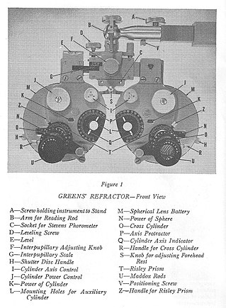
A phoropter or refractor is an ophthalmic testing device. It is commonly used by eye care professionals during an eye examination, and contains different lenses used for refraction of the eye during sight testing, to measure an individual's refractive error and determine their eyeglass prescription. It also is used to measure the patients' phorias and ductions, which are characteristics of binocularity.

In optics, defocus is the aberration in which an image is simply out of focus. This aberration is familiar to anyone who has used a camera, videocamera, microscope, telescope, or binoculars. Optically, defocus refers to a translation of the focus along the optical axis away from the detection surface. In general, defocus reduces the sharpness and contrast of the image. What should be sharp, high-contrast edges in a scene become gradual transitions. Fine detail in the scene is blurred or even becomes invisible. Nearly all image-forming optical devices incorporate some form of focus adjustment to minimize defocus and maximize image quality.

Astigmatism is a type of refractive error due to rotational asymmetry in the eye's refractive power. This results in distorted or blurred vision at any distance. Other symptoms can include eyestrain, headaches, and trouble driving at night. Astigmatism often occurs at birth and can change or develop later in life. If it occurs in early life and is left untreated, it may result in amblyopia.

Emmetropia is the state of vision in which a faraway object at infinity is in sharp focus with the ciliary muscle in a relaxed state. That condition of the normal eye is achieved when the refractive power of the cornea and eye lens and the axial length of the eye balance out, which focuses rays exactly on the retina, resulting in perfectly sharp distance vision. A human eye in a state of emmetropia requires no corrective lenses for distance; the vision scores well on a visual acuity test.

A cylindrical lens is a lens which focuses light into a line instead of a point, as a spherical lens would. The curved face or faces of a cylindrical lens are sections of a cylinder, and focus the image passing through it into a line parallel to intersection of the surface of the lens and a plane tangent to it along the cylinder's axis. The lens converges or diverges the image in the direction perpendicular to this line, and leaves it unaltered in the direction parallel to its cylinder's axis.

A pinhole occluder is an opaque disk with one or more small holes through it, used by ophthalmologists, orthoptists and optometrists to test visual acuity. The occluder is a simple way to focus light, as in a pinhole camera, temporarily removing the effects of refractive errors such as myopia. Because light passes only through the center of the eye's lens, defects in the shape of the lens have no effect while the occluder is used. In this way, the ophthalmologist, orthoptist or optometrist can estimate the maximum improvement in a patient's vision that can be attained by lenses to correct errors of refraction. This can be used to distinguish visual defects caused by refractive error, which improve when the occluder is used, from other problems, which do not. The pinhole occluder can also be used in testing visual acuity in mydriatic patients. In this case, the pinhole occluder compensates for the inability to contract the iris assisting the eye in obtaining a retinal projection similar to that of a non-cycloplegic eye.
The eye, like any other optical system, suffers from a number of specific optical aberrations. The optical quality of the eye is limited by optical aberrations, diffraction and scatter. Correction of spherocylindrical refractive errors has been possible for nearly two centuries following Airy's development of methods to measure and correct ocular astigmatism. It has only recently become possible to measure the aberrations of the eye and with the advent of refractive surgery it might be possible to correct certain types of irregular astigmatism.

A duochrome test is a test commonly used to refine the final sphere in refraction, which makes use of the longitudinal chromatic aberration of the eye. Because of the chromatic aberration of the eye, the shorter wavelengths (green) are focused in front of the longer red wavelengths. It is assumed that best vision is attained when the yellow wavelengths are focused on the retina.
The optometer was a device used for measuring the necessary spherical and/or cylindrical corrections to be prescribed for eyeglasses, from the middle of the 18th century until around 1922, when modern instruments were developed. The term, coined in 1738 by W. Porterfield to describe his Scheiner slit optometer, and used for 200 years to describe many different inventions to measure refractive error of the eye, has completely fallen out of usage today as the task of measuring eyes for spectacles is done with modern instruments, such as the phoropter.

A trial frame is a tool used by ophthalmic professionals like ophthalmologists and optometrists. It is basically an adjustable spectacle frame with multiple cells, used to hold corrective lenses, and other accessories in subjective refraction and retinoscopy.

The Jackson cross cylinder (JCC) is an instrument used by ophthalmologists, orthoptists and optometrists in their routine eye examination, particularly in determination of corrective lens power in patients with astigmatism. It is also used for testing near point of the eye.


