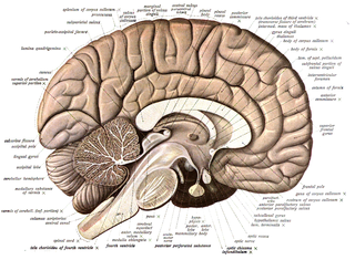
Neuroanatomy is the study of the structure and organization of the nervous system. In contrast to animals with radial symmetry, whose nervous system consists of a distributed network of cells, animals with bilateral symmetry have segregated, defined nervous systems. Their neuroanatomy is therefore better understood. In vertebrates, the nervous system is segregated into the internal structure of the brain and spinal cord and the routes of the nerves that connect to the rest of the body. The delineation of distinct structures and regions of the nervous system has been critical in investigating how it works. For example, much of what neuroscientists have learned comes from observing how damage or "lesions" to specific brain areas affects behavior or other neural functions.

The mushroom bodies or corpora pedunculata are a pair of structures in the brain of insects, other arthropods, and some annelids. They are known to play a role in olfactory learning and memory. In most insects, the mushroom bodies and the lateral horn are the two higher brain regions that receive olfactory information from the antennal lobe via projection neurons. They were first identified and described by French biologist Félix Dujardin in 1850.

Range fractionation is a term used in biology to describe the way by which a group of sensory neurons are able to encode varying magnitudes of a stimulus. Sense organs are usually composed of many sensory receptors measuring the same property. These sensory receptors show a limited degree of precision due to an upper limit in firing rate. If the receptors are endowed with distinct transfer functions in such a way that the points of highest sensitivity are scattered along the axis of the quality being measured, the precision of the sense organ as a whole can be increased.
A simple eye refers to a form of eye or an optical arrangement composed of a single lens and without an elaborate retina such as occurs in most vertebrates. In this sense "simple eye" is distinct from a multi-lensed "compound eye", and is not necessarily at all simple in the usual sense of the word.

Octopamine (molecular formula C8H11NO2; also known as OA, and also norsynephrine, para-octopamine and others) is an organic chemical closely related to norepinephrine, and synthesized biologically by a homologous pathway. Octopamine is often considered the major "fight-or-flight" neurohormone of invertebrates. Its name is derived from the fact that it was first identified in the salivary glands of the octopus.
Johnston's organ is a collection of sensory cells found in the pedicel of the antennae in the class Insecta. Johnston's organ detects motion in the flagellum. It consists of scolopidia arrayed in a bowl shape, each of which contains a mechanosensory chordotonal neuron. The number of scolopidia varies between species. In homopterans, the Johnston's organs contain 25 - 79 scolopidia. The presence of Johnston's organ is a defining characteristic which separates the class Insecta from the other hexapods belonging to the group Entognatha. Johnston's organ was named after the physician Christopher Johnston, father of the physician and Assyriologist Christopher Johnston.
The antennal lobe is the primary olfactory brain area in insects. The antennal lobe is a sphere-shaped deutocerebral neuropil in the brain that receives input from the olfactory sensory neurons in the antennae and mouthparts. Functionally, it shares some similarities with the olfactory bulb in vertebrates. The anatomy and physiology function of the insect brain can be studied by dissecting open the insect brain and imaging or carrying out in vivo electrophysiological recordings from it.

The ventral nerve cord is a major structure of the invertebrate central nervous system. It is the functional equivalent of the vertebrate spinal cord. The ventral nerve cord coordinates neural signaling from the brain to the body and vice versa, integrating sensory input and locomotor output. Because arthropods have an open circulatory system,decapitated insects can still walk, groom, and mate - illustrating that the circuitry of the ventral nerve cord is sufficient to perform complex motor programs without brain input.

Satellite glial cells, formerly called amphicytes, are glial cells that cover the surface of neuron cell bodies in ganglia of the peripheral nervous system. Thus, they are found in sensory, sympathetic, and parasympathetic ganglia. Both satellite glial cells (SGCs) and Schwann cells are derived from the neural crest of the embryo during development. SGCs have been found to play a variety of roles, including control over the microenvironment of sympathetic ganglia. They are thought to have a similar role to astrocytes in the central nervous system (CNS). They supply nutrients to the surrounding neurons and also have some structural function. Satellite cells also act as protective, cushioning cells. Additionally, they express a variety of receptors that allow for a range of interactions with neuroactive chemicals. Many of these receptors and other ion channels have recently been implicated in health issues including chronic pain and herpes simplex. There is much more to be learned about these cells, and research surrounding additional properties and roles of the SGCs is ongoing.
The suboesophageal ganglion of arthropods and in particular insects is part of the arthropod central nervous system (CNS). As indicated by its name, it is located below theoesophagus, inside the head. As part of the ventral nerve cord, it is connected to the brain and to the first thoracic ganglion. Its nerves innervate the sensory organs and muscles of the mouthparts and the salivary glands. Neurons in the suboesophageal ganglion control movement of the head and neck as well.

The (pan)arthropod head problem is a long-standing zoological dispute concerning the segmental composition of the heads of the various arthropod groups, and how they are evolutionarily related to each other. While the dispute has historically centered on the exact make-up of the insect head, it has been widened to include other living arthropods such as chelicerates, myriapods, crustaceans; and fossil forms, such as the many arthropods known from exceptionally preserved Cambrian faunas. While the topic has classically been based on insect embryology, in recent years a great deal of developmental molecular data has become available. Dozens of more or less distinct solutions to the problem, dating back to at least 1897, have been published, including several in the 2000s.
Insect physiology includes the physiology and biochemistry of insect organ systems.
The evolution of nervous systems dates back to the first development of nervous systems in animals. Neurons developed as specialized electrical signaling cells in multicellular animals, adapting the mechanism of action potentials present in motile single-celled and colonial eukaryotes. Primitive systems, like those found in protists, use chemical signalling for movement and sensitivity; data suggests these were precursors to modern neural cell types and their synapses. When some animals started living a mobile lifestyle and eating larger food particles externally, they developed ciliated epithelia, contractile muscles and coordinating & sensitive neurons for it in their outer layer.
The lateral horn is one of the two areas of the insect brain where projection neurons of the antennal lobe send their axons. The other area is the mushroom body. Several morphological classes of neurons in the lateral horn receive olfactory information through the projection neurons.

Insect olfaction refers to the function of chemical receptors that enable insects to detect and identify volatile compounds for foraging, predator avoidance, finding mating partners and locating oviposition habitats. Thus, it is the most important sensation for insects. Most important insect behaviors must be timed perfectly which is dependent on what they smell and when they smell it. For example, olfaction is essential for locating host plants and hunting prey in many species of insects, such as the moth Deilephila elpenor and the wasp Polybia sericea, respectively.
Lateral accessory lobes, or LALs are paired, symmetrical, systems of synaptic neuropils that exist in the brains of insects and other arthropods. Lateral accessory lobes are located inferiorly and laterally from ellipsoid body, anteriorly and laterally from the bulb. In the frontal section of the arthropod brain the LALs are projected as two triangles, called lateral triangles. The LALs have roughly pyramidal shape.
The protocerebrum is the first segment of the panarthropod brain.

Hair-plates are a type of proprioceptor found in the folds of insect joints. They consist of a cluster of hairs, in which each hair is innervated by a single mechanosensory neuron. Functionally, hair-plates operate as "limit-detectors" by signaling the extreme ranges of motion of a joint.

Reinhard F. Stocker is a Swiss biologist. He pioneered the analysis of the sense of smell and taste in higher animals, using the fly Drosophila melanogaster as a study case. He provided a detailed account of the anatomy and development of the olfactory system, in particular across metamorphosis, for which he received the Théodore-Ott-Prize of the Swiss Academy of Medical Sciences in 2007, and pioneered the use of larval Drosophila for the brain and behavioural sciences.

