Surgical staple

Last updated
34 surgical staples closing scalp following craniotomy 34 surgical staples, craniotomy.jpg
34 surgical staples closing scalp following craniotomy
Projectional radiograph of surgical staples Medical-staples.jpg
Projectional radiograph of surgical staples

Surgical staples are specialized staples used in surgery in place of sutures to close skin wounds or connect or remove parts of the bowels or lungs. The use of staples over sutures reduces the local inflammatory response, width of the wound, and time it takes to close. [1]

Contents

A more recent development, from the 1990s, uses clips instead of staples for some applications; this does not require the staple to penetrate. [2]

History

The technique was pioneered by "father of surgical stapling", Hungarian surgeon Hümér Hültl. [3] [4] Hultl's prototype stapler of 1908 weighed 8 pounds (3.6 kg), and required two hours to assemble and load.

The technology was refined in the 1950s in the Soviet Union, allowing for the first commercially produced re-usable stapling devices for creation of bowel and anastomeses. [4] Mark M. Ravitch brought a sample of stapling device after attending a surgical conference in USSR, and introduced it to entrepreneur Leon C. Hirsch, who founded the United States Surgical Corporation in 1964 to manufacture surgical staplers under its Auto Suture brand. [5] Until the late 1970s USSC had the market essentially to itself, but in 1977 Johnson & Johnson's Ethicon brand entered the market and today both are widely used, along with competitors from the Far East. USSC was bought by Tyco Healthcare in 1998, which became Covidien on June 29, 2007.

Safety and patency of mechanical (stapled) bowel anastomoses has been widely studied. It is generally the case in such studies that sutured anastomoses are either comparable or less prone to leakage. [6] It is possible that this is the result of recent advances in suture technology, along with increasingly risk-conscious surgical practice. Certainly modern synthetic sutures are more predictable and less prone to infection than catgut, silk and linen, which were the main suture materials used up to the 1990s.

One key feature of intestinal staplers is that the edges of the stapler act as a haemostat, compressing the edges of the wound and closing blood vessels during the stapling process. Recent studies have shown that with current suturing techniques there is no significant difference in outcome between hand sutured and mechanical anastomoses (including clips), but mechanical anastomoses are significantly quicker to perform. [7] [2]

In patients that are subjected to pulmonary resections where lung tissue is sealed with staplers, there is often postoperative air leakage. [8] Alternative techniques to seal lung tissue are currently investigated. [9]

Types and applications

Laparoscopic cholecystectomy. Gallbladderop.jpg
Laparoscopic cholecystectomy.
Close-up demonstration of a surgical skin stapler. Skin stapler closeup.jpg
Close-up demonstration of a surgical skin stapler.

The first commercial staplers were made of stainless steel with titanium staples loaded into reloadable staple cartridges.

Modern surgical staplers are either disposable and made of plastic, or reusable and made of stainless steel. Both types are generally loaded using disposable cartridges.

The staple line may be straight, curved or circular. Circular staplers are used for end-to-end anastomosis after bowel resection or, somewhat more controversially, in esophagogastric surgery. [10] The instruments may be used in either open or laparoscopic surgery, different instruments are used for each application. Laparoscopic staplers are longer, thinner, and may be articulated to allow for access from a restricted number of trocar ports.

Some staplers incorporate a knife, to complete excision and anastomosis in a single operation. Staplers are used to close both internal and skin wounds. Skin staples are usually applied using a disposable stapler, and removed with a specialized staple remover. Staplers are also used in vertical banded gastroplasty surgery (popularly known as "stomach stapling").

Vascular stapler for reducing warm ischemia in organ transplantation. With this model each stapler end can be mounted on donor and recipient by independent surgical teams without care for reciprocal orientation, being the maximal possible vascular axis torsion <=30deg. Activating guide-wire is connected just immediately before firing (video) Vascular stapler.jpg
Vascular stapler for reducing warm ischemia in organ transplantation. With this model each stapler end can be mounted on donor and recipient by independent surgical teams without care for reciprocal orientation, being the maximal possible vascular axis torsion ≤30°. Activating guide-wire is connected just immediately before firing (video)

While devices for circular end-to-end anastomosis of digestive tract are widely used, in spite of intensive research [11] [12] [13] [14] [15] circular staplers for vascular anastomosis never had yet significant impact on standard hand (Carrel) suture technique. Apart from the different modality of coupling of vascular (everted) in respect to digestive (inverted) stumps, the main basic reason could be that, particularly for small vessels, the manuality and precision required just for positioning on vascular stumps and actioning any device cannot be significantly inferior to that required to carry out the standard hand suture, then making of little utility the use of any device. An exception to that however could be organ transplantation where these two phases, i.e.device positioning at the vascular stumps and device actioning, can be carried out in different time, by different surgical team, in safe conditions when the time required does not influence donor organ preservation, i.e. at the back table in cold ischemia condition for the donor organ and after native organ removal in the recipient. This is finalized to make as brief as possible the donor organ dangerous warm ischemia phase that can be contained in the couple of minutes or less necessary just to connect the device's ends and actioning the stapler.

Although most surgical staples are made of titanium, stainless steel is more often used in some skin staples and clips. Titanium produces less reaction with the immune system and, being non-ferrous, does not interfere significantly with MRI scanners, although some imaging artifacts may result. Synthetic absorbable (bioabsorbable) staples are also now becoming available, based on polyglycolic acid, as with many synthetic absorbable sutures.

Removal of skin staples

Where skin staples are used to seal a skin wound it will be necessary to remove the staples after an appropriate healing period, usually between 5 and 10 days, depending on the location of the wound and other factors. The skin staple remover is a small manual device which consists of a shoe or plate that is sufficiently narrow and thin to insert under the skin staple. The active part is a small blade that, when hand-pressure is exerted, pushes the staple down through a slot in the shoe, deforming the staple into an 'M' shape to facilitate its removal. In an emergency it is possible to remove staples with a pair of artery forceps. [16] Skin staple removers are manufactured in many shapes and forms, [17] some disposable and some reusable.

See also

Related Research Articles

<span class="mw-page-title-main">Coronary artery bypass surgery</span> Surgical procedure to restore normal blood flow to an obstructed coronary artery

Coronary artery bypass surgery, also known as coronary artery bypass graft, is a surgical procedure to treat coronary artery disease (CAD), the buildup of plaques in the arteries of the heart. It can relieve chest pain caused by CAD, slow the progression of CAD, and increase life expectancy. It aims to bypass narrowings in heart arteries by using arteries or veins harvested from other parts of the body, thus restoring adequate blood supply to the previously ischemic heart.

<span class="mw-page-title-main">Aneurysm</span> Bulge in the wall of a blood vessel

An aneurysm is an outward bulging, likened to a bubble or balloon, caused by a localized, abnormal, weak spot on a blood vessel wall. Aneurysms may be a result of a hereditary condition or an acquired disease. Aneurysms can also be a nidus for clot formation (thrombosis) and embolization. As an aneurysm increases in size, the risk of rupture, which leads to uncontrolled bleeding, increases. Although they may occur in any blood vessel, particularly lethal examples include aneurysms of the circle of Willis in the brain, aortic aneurysms affecting the thoracic aorta, and abdominal aortic aneurysms. Aneurysms can arise in the heart itself following a heart attack, including both ventricular and atrial septal aneurysms. There are congenital atrial septal aneurysms, a rare heart defect.

<span class="mw-page-title-main">Anastomosis</span> Connection or opening between two things

An anastomosis is a connection or opening between two things that are normally diverging or branching, such as between blood vessels, leaf veins, or streams. Such a connection may be normal or abnormal ; it may be acquired or innate ; and it may be natural or artificial. The reestablishment of an anastomosis that had become blocked is called a reanastomosis. Anastomoses that are abnormal, whether congenital or acquired, are often called fistulas.

<span class="mw-page-title-main">Stapler</span> Tool used to fasten paper or flesh together

A stapler is a mechanical device that joins pages of paper or similar material by driving a thin metal staple through the sheets and folding the ends. Staplers are widely used in government, business, offices, work places, homes and schools.

<span class="mw-page-title-main">Vascular surgery</span> Medical specialty, operative procedures for the treatment of vascular disorders

Vascular surgery is a surgical subspecialty in which vascular diseases involving the arteries, veins, or lymphatic vessels, are managed by medical therapy, minimally-invasive catheter procedures and surgical reconstruction. The specialty evolved from general and cardiovascular surgery where it refined the management of just the vessels, no longer treating the heart or other organs. Modern vascular surgery includes open surgery techniques, endovascular techniques and medical management of vascular diseases - unlike the parent specialities. The vascular surgeon is trained in the diagnosis and management of diseases affecting all parts of the vascular system excluding the coronaries and intracranial vasculature. Vascular surgeons also are called to assist other physicians to carry out surgery near vessels, or to salvage vascular injuries that include hemorrhage control, dissection, occlusion or simply for safe exposure of vascular structures.

<span class="mw-page-title-main">Microsurgery</span> Surgery requiring an operating microscope

Microsurgery is a general term for surgery requiring an operating microscope. The most obvious developments have been procedures developed to allow anastomosis of successively smaller blood vessels and nerves which have allowed transfer of tissue from one part of the body to another and re-attachment of severed parts. Microsurgical techniques are utilized by several specialties today, such as general surgery, ophthalmology, orthopedic surgery, gynecological surgery, otolaryngology, neurosurgery, oral and maxillofacial surgery, endodontic microsurgery, plastic surgery, podiatric surgery and pediatric surgery.

<span class="mw-page-title-main">Surgical instrument</span> Tools designed for use during surgery

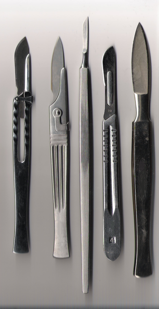
A surgical instrument is a medical device for performing specific actions or carrying out desired effects during a surgery or operation, such as modifying biological tissue, or to provide access for viewing it. Over time, many different kinds of surgical instruments and tools have been invented. Some surgical instruments are designed for general use in all sorts of surgeries, while others are designed for only certain specialties or specific procedures.

A vascular bypass is a surgical procedure performed to redirect blood flow from one area to another by reconnecting blood vessels. Often, this is done to bypass around a diseased artery, from an area of normal blood flow to another relatively normal area. It is commonly performed due to inadequate blood flow (ischemia) caused by atherosclerosis, as a part of organ transplantation, or for vascular access in hemodialysis. In general, someone's own vein (autograft) is the preferred graft material for a vascular bypass, but other types of grafts such as polytetrafluoroethylene (Teflon), polyethylene terephthalate (Dacron), or a different person's vein (allograft) are also commonly used. Arteries can also serve as vascular grafts. A surgeon sews the graft to the source and target vessels by hand using surgical suture, creating a surgical anastomosis.

<span class="mw-page-title-main">Surgical anastomosis</span> Surgical technique

A surgical anastomosis is a surgical technique used to make a new connection between two body structures that carry fluid, such as blood vessels or bowel. For example, an arterial anastomosis is used in vascular bypass and a colonic anastomosis is used to restore colonic continuity after the resection of colon cancer.

<span class="mw-page-title-main">Endovascular aneurysm repair</span> Surgery used to treat abdominal aortic aneurysm

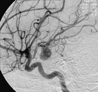
Endovascular aneurysm repair (EVAR) is a type of minimally-invasive endovascular surgery used to treat pathology of the aorta, most commonly an abdominal aortic aneurysm (AAA). When used to treat thoracic aortic disease, the procedure is then specifically termed TEVAR for "thoracic endovascular aortic/aneurysm repair." EVAR involves the placement of an expandable stent graft within the aorta to treat aortic disease without operating directly on the aorta. In 2003, EVAR surpassed open aortic surgery as the most common technique for repair of AAA, and in 2010, EVAR accounted for 78% of all intact AAA repair in the United States.

Cardiothoracic anesthesiology is a subspeciality of the medical practice of anesthesiology, devoted to the preoperative, intraoperative, and postoperative care of adult and pediatric patients undergoing cardiothoracic surgery and related invasive procedures.

<span class="mw-page-title-main">John Benjamin Murphy</span> American surgeon

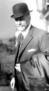
John Benjamin Murphy, born John Murphy was an American physician and abdominal surgeon noted for advocating early surgical intervention in appendicitis appendectomy, and several eponyms: Murphy’s button, Murphy drip, Murphy’s punch, Murphy’s test, and Murphy-Lane bone skid. He is best remembered for the eponymous clinical sign that is used in evaluating patients with acute cholecystitis. His career spanned general surgery, orthopedics, neurosurgery, and cardiothoracic surgery, which helped him to gain international prominence in the surgical profession. Mayo Clinic co-founder William James Mayo called him "the surgical genius of our generation".

<span class="mw-page-title-main">Hybrid cardiac surgery</span>

A hybrid cardiac surgical procedure in a narrow sense is defined as a procedure that combines a conventional, more invasive surgical part with an interventional part, using some sort of catheter-based procedure guided by fluoroscopy imaging in a hybrid operating room (OR) without interruption. The hybrid technique has a reduced risk of surgical complications and has shown decreased recovery time. It can be used to treat numerous heart diseases and conditions and with the increasing complexity of each case, the hybrid surgical technique is becoming more common.

<span class="mw-page-title-main">Félicien M. Steichen</span> American surgeon (1926–2011)

Félicien M. Steichen was a Luxembourgish-born American surgeon and Professor of Surgery. He was a pioneer in the development and use of surgical staples. He and Mark M. Ravitch are considered the fathers of modern surgical stapling. Steichen was also noted for his contributions to the development of minimally-invasive surgery.

<span class="mw-page-title-main">Open aortic surgery</span> Surgical technique

Open aortic surgery (OAS), also known as open aortic repair (OAR), describes a technique whereby an abdominal, thoracic or retroperitoneal surgical incision is used to visualize and control the aorta for purposes of treatment, usually by the replacement of the affected segment with a prosthetic graft. OAS is used to treat aneurysms of the abdominal and thoracic aorta, aortic dissection, acute aortic syndrome, and aortic ruptures. Aortobifemoral bypass is also used to treat atherosclerotic disease of the abdominal aorta below the level of the renal arteries. In 2003, OAS was surpassed by endovascular aneurysm repair (EVAR) as the most common technique for repairing abdominal aortic aneurysms in the United States.

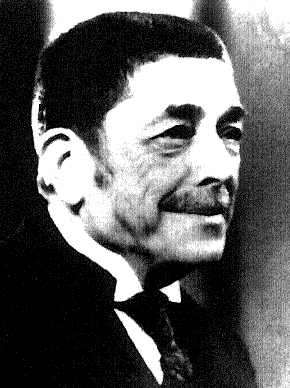
Vasilii Ivanovich Kolesov was one of the pioneers of global cardiac surgery. He was the first to perform successful internal coronary artery bypass surgery using mammary artery–coronary artery anastomosis in 1964. Also in 1964, he performed the first successful coronary bypass using a standard suture technique. Kolesov was a recipient of the USSR State Prize and Honoured Worker of Science of the RSFSR (1964).

<span class="mw-page-title-main">Hümér Hültl</span> Hungarian surgeon

Hümér Hültl was a Hungarian surgeon, noted for his work with surgical staples.

<span class="mw-page-title-main">Anil Bhan</span> Indian cardiologist

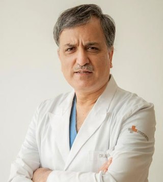
Dr. Anil Bhan is the Chairman of Cardiac Surgery Heart Institute, Medanta Hospital, Gurugram, India. He graduated from Medical College Srinagar. He has the largest experience in aortic surgery in India since he has designed and developed more than 50 surgical instruments in the field of cardiac surgery. He was one of the team members to perform the first successful heart transplant in India in1994. He served as a co-founder of Max Heart and Vascular Institute, Saket, New Delhi, Director and Chief Co-Ordinator, Cardio thoracic and Vascular Surgery, MHVI, Saket.Additional Professor, Cardiothoracic Surgery and Vascular Surgery, AIIMS, New Delhi.

<span class="mw-page-title-main">George E. Green (doctor)</span> American cardiac surgeon (born 1932)

George E. Green is an American cardiac surgeon best known for pioneering and implementing the first surgical procedure of the left coronary artery bypass graft using the internal thoracic artery sutured to the left anterior descending coronary artery to bypass obstruction to the heart circulation in the late 1960s. He applied these techniques in 1968 at New York University Medical Center. In 1970 he was hired to establish St. Luke's Hospital's cardiac surgery program in Manhattan, New York, which by 1982 was seeing approximately 1,800 cases a year, the biggest program in the state. Green has lectured internationally on the topic, and has written numerous reports on internal thoracic artery grafting, as well as co-authoring Surgical Revascularization of the Heart: The Internal Thoracic Arteries.

<span class="mw-page-title-main">Antrectomy</span> Type of gastric resection surgery

Antrectomy, also called distal gastrectomy, is a type of gastric resection surgery that involves the removal of the stomach antrum to treat gastric diseases causing the damage, bleeding, or blockage of the stomach. This is performed using either the Billroth I (BI) or Billroth II (BII) reconstruction method. Quite often, antrectomy is used alongside vagotomy to maximise its safety and effectiveness. Modern antrectomies typically have a high success rate and low mortality rate, but the exact numbers depend on the specific conditions being treated.

References

  1. Iavazzo, Christos; Gkegkes, Ioannis D.; Vouloumanou, Evridiki K.; Mamais, Ioannis; Peppas, George; Falagas, Matthew E. (September 2011). "Sutures versus staples for the management of surgical wounds: a meta-analysis of randomized controlled trials". The American Surgeon. 77 (9): 1206–1221. doi: 10.1177/000313481107700935 . ISSN   1555-9823. PMID   21944632. S2CID   40578006.
  2. 1 2 Chughtai, T.; Chen, L. Q.; Salasidis, G.; Nguyen, D.; Tchervenkov, C.; Morin, J. F. (November 2000). "Clips versus suture technique: is there a difference?". The Canadian Journal of Cardiology. 16 (11): 1403–1407. ISSN   0828-282X. PMID   11109037.
  3. Non-suture methods of vascular anastomosis, British Journal of Surgery, 19 Feb 2003: Volume 90, Issue 3, Pages 261 - 271
  4. 1 2 Konstantinov, Igor E (July 2004). "Circular vascular stapling in coronary surgery". The Annals of Thoracic Surgery. 78 (1): 369–373. doi: 10.1016/j.athoracsur.2003.11.050 . PMID   15223474.
  5. History of United States Surgical Corporation
  6. Brundage Susan I (2001). "Stapled versus Sutured Gastrointestinal Anastomoses in the Trauma Patient: A Multicenter Trial". Journal of Trauma-Injury Infection & Critical Care. 51 (6): 1054–1061. doi:10.1097/00005373-200112000-00005. PMID   11740250.
  7. Catena, Fausto; Donna, Michele La; Gagliardi, Stefano; Avanzolini, Andrea; Taffurelli, Mario (2004). "Stapled Versus Hand-Sewn Anastomoses in Emergency Intestinal Surgery: Results of a Prospective Randomized Study". Surgery Today. 34 (2): 123–126. doi:10.1007/s00595-003-2678-0. PMID   14745611. S2CID   6386495.
  8. Venuta, F; Rendina, EA; De Giacomo, T; Flaishman, I; Guarino, E; Ciccone, AM; Ricci, C (April 1998). "Technique to reduce air leaks after pulmonary lobectomy". European Journal of Cardio-Thoracic Surgery. 13 (4): 361–4. doi: 10.1016/S1010-7940(98)00038-4 . PMID   9641332.
  9. Guedes, Rogério Luizari; Höglund, Odd Viking; Brum, Juliana Sperotto; Borg, Niklas; Dornbusch, Peterson Triches (3 January 2018). "Resorbable Self-Locking Implant for Lung Lobectomy Through Video-Assisted Thoracoscopic Surgery: First Live Animal Application". Surgical Innovation. 25 (2): 158–164. doi:10.1177/1553350617751293. PMID   29298608. S2CID   4965005.
  10. European Journal of Cardio-Thoracic Surgery, Volume 25, Issue 6, June 2004, Pages 1097-1101 [ dead link ]
  11. Kolesov VI, Kolesov EV, Gurevich IY, Leosko VA (1970). "Vasosuturing apparatuses in surgery of coronary arteries". Med Tekhnika. 6: 24–8.{{cite journal}}: CS1 maint: multiple names: authors list (link)
  12. Kolesov VI & Kolesov EV. (1991) Twenty years' results with internal thoracic artery-coronary artery anastomosis [letter]. J Thorac Cardiovasc Surg. 101:360–1
  13. Nazari S et al. A new vascular stapler for pulmonary artery anastomosis in experimental single lung trasnplantation.Video, Proceedings of the 4th Annual Meeting of The Association for Cardio-Thoracic Surgery, Naples, Sept 16-19, 1990
  14. "Evaluation of an aortic stapler for an open aortic anastomosis". The Journal of Cardiovascular Surgery (Torino). 48 (5): 659–65. Oct 2007 via Minerva Medica.
  15. Shifrin, E.G.; Moore, W.S.; Bell, P.R.F.; Kolvenbach, R.; Daniline, E.I. (Apr 2007). "Intravascular Stapler for "Open" Aortic Surgery: Preliminary Results". European Journal of Vascular and Endovascular Surgery. 33 (4): 408–11. doi: 10.1016/j.ejvs.2006.10.019 . PMID   17137806.
  16. Teoh, MK; Bird, DA (1 September 1987), "Removal of skin staples in an emergency", Ann R Coll Surg Engl, 69 (5): 222–4, PMC   2498551 , PMID   3314634
  17. "Skin staple removers - Google Search".