Telomeres in the cell cycle

Last updated

Telomeres, the caps on the ends of eukaryotic chromosomes, play critical roles in cellular aging and cancer. An important facet to how telomeres function in these roles is their involvement in cell cycle regulation.

Contents

Eukaryotic cells

Because eukaryotic chromosomes are linear and because DNA replication by DNA polymerase requires the presence of an RNA primer that is later degraded, eukaryotic cells face the end-replication problem. [1] This problem makes eukaryotic cells unable to copy the last few bases on the 3’ end of the template DNA strand, leading to chromosome—and, therefore, telomere—shortening every S phase. [2] Measurements of telomere lengths across cell types at various ages suggest that this gradual chromosome shortening results in a gradual reduction in telomere length at a rate of approximately 25 nucleotides per year. [3]

Cell cycle enablers and regulators

The telomere-shelterin complexes that cap all eukaryotic chromosomes ensure that healthy cells can progress through the cell cycle by preventing the cellular DNA damage response from identifying chromosome ends as double-stranded breaks (DSBs). [4] [5] Without a protective cap, chromosome ends would appear identical to intrachromosomal DSBs. These DSBs activate a DNA damage response pathway that halts the cell cycle until the breaks are repaired. This checkpoint pathway is initiated in S. cerevisiae by recruitment of protein kinases Mec1 and Tel1 and in mammals by recruitment of protein kinases ATR and ATM. [6] Regarding DSB repair, eukaryotes generally use two strategies: non-homologous end joining (NHEJ), which involves rapid reattachment of the broken ends; and homologous recombination (HR), which involves the use of a homologous DNA sequence to repair the break. Because HR requires a homologous sequence, its use is restricted to S/G2 phase. [7] (Interestingly, as with many other aspects of the cell cycle, [8] cyclin-dependent kinases are responsible for downregulating NHEJ during S/G2 phase to ensure use of the more accurate HR. [9] ) As shown in Figure 1A, telomere-shelterin complexes contain motifs that inhibit the DNA damage checkpoint, NHEJ, and HR.

Initial work on the role of telomere-bound protein complexes in S. cerevisiae elucidated the mechanism by which these complexes prevent checkpoint activation and DSB repair of chromosome ends. The two major protein complexes that bind to telomeric DNA in S. cerevisiae are:

The CST complex and Rif1 prevent Mec1 recruitment, thereby preventing checkpoint activation. [5] [10] Meanwhile, Rif2 and Rap1 inhibit NHEJ: knocking out Rif2 or Rap1 results in longer telomeres as measured by PCR, indicating that NHEJ occurred. [11] These knockout strains (unlike strains lacking functional CST or Rif1) continue to cycle, further suggesting that Rif2 and Rap1 are not involved in inhibiting checkpoint activation. [12]

Analogously, proteins that bind to human telomeres as part of the shelterin complex enable cell cycle progress and prevent erroneous DSB repair. POT1 protein binds to ssDNA, prevents checkpoint activation through inhibiting ATR recruitment, and prevents HR; [13] [14] RAP1, a GTPase, binds to dsDNA and prevents HR; [15] and TRF2 protein (also known as TERF2) binds to dsDNA, prevents checkpoint activation through inhibiting ATM recruitment, and prevents NHEJ. [16] [17] TRF2 is unique among these proteins in its role in the formation and maintenance of T-loops: lariat structures formed by the folding of the ssDNA overhang back onto the dsDNA. T-loops may further inhibit the binding of checkpoint activation proteins. [18]

Figure 1. (A) Telomere-bound proteins involved in preventing the activation of the DNA damage response checkpoint and of DSB repair mechanisms in S. cerevisiae (top) and in humans (bottom). (B) Overview of the normal function of telomere-shelterin complexes and the pathways activated by telomere shortening. Shelterin-telomere complexes.png
Figure 1.(A) Telomere-bound proteins involved in preventing the activation of the DNA damage response checkpoint and of DSB repair mechanisms in S. cerevisiae (top) and in humans (bottom). (B) Overview of the normal function of telomere-shelterin complexes and the pathways activated by telomere shortening.

As telomeres shorten as a natural consequence of repeated cell division or due to other factors, such as oxidative stress, [19] shelterin proteins lose the ability to bind to telomeric DNA. When telomeres reach a critically short length, sufficient shelterin proteins to inhibit checkpoint activation are not available, although NHEJ and HR generally remain inhibited at this point. [5] This loss of inhibition is one reason why telomere shortening causes senescence (Figure 1B).

Telomeres and cell cycle deregulation

Almost all cancer cells have shortened telomeres. [20] This may seem counter-intuitive, as short telomeres should activate the ATR/ATM DNA damage checkpoint and thereby prevent division. Resolving the question of why cancer cells have short telomeres led to the development of a two-stage model for how cancer cells subvert telomeric regulation of the cell cycle. First, the DNA damage checkpoint must be inactivated to allow cells to continue dividing even when telomeres pass the critical length threshold. This requirement follows not only from the discussion above but also from in vivo evidence showing the function of this checkpoint in precancerous lesions and its dysfunction in late-stage tumors. [21] Second, to survive after disabling the DNA damage checkpoint, precancerous cells must activate mechanisms to extend their telomeres. [22] [23] As a result of the continued division past the point of normal senescence, the telomeres of these cells become too short to prevent NHEJ(Non Homologous End Joining) and HR(Homologous Recombination) of chromosome ends, causing a state known as crisis. [24] The application of these DSB (double strand breaks)repair mechanisms to chromosome ends leads to genetic instability, and while this instability can promote carcinogenesis, it induces apoptosis if experienced for too long. [23] To survive and replicate, precancerous cells must stabilize their telomere lengths. This occurs through telomerase activation or the activation of a telomere-recombination pathway (i.e., the ALT pathway). [22] [25] Thus, cancer cells have short telomeres because they progress through an intermediate stage of telomere shortening—caused by division after DNA damage checkpoint inactivation—before enabling mechanisms for maintaining telomere length.

Since the late 1990s, researchers have proposed using telomerase inhibitors as cancer treatments. [26] [27] [28] While such inhibitors have been seriously considered for cancer therapy since the late 2000s, they are not commonly used. [29] Two concerns with applying telomerase inhibitors in cancer treatment are that effective treatment requires continuous, long-term drug application and that off-target effects are common. [30] For example, the telomerase inhibitor imetelstat, first proposed in 2003, [31] [32] has been held up in clinical trials due to hematological toxicity. [30] Despite these concerns, the development of telomerase-based cancer treatments remains an active research area.

Cell cycle timing of telomere elongation

Although telomerase activation does not occur during the cell cycle of normal somatic human cells, the association between telomere elongation (especially elongation by telomerase) and tumor development emphasizes the importance of understanding when such elongation can occur during the cell cycle. Work with S. cerevisiae has identified telomerase activity as restricted to late S phase. [33] [34] Researchers generated S. cerevisiae strains with galactose-inducible shortened telomeres. [33] They then used α factor to block cells with induced short telomeres in late G1 phase and measured the change in telomere length when the cells were released under a variety of conditions. They found that when the cells were released and concurrently treated with nocodazole, a G2/M phase cell cycle inhibitor, telomere length increased for the first few hours and then remained constant. In comparison, when cells were released and allowed to cycle, telomere length increased linearly with time. [34] These data suggest that telomere elongation occurs only in S phase. Additional experiments with greater time resolution support this hypothesis and narrow the timeframe to late S phase. Researchers tied telomere elongation in these experiments to telomerase activity by observing that in an S. cerevisiae strain with a dysfunctional ALT pathway telomere elongation still occurs. [34]

Related Research Articles

Telomerase Telomere-restoring protein active in the most rapidly dividing cells

Telomerase, also called terminal transferase, is a ribonucleoprotein that adds a species-dependent telomere repeat sequence to the 3' end of telomeres. A telomere is a region of repetitive sequences at each end of the chromosomes of most eukaryotes. Telomeres protect the end of the chromosome from DNA damage or from fusion with neighbouring chromosomes. The fruit fly Drosophila melanogaster lacks telomerase, but instead uses retrotransposons to maintain telomeres.

DNA repair Cellular mechanism

DNA repair is a collection of processes by which a cell identifies and corrects damage to the DNA molecules that encode its genome. In human cells, both normal metabolic activities and environmental factors such as radiation can cause DNA damage, resulting in tens of thousands of individual molecular lesions per cell per day. Many of these lesions cause structural damage to the DNA molecule and can alter or eliminate the cell's ability to transcribe the gene that the affected DNA encodes. Other lesions induce potentially harmful mutations in the cell's genome, which affect the survival of its daughter cells after it undergoes mitosis. As a consequence, the DNA repair process is constantly active as it responds to damage in the DNA structure. When normal repair processes fail, and when cellular apoptosis does not occur, irreparable DNA damage may occur, including double-strand breaks and DNA crosslinkages. This can eventually lead to malignant tumors, or cancer as per the two hit hypothesis.

Non-homologous end joining

Non-homologous end joining (NHEJ) is a pathway that repairs double-strand breaks in DNA. NHEJ is referred to as "non-homologous" because the break ends are directly ligated without the need for a homologous template, in contrast to homology directed repair, which requires a homologous sequence to guide repair. The term "non-homologous end joining" was coined in 1996 by Moore and Haber.

Homologous recombination Type of genetic recombination

Homologous recombination is a type of genetic recombination in which genetic information is exchanged between two similar or identical molecules of double-stranded or single-stranded nucleic acids. It is widely used by cells to accurately repair harmful breaks that occur on both strands of DNA, known as double-strand breaks (DSB), in a process called homologous recombinational repair (HRR). Homologous recombination also produces new combinations of DNA sequences during meiosis, the process by which eukaryotes make gamete cells, like sperm and egg cells in animals. These new combinations of DNA represent genetic variation in offspring, which in turn enables populations to adapt during the course of evolution. Homologous recombination is also used in horizontal gene transfer to exchange genetic material between different strains and species of bacteria and viruses.

Dyskeratosis congenita Medical condition

Dyskeratosis congenita (DKC),also known as Zinsser-Engman-Cole syndrome, is a rare progressive congenital disorder with a highly variable phenotype. The entity was classically defined by the triad of abnormal skin pigmentation, nail dystrophy, and leukoplakia of the oral mucosa, but these components do not always occur. DKC is characterized by short telomeres. Some of the manifestations resemble premature ageing. The disease initially mainly affects the skin, but a major consequence is progressive bone marrow failure which occurs in over 80%, causing early mortality.

Ku (protein)

Ku is a dimeric protein complex that binds to DNA double-strand break ends and is required for the non-homologous end joining (NHEJ) pathway of DNA repair. Ku is evolutionarily conserved from bacteria to humans. The ancestral bacterial Ku is a homodimer. Eukaryotic Ku is a heterodimer of two polypeptides, Ku70 (XRCC6) and Ku80 (XRCC5), so named because the molecular weight of the human Ku proteins is around 70 kDa and 80 kDa. The two Ku subunits form a basket-shaped structure that threads onto the DNA end. Once bound, Ku can slide down the DNA strand, allowing more Ku molecules to thread onto the end. In higher eukaryotes, Ku forms a complex with the DNA-dependent protein kinase catalytic subunit (DNA-PKcs) to form the full DNA-dependent protein kinase, DNA-PK. Ku is thought to function as a molecular scaffold to which other proteins involved in NHEJ can bind, orienting the double-strand break for ligation.

Heterogeneous nuclear ribonucleoproteins (hnRNPs) are complexes of RNA and protein present in the cell nucleus during gene transcription and subsequent post-transcriptional modification of the newly synthesized RNA (pre-mRNA). The presence of the proteins bound to a pre-mRNA molecule serves as a signal that the pre-mRNA is not yet fully processed and therefore not ready for export to the cytoplasm. Since most mature RNA is exported from the nucleus relatively quickly, most RNA-binding protein in the nucleus exist as heterogeneous ribonucleoprotein particles. After splicing has occurred, the proteins remain bound to spliced introns and target them for degradation.

Ataxia telangiectasia and Rad3 related Protein kinase that detects DNA damage and halts cell division

Serine/threonine-protein kinase ATR also known as ataxia telangiectasia and Rad3-related protein (ATR) or FRAP-related protein 1 (FRP1) is an enzyme that, in humans, is encoded by the ATR gene. It is a large kinase of about 301.66 kDa. ATR belongs to the phosphatidylinositol 3-kinase-related kinase protein family. ATR is activated in response to single strand breaks, and works with ATM to ensure genome integrity.

TERF2

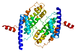
Telomeric repeat-binding factor 2 is a protein that is present at telomeres throughout the cell cycle. It is also known as TERF2, TRF2, and TRBF2, and is encoded in humans by the TERF2 gene. It is a component of the shelterin nucleoprotein complex and a second negative regulator of telomere length, playing a key role in the protective activity of telomeres. It was first reported in 1997 in the lab of Titia de Lange, where a DNA sequence similar, but not identical, to TERF1 was discovered, with respect to the Myb-domain. De Lange isolated the new Myb-containing protein sequence and called it TERF2.

TERF1 Protein-coding gene in humans

Telomeric repeat-binding factor 1 is a protein that in humans is encoded by the TERF1 gene.

PINX1

PIN2/TERF1-interacting telomerase inhibitor 1, also known as PINX1, is a human gene. PINX1 is also known as PIN2 interacting protein 1. PINX1 is a telomerase inhibitor and a possible tumor suppressor.

The MRN complex is a protein complex consisting of Mre11, Rad50 and Nbs1. In eukaryotes, the MRN/X complex plays an important role in the initial processing of double-strand DNA breaks prior to repair by homologous recombination or non-homologous end joining. The MRN complex binds avidly to double-strand breaks both in vitro and in vivo and may serve to tether broken ends prior to repair by non-homologous end joining or to initiate DNA end resection prior to repair by homologous recombination. The MRN complex also participates in activating the checkpoint kinase ATM in response to DNA damage. Production of short single-strand oligonucleotides by Mre11 endonuclease activity has been implicated in ATM activation by the MRN complex.

Meiotic recombination checkpoint

The meiotic recombination checkpoint monitors meiotic recombination during meiosis, and blocks the entry into metaphase I if recombination is not efficiently processed.

Telomere-binding proteins function to bind telomeric DNA in various species. In particular, telomere-binding protein refers to TTAGGG repeat binding factor-1 (TERF1) and TTAGGG repeat binding factor-2 (TERF2). Telomere sequences in humans are composed of TTAGGG sequences which provide protection and replication of chromosome ends to prevent degradation. Telomere-binding proteins can generate a T-loop to protect chromosome ends. TRFs are double-stranded proteins which are known to induce bending, looping, and pairing of DNA which aids in the formation of T-loops. They directly bind to TTAGGG repeat sequence in the DNA. There are also subtelomeric regions present for regulation. However, in humans, there are six subunits forming a complex known as shelterin.

Shelterin is a protein complex known to protect telomeres in many eukaryotes from DNA repair mechanisms, as well as to regulate telomerase activity. In mammals and other vertebrates, telomeric DNA consists of repeating double-stranded 5'-TTAGGG-3' (G-strand) sequences along with the 3'-AATCCC-5' (C-strand) complement, ending with a 50-400 nucleotide 3' (G-strand) overhang. Much of the final double-stranded portion of the telomere forms a T-loop (Telomere-loop) that is invaded by the 3' (G-strand) overhang to form a small D-loop (Displacement-loop).

Chromosomal instability (CIN) is a type of genomic instability in which chromosomes are unstable, such that either whole chromosomes or parts of chromosomes are duplicated or deleted. More specifically, CIN refers to the increase in rate of addition or loss of entire chromosomes or sections of them. The unequal distribution of DNA to daughter cells upon mitosis results in a failure to maintain euploidy leading to aneuploidy. In other words, the daughter cells do not have the same number of chromosomes as the cell they originated from. Chromosomal instability is the most common form of genetic instability and cause of aneuploidy.

Titia de Lange Dutch geneticist

Titia de Lange is the Director of the Anderson Center for Cancer Research, the Leon Hess professor and the head of Laboratory Cell Biology and Genetics at Rockefeller University.

Telomeric repeat–containing RNA Long non-coding RNA transcribed from telomeres

Telomeric repeat-containing RNA (TERRA) is a long non-coding RNA transcribed from telomeres - repetitive nucleotide regions found on the ends of chromosomes that function to protect DNA from deterioration or fusion with neighboring chromosomes. TERRA has been shown to be ubiquitously expressed in almost all cell types containing linear chromosomes - including humans, mice, and yeasts. While the exact function of TERRA is still an active area of research, it is generally believed to play a role in regulating telomerase activity as well as maintaining the heterochromatic state at the ends of chromosomes. TERRA interaction with other associated telomeric proteins has also been shown to help regulate telomere integrity in a length-dependent manner.

Cell cycle withdrawal refers to the natural stoppage of cell cycle during cell division. When cells divide, there are many internal or external factors that would lead to a stoppage of division. This stoppage could be permanent or temporary, and could occur in any one of the four cycle phases, depending on the status of cells or the activities they are undergoing. During the process, all cell duplication process, including mitosis, meiosis as well as DNA replication, will be paused. The mechanisms involve the proteins and DNA sequences inside cells.

DNA end resection

DNA end resection, also called 5′–3′ degradation, is a biochemical process where the blunt end of a section of double-stranded DNA is modified by cutting away some nucleotides from the 5' end to produce a 3' single-stranded sequence.

References

  1. Levy, Michael Z.; Allsopp, Richard C.; Futcher, A.Bruce; Greider, Carol W.; Harley, Calvin B. (June 1996). "Telomere end-replication problem and cell aging". Journal of Molecular Biology. 225 (4): 951–960. doi:10.1016/0022-2836(92)90096-3. ISSN   0022-2836. PMID   1613801.
  2. Aubert, Geraldine; Lansdorp, Peter M. (April 2008). "Telomeres and Aging". Physiological Reviews. 88 (2): 557–579. doi:10.1152/physrev.00026.2007. ISSN   0031-9333. PMID   18391173.
  3. Daniali, Lily; Benetos, Athanase; Susser, Ezra; Kark, Jeremy D.; Labat, Carlos; Kimura, Masayuki; Desai, Kunj K.; Granick, Mark; Aviv, Abraham (2013-07-24). "Erratum: Corrigendum: Telomeres shorten at equivalent rates in somatic tissues of adults". Nature Communications. 4 (1). doi: 10.1038/ncomms2976 . ISSN   2041-1723.
  4. Martínez, Paula; Blasco, Maria A. (2010-09-16). "Role of shelterin in cancer and aging". Aging Cell. 9 (5): 653–666. doi: 10.1111/j.1474-9726.2010.00596.x . ISSN   1474-9718. PMID   20569239.
  5. 1 2 3 4 Gobbini, Elisa; Trovesi, Camilla; Cassani, Corinne; Longhese, Maria Pia (28 July 2014). "Telomere uncapping at the crossroad between cell cycle arrest and carcinogenesis". Molecular & Cellular Oncology. 1 (1): e29901. doi:10.4161/mco.29901. PMC   4905175 . PMID   27308311.
  6. Burma, Sandeep; Chen, Benjamin P.C.; Chen, David J. (September 2006). "Role of non-homologous end joining (NHEJ) in maintaining genomic integrity". DNA Repair. 5 (9–10): 1042–1048. doi:10.1016/j.dnarep.2006.05.026. ISSN   1568-7864. PMID   16822724.
  7. Ciccia, Alberto; Elledge, Stephen J. (October 2010). "The DNA Damage Response: Making It Safe to Play with Knives". Molecular Cell. 40 (2): 179–204. doi:10.1016/j.molcel.2010.09.019. ISSN   1097-2765. PMC   2988877 . PMID   20965415.
  8. Murray, Andrew W.; Kirschner, Marc W. (May 1989). "Cyclin synthesis drives the early embryonic cell cycle". Nature. 339 (6222): 275–280. Bibcode:1989Natur.339..275M. doi:10.1038/339275a0. ISSN   0028-0836. PMID   2566917.
  9. Ira, Grzegorz; Pellicioli, Achille; Balijja, Alitukiriza; Wang, Xuan; Fiorani, Simona; Carotenuto, Walter; Liberi, Giordano; Bressan, Debra; Wan, Lihong; Hollingsworth, Nancy M.; Haber, James E. (October 2004). "DNA end resection, homologous recombination and DNA damage checkpoint activation require CDK1". Nature. 431 (7011): 1011–1017. Bibcode:2004Natur.431.1011I. doi:10.1038/nature02964. ISSN   0028-0836. PMC   4493751 . PMID   15496928.
  10. Ribeyre, Cyril; Shore, David (2012-02-12). "Anticheckpoint pathways at telomeres in yeast". Nature Structural & Molecular Biology. 19 (3): 307–313. doi:10.1038/nsmb.2225. ISSN   1545-9993. PMID   22343724.
  11. Marcand, S.; Pardo, B.; Gratias, A.; Cahun, S.; Callebaut, I. (2008-05-01). "Multiple pathways inhibit NHEJ at telomeres". Genes & Development. 22 (9): 1153–1158. doi:10.1101/gad.455108. ISSN   0890-9369. PMC   2335312 . PMID   18451106.
  12. Bonetti, Diego; Clerici, Michela; Anbalagan, Savani; Martina, Marina; Lucchini, Giovanna; Longhese, Maria Pia (2010-05-27). "Shelterin-Like Proteins and Yku Inhibit Nucleolytic Processing of Saccharomyces cerevisiae Telomeres". PLoS Genetics. 6 (5): e1000966. doi:10.1371/journal.pgen.1000966. ISSN   1553-7404. PMC   2877729 . PMID   20523746.
  13. Hockemeyer, Dirk; Sfeir, Agnel J; Shay, Jerry W; Wright, Woodring E; de Lange, Titia (2005-06-23). "POT1 protects telomeres from a transient DNA damage response and determines how human chromosomes end". The EMBO Journal. 24 (14): 2667–2678. doi:10.1038/sj.emboj.7600733. ISSN   0261-4189. PMC   1176460 . PMID   15973431.
  14. Wu, Ling; Multani, Asha S.; He, Hua; Cosme-Blanco, Wilfredo; Deng, Yu; Deng, Jian Min; Bachilo, Olga; Pathak, Sen; Tahara, Hidetoshi; Bailey, Susan M.; Deng, Yibin (July 2006). "Pot1 Deficiency Initiates DNA Damage Checkpoint Activation and Aberrant Homologous Recombination at Telomeres". Cell. 126 (1): 49–62. doi: 10.1016/j.cell.2006.05.037 . ISSN   0092-8674. PMID   16839876.
  15. Sfeir, A.; Kabir, S.; van Overbeek, M.; Celli, G. B.; de Lange, T. (2010-03-25). "Loss of Rap1 Induces Telomere Recombination in the Absence of NHEJ or a DNA Damage Signal". Science. 327 (5973): 1657–1661. Bibcode:2010Sci...327.1657S. doi:10.1126/science.1185100. ISSN   0036-8075. PMC   2864730 . PMID   20339076.
  16. Karlseder, J. (1999-02-26). "p53- and ATM-Dependent Apoptosis Induced by Telomeres Lacking TRF2". Science. 283 (5406): 1321–1325. doi:10.1126/science.283.5406.1321. ISSN   0036-8075. PMID   10037601.
  17. Celli, Giulia B.; de Lange, Titia (2005-06-19). "DNA processing is not required for ATM-mediated telomere damage response after TRF2 deletion". Nature Cell Biology. 7 (7): 712–718. doi:10.1038/ncb1275. ISSN   1465-7392. PMID   15968270.
  18. Doksani, Ylli; Wu, John Y.; de Lange, Titia; Zhuang, Xiaowei (October 2013). "Super-Resolution Fluorescence Imaging of Telomeres Reveals TRF2-Dependent T-loop Formation". Cell. 155 (2): 345–356. doi:10.1016/j.cell.2013.09.048. ISSN   0092-8674. PMC   4062873 . PMID   24120135.
  19. von Zglinicki, Thomas (July 2002). "Oxidative stress shortens telomeres". Trends in Biochemical Sciences. 27 (7): 339–344. doi:10.1016/s0968-0004(02)02110-2. ISSN   0968-0004. PMID   12114022.
  20. Shay, Jerry W.; Wright, Woodring E. (December 2011). "Role of telomeres and telomerase in cancer". Seminars in Cancer Biology. 21 (6): 349–353. doi:10.1016/j.semcancer.2011.10.001. ISSN   1044-579X. PMC   3370415 . PMID   22015685.
  21. Wang, Haiyong; Zhang, Xiaoshan; Teng, Lisong; Legerski, Randy J. (June 2015). "DNA damage checkpoint recovery and cancer development". Experimental Cell Research. 334 (2): 350–358. doi:10.1016/j.yexcr.2015.03.011. ISSN   0014-4827. PMID   25842165.
  22. 1 2 Chin, Lynda; Artandi, Steven E; Shen, Qiong; Tam, Alice; Lee, Shwu-Luan; Gottlieb, Geoffrey J; Greider, Carol W; DePinho, Ronald A (May 1999). "p53 Deficiency Rescues the Adverse Effects of Telomere Loss and Cooperates with Telomere Dysfunction to Accelerate Carcinogenesis". Cell. 97 (4): 527–538. doi: 10.1016/s0092-8674(00)80762-x . ISSN   0092-8674. PMID   10338216.
  23. 1 2 Rudolph, Karl Lenhard; Millard, Melissa; Bosenberg, Marcus W.; DePinho, Ronald A. (June 2001). "Telomere dysfunction and evolution of intestinal carcinoma in mice and humans". Nature Genetics. 28 (2): 155–159. doi:10.1038/88871. ISSN   1061-4036. PMID   11381263.
  24. Counter, C.M.; Avilion, A.A.; LeFeuvre, C.E.; Stewart, N.G.; Greider, C.W.; Harley, C.B.; Bacchetti, S. (May 1992). "Telomere shortening associated with chromosome instability is arrested in immortal cells which express telomerase activity". The EMBO Journal. 11 (5): 1921–1929. doi:10.1002/j.1460-2075.1992.tb05245.x. ISSN   0261-4189.
  25. Lundblad, Victoria; Blackburn, Elizabeth H. (April 1993). "An alternative pathway for yeast telomere maintenance rescues est1− senescence". Cell. 73 (2): 347–360. doi:10.1016/0092-8674(93)90234-h. ISSN   0092-8674. PMID   8477448.
  26. Saretzki, Gabriele (May 2003). "Telomerase inhibition as cancer therapy". Cancer Letters. 194 (2): 209–219. doi:10.1016/s0304-3835(02)00708-5. ISSN   0304-3835. PMID   12757979.
  27. Kim, N.W. (April 1997). "Clinical implications of telomerase in cancer". European Journal of Cancer. 33 (5): 781–786. doi:10.1016/s0959-8049(97)00057-9. ISSN   0959-8049. PMID   9282117.
  28. Autexier, Chantal; Greider, Carol (October 1996). "Telomerase and cancer: revisiting the telomere hypothesis". Trends in Biochemical Sciences. 21 (10): 387–391. doi:10.1016/s0968-0004(96)10042-6. ISSN   0968-0004.
  29. Ivancich, Marko; Schrank, Zachary; Wojdyla, Luke; Leviskas, Brandon; Kuckovic, Adijan; Sanjali, Ankita; Puri, Neelu (2017-02-19). "Treating Cancer by Targeting Telomeres and Telomerase". Antioxidants. 6 (1): 15. doi:10.3390/antiox6010015. ISSN   2076-3921. PMC   5384178 . PMID   28218725.
  30. 1 2 Jafri, Mohammad A.; Ansari, Shakeel A.; Alqahtani, Mohammed H.; Shay, Jerry W. (2016-06-20). "Roles of telomeres and telomerase in cancer, and advances in telomerase-targeted therapies". Genome Medicine. 8 (1): 69. doi:10.1186/s13073-016-0324-x. ISSN   1756-994X. PMC   4915101 . PMID   27323951.
  31. Gryaznov, Sergei; Asai, Akira; Oshima, Yuko; Yamamoto, Yoshihiro; Pongracz, Krisztina; Pruzan, Ronald; Wunder, Ellen; Piatyszek, Mieczyslaw; Li, Shihong; Chin, Allison; Harley, Calvin; Akinaga, Shiro; Yamashita, Yoshinori (October 2003). "Oligonucleotide N3′ → P5′ Thio-phosphoramidate Telomerase Template Antagonists as Potential Anticancer Agents". Nucleosides, Nucleotides and Nucleic Acids. 22 (5–8): 577–581. doi:10.1081/NCN-120021958. PMID   14565232.
  32. Röth, Alexander; Harley, Calvin B.; Baerlocher, Gabriela M. (2010). "Imetelstat (GRN163L) - Telomerase-Based Cancer Therapy". Small Molecules in Oncology. Recent Results in Cancer Research. 184. pp. 221–234. doi:10.1007/978-3-642-01222-8_16. ISBN   978-3-642-01221-1. PMID   20072842.
  33. 1 2 Marcand, S.; Brevet, V; Gilson, E (15 June 1999). "Progressive cis-inhibition of telomerase upon telomere elongation". The EMBO Journal. 18 (12): 3509–3519. doi:10.1093/emboj/18.12.3509. PMC   1171430 . PMID   10369690.
  34. 1 2 3 Marcand, Stéphane; Brevet, Vanessa; Mann, Carl; Gilson, Eric (April 2000). "Cell cycle restriction of telomere elongation". Current Biology. 10 (8): 487–490. doi: 10.1016/s0960-9822(00)00450-4 . PMID   10801419.