Unihemispheric slow-wave sleep

Last updated
A young house sparrow (Passer domesticus) exhibits unihemispheric slow-wave sleep. Half Sleeping Bird.jpg
A young house sparrow ( Passer domesticus ) exhibits unihemispheric slow-wave sleep.

Unihemispheric slow-wave sleep (USWS) is sleep where one half of the brain rests while the other half remains alert. This is in contrast to normal sleep where both eyes are shut and both halves of the brain show unconsciousness. In USWS, also known as asymmetric slow-wave sleep, one half of the brain is in deep sleep, a form of non-rapid eye movement sleep and the eye corresponding to this half is closed while the other eye remains open. When examined by electroencephalography (EEG), the characteristic slow-wave sleep tracings are seen from one side while the other side shows a characteristic tracing of wakefulness. [1] The phenomenon has been observed in a number of terrestrial, aquatic and avian species.

Contents

Unique physiology, including the differential release of the neurotransmitter acetylcholine, has been linked to the phenomenon. [1] USWS offers a number of benefits, including the ability to rest in areas of high predation or during long migratory flights. The behaviour remains an important research topic because USWS is possibly the first animal behaviour which uses different regions of the brain to simultaneously control sleep and wakefulness. [2] The greatest theoretical importance of USWS is its potential role in elucidating the function of sleep by challenging various current notions. Researchers have looked to animals exhibiting USWS to determine if sleep must be essential; otherwise, species exhibiting USWS would have eliminated the behaviour altogether through evolution. [3]

The amount of time spent sleeping during the unihemispheric slow-wave stage is considerably less than the bilateral slow-wave sleep. In the past, aquatic animals, such as dolphins and seals, had to regularly surface in order to breathe and regulate body temperature. USWS might have been generated by the need to perform these vital activities simultaneously with sleep. [4]

On land, birds can switch between sleeping with both hemispheres to one hemisphere. Due to their poorly webbed feet and long wings, which are not completely waterproof, it is not energetically efficient for them to make rest stops or land on water, only to take flight again. Using unihemispheric slow-wave sleep, birds are able to maintain environmental awareness and aerodynamic control of wings while obtaining the necessary sleep they need to sustain attention during wakefulness. Their sleep is more asymmetric in flight than on land, and they sleep mostly while circling air currents during flight. The eye connected to the awake hemisphere of their brain is the one facing the direction of flight. Once they land, they pay off their sleep debt, as their REM sleep duration significantly decreases and slow-wave sleep increases. [5]

Despite the reduced sleep quantity, species having USWS do not present limits at a behavioral or healthy level. Cetaceans, such as dolphins, show preserved health as well as great memory skills. Indeed, cetaceans, seals, and birds compensate for the lack of complete sleep with efficient immune systems, preserved brain plasticity, thermoregulation, and restoration of brain metabolism. [4]

Physiology

Polysomnogram demonstrating slow-wave sleep.
High amplitude EEG is highlighted in red. Sleep EEG Stage 4.jpg
Polysomnogram demonstrating slow-wave sleep.
High amplitude EEG is highlighted in red.

Slow-wave sleep (SWS), also known as Stage 3, is characterized by a lack of movement and difficulty of arousal. Slow-wave sleep occurring in both hemispheres is referred to as bihemispheric slow-wave sleep (BSWS) and is common among most animals. Slow-wave sleep contrasts with rapid eye movement sleep (REM), which can only occur simultaneously in both hemispheres. [6] In most animals, slow-wave sleep is characterized by high amplitude, low frequency EEG readings. This is also known as the desynchronized state of the brain, or deep sleep.

In USWS, only one hemisphere exhibits the deep sleep EEG while the other hemisphere exhibits an EEG typical of wakefulness with a low amplitude and high frequency. There also exist instances in which hemispheres are in transitional stages of sleep, but they have not been the subject of study due to their ambiguous nature. [7] USWS represents the first known behavior in which one part of the brain controls sleep while another part controls wakefulness. [2]

Generally, when the whole amount of sleeping of each hemisphere is summed, both hemispheres get equal amounts of USWS. However, when every single session is taken into account, a large asymmetry of USWS episodes can be observed. This information suggests that at one time the neural circuit is more active in one hemisphere than on the other one and vice versa the following time. [4]

According to Fuller, [4] awakening is characterized by high activity of neural groups that promote awakening: they activate the cortex as well as subcortical structures and simultaneously inhibit neural groups which promotes sleep. Therefore, sleep is defined by the opposite mechanism. It can be assumed that cetaceans show a similar structure, but the neural groups are stimulated according to the need of each hemisphere. So, neural mechanisms that promote sleep are predominant in the sleeping hemisphere, while the ones that promote awakening are more active in the non-sleeping hemisphere. [4]

Role of acetylcholine

Due to the origin of USWS in the brain, neurotransmitters are believed to be involved in its regulation. The neurotransmitter acetylcholine has been linked to hemispheric activation in northern fur seals. Researchers studied seals in controlled environments by observing behaviour as well as through surgically implanted EEG electrodes. [1] Acetylcholine is released in nearly the same amounts per hemisphere in bilateral slow-wave sleep. However, in USWS, the maximal release of the cortical acetylcholine neurotransmitter is lateralized to the hemisphere exhibiting an EEG trace resembling wakefulness. The hemisphere exhibiting SWS is marked by the minimal release of acetylcholine. This model of acetylcholine release has been further discovered in additional species such as the bottlenose dolphin. [1]

Eye opening

In domestic chicks and other species of birds exhibiting USWS, one eye remained open contra-lateral (on the opposite side) to the "awake" hemisphere. The closed eye was shown to be opposite the hemisphere engaging in slow-wave sleep. Learning tasks, such as those including predator recognition, demonstrated the open eye could be preferential. [8] This has also been shown to be the favored behavior of belugas, although inconsistencies have arisen directly relating the sleeping hemisphere and open eye. [9] Keeping one eye open aids birds in engaging in USWS while mid-flight as well as helping them observe predators in their vicinity. [10] Also crocodilians have been shown to sleep with one eye open. [11]

Given that USWS is preserved also in blind animals or during a lack of visual stimuli, it cannot be considered as a consequence of keeping an eye open while sleeping. Furthermore, the open eye in dolphins does not forcibly activate the contralateral hemisphere. Although unilateral vision plays a considerable role in keeping active the contralateral hemisphere, it is not the motive power of USWS. Consequently, USWS might be generated by endogenous mechanisms. [4]

Thermoregulation

Brain temperature has been shown to drop when a sleeping EEG is exhibited in one or both hemispheres. This decrease in temperature has been linked to a method to thermoregulate and conserve energy while maintaining the vigilance of USWS. The thermoregulation has been demonstrated in dolphins and is believed to be conserved among species exhibiting USWS. [12]

Anatomical variations

Smaller corpus callosum

USWS requires hemispheric separation to isolate the cerebral hemispheres enough to ensure that the one can engage in SWS while the other is awake. The corpus callosum is the anatomical structure in the mammalian brain which allows for interhemispheric communication. Cetaceans have been observed to have a smaller corpus callosum when compared to other mammals. Similarly, birds lack a corpus callosum altogether and have only few means of interhemispheric connections. Other evidence contradicts this potential role; sagittal transsections of the corpus callosum have been found to result in strictly bihemispheric sleep. As a result, it seems this anatomical difference, though well correlated, does not directly explain the existence of USWS. [7]

Noradrenergic diffuse modulatory system variations

A promising method of identifying the neuroanatomical structures responsible for USWS is continuing comparisons of brains that exhibit USWS with those that do not. Some studies have shown induced asynchronous SWS in non-USWS-exhibiting animals as a result of sagittal transactions of subcortical regions, including the lower brainstem, while leaving the corpus callosum intact. Other comparisons found that mammals exhibiting USWS have a larger posterior commissure and increased decussation of ascending fibres from the locus coeruleus in the brainstem. This is consistent with the fact that one form for neuromodulation, the noradrenergic diffuse modulatory system present in the locus coeruleus, is involved in regulating arousal, attention, and sleep-wake cycles. [7]

During USWS the proportion of noradrenergic secretion is asymmetric. It is indeed high in the awaken hemisphere and low in the sleeping one. The continuous discharge of noradrenergic neurons stimulates heat production: the awake hemisphere of dolphins shows a higher, but stable, temperature. On the contrary, the sleeping hemisphere reports a slightly lower temperature compared to the other hemisphere. According to researchers, the difference in hemispheric temperatures may play a role in shifting between the SWS and awaken status. [4]

Complete crossing of the optic nerve

Complete crossing (decussation) of the nerves at the optic chiasm in birds has also stimulated research. Complete decussation of the optic tract has been seen as a method of ensuring the open eye strictly activates the contralateral hemisphere. Some evidence indicates that this alone is not enough as blindness would theoretically prevent USWS if retinal nerve stimuli were the sole player. However, USWS is still exhibited in blinded birds despite the absence of visual input. [7]

Benefits

Many species of birds and marine mammals have advantages due to their unihemispheric slow-wave sleep capability, including, but not limited to, increased ability to evade potential predators and the ability to sleep during migration. Unihemispheric sleep allows visual vigilance of the environment, preservation of movement, and in cetaceans, control of the respiratory system. [9]

Adaptation to high-risk predation

Most species of birds are able to detect approaching predators during unihemispheric slow-wave sleep. During flight, birds maintain visual vigilance by utilizing USWS and by keeping one eye open. The utilization of unihemispheric slow-wave sleep by avian species is directly proportional to the risk of predation. In other words, the usage of USWS of certain species of birds increases as the risk of predation increases. [2]

Survival of the fittest adaptation

The evolution of both cetaceans and birds may have involved some mechanisms for the purpose of increasing the likelihood of avoiding predators. [6] Certain species, especially of birds, that acquired the ability to perform unihemispheric slow-wave sleep had an advantage and were more likely to escape their potential predators over other species that lacked the ability.

Regulation based on surroundings

Birds can sleep more efficiently with both hemispheres sleeping simultaneously (bihemispheric slow-wave sleep) when in safe conditions, but will increase the usage of USWS if they are in a potentially more dangerous environment. It is more beneficial to sleep using both hemispheres; however, the positives of unihemispheric slow-wave sleep prevail over its negatives under extreme conditions. While in unihemispheric slow-wave sleep, birds will sleep with one open eye towards the direction from which predators are more likely to approach. When birds do this in a flock, it's called the "group edge effect". [2]

The mallard is one bird that has been used experimentally to illustrate the "group edge effect". Birds positioned at the edge of the flock are most alert, scanning often for predators. These birds are more at risk than the birds in the center of the flock and are required to be on the lookout for both their own safety and the safety of the group as a whole. They have been observed spending more time in unihemispheric slow-wave sleep than the birds in the center. Since USWS allows for the one eye to be open, the cerebral hemisphere that undergoes slow-wave sleep varies depending on the position of the bird relative to the rest of the flock. If the bird's left side is facing outward, the left hemisphere will be in slow-wave sleep; if the bird's right side is facing outward, the right hemisphere will be in slow-wave sleep. This is because the eyes are contralateral to the left and right hemispheres of the cerebral cortex. The open eye of the bird is always directed towards the outside of the group, in the direction from which predators could potentially attack. [2]

Surfacing for air and pod cohesion

Unihemispheric slow-wave sleep seems to allow the simultaneous sleeping and surfacing to breathe of aquatic mammals including both dolphins and seals. [7] Bottlenose dolphins are one specific species of cetaceans that have been proven experimentally to use USWS in order to maintain both swimming patterns and the surfacing for air while sleeping. [13]

In addition, a reversed version of the "group edge effect" has been observed in pods of Pacific white-sided dolphins. Dolphins swimming on the left side of the pod have their right eyes open while dolphins swimming on the right side of the pod have their left eyes open. Unlike in some species of birds, the open eyes of these cetaceans are facing the inside of the group, not the outside. The dangers of possible predation do not play a significant role during USWS in Pacific white-sided dolphins. It has been suggested that this species utilizes this reversed version of the "group edge effect" in order to maintain pod formation and cohesion while maintaining unihemispheric slow-wave sleep. [10]

Rest during long bird flights

While migrating, birds may undergo unihemispheric slow-wave sleep in order to simultaneously sleep and visually navigate flight. Certain species may thus avoid a need to make frequent stops along the way. Certain bird species are more likely to utilize USWS during soaring flight, but it is possible for birds to undergo USWS in flapping flight as well. Much is still unknown about the usage of unihemispheric slow-wave sleep, since the inter-hemispheric EEG asymmetry that is viewed in idle birds may not be equivalent to that of birds that are flying. [10]

Species exhibiting USWS

Although humans show reduced left-hemisphere delta waves during slow-wave sleep in an unfamiliar bedchamber, this is not wakeful alertness of USWS. [14]

Aquatic mammals

Cetaceans

Of all the cetacean species, USWS has been found to be exhibited in the following species

Pinnipeds

Though pinnipeds are capable of sleeping on either land or water, it has been found that pinnipeds that exhibit USWS do so at a higher rate while sleeping in water. Though no USWS has been observed in true seals, four different species of eared seals have been found to exhibit USWS including

Significant research has been done illustrating that the northern fur seal can alternate between BSWS and USWS depending on its location while sleeping. While on land, 69% of all SWS is present bilaterally; however, when sleep takes place in water, 68% of all SWS is found with interhemispheric EEG asymmetry, indicating USWS.

Sirenia

In the final order of aquatic mammals, sirenia, experiments have only exhibited USWS in the Amazonian manatee (Trichechus inunguis). [7]

Birds

Common swift Apus apus -Barcelona, Spain-8 (1).jpg
Common swift

The common swift (Apus apus) was the best candidate for research aimed at determining whether or not birds exhibiting USWS can sleep in flight. The selection of the common swift as a model stemmed from observations elucidating the fact that the common swift left its nest at night, only returning in the early morning. Still, evidence for USWS is strictly circumstantial and based on the notion that if swifts must sleep to survive, they must do so via aerial roosting as little time is spent sleeping in a nest. [10]

Multiple other species of birds have also been found to exhibit USWS including

Future research

Recent studies have illustrated that the white-crowned sparrow, as well as other passerines, have the capability of sleeping most significantly during the migratory season while in flight. However, the sleep patterns in this study were observed during migratory restlessness in captivity and might not be analogous to those of free-flying birds. Free-flying birds might be able to spend some time sleeping while in non-migratory flight as well when in the unobstructed sky as opposed to in controlled captive conditions. To truly determine if birds can sleep in flight, recordings of brain activity must take place during flight instead of after landing. A method of recording brain activity in pigeons during flight has recently proven promising in that it could obtain an EEG of each hemisphere but for relatively short periods of time. Coupled with simulated wind tunnels in a controlled setting, these new methods of measuring brain activity could elucidate the truth behind whether or not birds sleep during flight. [10]

Additionally, based on research elucidating the role of acetylcholine in control of USWS, additional neurotransmitters are being researched to understand their roles in the asymmetric sleep model. [1]

See also

Related Research Articles

<span class="mw-page-title-main">Dolphin</span> Marine mammals, closely related to whales and porpoises

A dolphin is an aquatic mammal in the clade Odontoceti. Dolphins belong to the families Delphinidae, Platanistidae, Iniidae, Pontoporiidae, and possibly extinct Lipotidae. There are 40 extant species named as dolphins.

<span class="mw-page-title-main">Rapid eye movement sleep</span> Phase of sleep characterized by random & rapid eye movements

Rapid eye movement sleep is a unique phase of sleep in mammals and birds, characterized by random rapid movement of the eyes, accompanied by low muscle tone throughout the body, and the propensity of the sleeper to dream vividly.

<span class="mw-page-title-main">Cetacean intelligence</span> Intellectual capacity of cetaceans

Cetacean intelligence is the overall intelligence and derived cognitive ability of aquatic mammals belonging in the infraorder Cetacea (cetaceans), including baleen whales, porpoises, and dolphins. In 2014, a study found for first time that the long-finned pilot whale has more neocortical neurons than any other mammal, including humans, examined to date.

<span class="mw-page-title-main">Delta wave</span> High amplitude low frequency brain wave

Delta waves are high amplitude neural oscillations with a frequency between 0.5 and 4 hertz. Delta waves, like other brain waves, can be recorded with electroencephalography (EEG) and are usually associated with the deep stage 3 of NREM sleep, also known as slow-wave sleep (SWS), and aid in characterizing the depth of sleep. Suppression of delta waves leads to inability of body rejuvenation, brain revitalization and poor sleep.

<span class="mw-page-title-main">Cetacean surfacing behaviour</span> Cetacean movement types

Cetacean surfacing behaviour is a grouping of movement types that cetaceans make at the water's surface in addition to breathing. Cetaceans have developed and use surface behaviours for many functions such as display, feeding and communication. All regularly observed members of the order Cetacea, including whales, dolphins and porpoises, show a range of surfacing behaviours.

Non-rapid eye movement sleep (NREM), also known as quiescent sleep, is, collectively, sleep stages 1–3, previously known as stages 1–4. Rapid eye movement sleep (REM) is not included. There are distinct electroencephalographic and other characteristics seen in each stage. Unlike REM sleep, there is usually little or no eye movement during these stages. Dreaming occurs during both sleep states, and muscles are not paralyzed as in REM sleep. People who do not go through the sleeping stages properly get stuck in NREM sleep, and because muscles are not paralyzed a person may be able to sleepwalk. According to studies, the mental activity that takes place during NREM sleep is believed to be thought-like, whereas REM sleep includes hallucinatory and bizarre content. NREM sleep is characteristic of dreamer-initiated friendliness, compared to REM sleep where it is more aggressive, implying that NREM is in charge of simulating friendly interactions. The mental activity that occurs in NREM and REM sleep is a result of two different mind generators, which also explains the difference in mental activity. In addition, there is a parasympathetic dominance during NREM. The reported differences between the REM and NREM activity are believed to arise from differences in the memory stages that occur during the two types of sleep.

Hypnagogia is the transitional state from wakefulness to sleep, also defined as the waning state of consciousness during the onset of sleep. Its opposite state is described as hypnopompia – the transitional state from sleep into wakefulness. Mental phenomena that may occur during this "threshold consciousness" phase include hypnagogic hallucinations, lucid dreaming, and sleep paralysis.

Sexsomnia, also known as sleep sex, is a distinct form of parasomnia, or an abnormal activity that occurs while an individual is asleep. Sexsomnia is characterized by an individual engaging in sexual acts while in non-rapid eye movement (NREM) sleep. Sexual behaviors that result from sexsomnia are not to be mistaken with normal nocturnal sexual behaviors, which do not occur during NREM sleep. Sexual behaviors that are viewed as normal during sleep and are accompanied by extensive research and documentation include nocturnal emissions, nocturnal erections, and sleep orgasms.

<span class="mw-page-title-main">K-complex</span>

A K-complex is a waveform that may be seen on an electroencephalogram (EEG). It occurs during stage 2 NREM sleep. It is the "largest event in healthy human EEG". They are more frequent in the first sleep cycles.

<span class="mw-page-title-main">Ventrolateral preoptic nucleus</span> Nucleus of the anterior hypothalamus

The ventrolateral preoptic nucleus (VLPO), also known as the intermediate nucleus of the preoptic area (IPA), is a small cluster of neurons situated in the anterior hypothalamus, sitting just above and to the side of the optic chiasm in the brain of humans and other animals. The brain's sleep-promoting nuclei, together with the ascending arousal system which includes components in the brainstem, hypothalamus and basal forebrain, are the interconnected neural systems which control states of arousal, sleep, and transitions between these two states. The VLPO is active during sleep, particularly during non-rapid eye movement sleep, and releases inhibitory neurotransmitters, mainly GABA and galanin, which inhibit neurons of the ascending arousal system that are involved in wakefulness and arousal. The VLPO is in turn innervated by neurons from several components of the ascending arousal system. The VLPO is activated by the endogenous sleep-promoting substances adenosine and prostaglandin D2. The VLPO is inhibited during wakefulness by the arousal-inducing neurotransmitters norepinephrine and acetylcholine. The role of the VLPO in sleep and wakefulness, and its association with sleep disorders – particularly insomnia and narcolepsy – is a growing area of neuroscience research.

<span class="mw-page-title-main">Slow-wave sleep</span> Period of sleep in humans and other animals

Slow-wave sleep (SWS), often referred to as deep sleep, is the third stage of non-rapid eye movement sleep (NREM), where electroencephalography activity is characterised by slow delta waves.

<span class="mw-page-title-main">Basal forebrain</span> Brain structures in the forebrain

Part of the human brain, the basal forebrain structures are located in the forebrain to the front of and below the striatum. They include the ventral basal ganglia, nucleus basalis, diagonal band of Broca, substantia innominata, and the medial septal nucleus. These structures are important in the production of acetylcholine, which is then distributed widely throughout the brain. The basal forebrain is considered to be the major cholinergic output of the central nervous system (CNS) centred on the output of the nucleus basalis. The presence of non-cholinergic neurons projecting to the cortex have been found to act with the cholinergic neurons to dynamically modulate activity in the cortex.

<span class="mw-page-title-main">Sleep in animals</span>

Sleep in animals refers to a behavioral and physiological state characterized by altered consciousness, reduced responsiveness to external stimuli, and homeostatic regulation observed in various animals. Sleep has been observed in mammals, birds, reptiles, amphibians, and some fish, and, in some form, in insects and even in simpler animals such as nematodes. The internal circadian clock promotes sleep at night for diurnal organisms and in the day for nocturnal organisms. Sleep patterns vary widely among species. It appears to be a requirement for all mammals and most other animals.

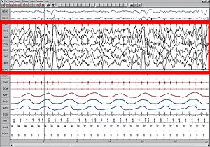
Ponto-geniculo-occipital waves or PGO waves are distinctive wave forms of propagating activity between three key brain regions: the pons, lateral geniculate nucleus, and occipital lobe; specifically, they are phasic field potentials. These waves can be recorded from any of these three structures during and immediately before REM sleep. The waves begin as electrical pulses from the pons, then move to the lateral geniculate nucleus residing in the thalamus, and end in the primary visual cortex of the occipital lobe. The appearances of these waves are most prominent in the period right before REM sleep, albeit they have been recorded during wakefulness as well. They are theorized to be intricately involved with eye movement of both wake and sleep cycles in many different animals.

Sleep onset is the transition from wakefulness into sleep. Sleep onset usually transits into non-rapid eye movement sleep but under certain circumstances it is possible to transit from wakefulness directly into rapid eye movement sleep.

<span class="mw-page-title-main">Sleep and memory</span> Relationship between sleep and memory

The relationship between sleep and memory has been studied since at least the early 19th century. Memory, the cognitive process of storing and retrieving past experiences, learning and recognition, is a product of brain plasticity, the structural changes within synapses that create associations between stimuli. Stimuli are encoded within milliseconds; however, the long-term maintenance of memories can take additional minutes, days, or even years to fully consolidate and become a stable memory that is accessible. Therefore, the formation of a specific memory occurs rapidly, but the evolution of a memory is often an ongoing process.

<span class="mw-page-title-main">Hypnogram</span> Graph of a persons sleep

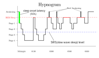
A hypnogram is a form of polysomnography; it is a graph that represents the stages of sleep as a function of time. It was developed as an easy way to present the recordings of the brain wave activity from an electroencephalogram (EEG) during a period of sleep. It allows the different stages of sleep: rapid eye movement sleep (REM) and non-rapid eye movement sleep (NREM) to be identified during the sleep cycle. NREM sleep can be further classified into NREM stage 1, 2 and 3. The previously considered 4th stage of NREM sleep has been included within stage 3; this stage is also called slow wave sleep (SWS) and is the deepest stage of sleep.

<span class="mw-page-title-main">Neuroscience of sleep</span> Study of the neuroscientific and physiological basis of the nature of sleep

The neuroscience of sleep is the study of the neuroscientific and physiological basis of the nature of sleep and its functions. Traditionally, sleep has been studied as part of psychology and medicine. The study of sleep from a neuroscience perspective grew to prominence with advances in technology and the proliferation of neuroscience research from the second half of the twentieth century.

The parafacial zone (PZ) is a brain structure located in the brainstem within the medulla oblongata believed to be heavily responsible for non-rapid eye movement (non-REM) sleep regulation, specifically for inducing slow-wave sleep.

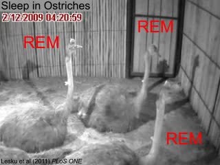
In birds, sleep consists of "periods of eye closure interrupted by short periods of eye-opening." During the short periods of eye-opening, electroencephalographic (EEG) studies indicate that the birds are still sleeping; the voltage level in the brain is identical. Birds restore their arousal thresholds during sleep. During their short eye-open periods, sleeping birds can mobilize almost instantaneously when threatened by a predator. Avian species have been found to rely on flock size and height for predatory precautions. Between the eye-opening and group sleep, these precautions allow sleep to be beneficial and safe.

References

  1. 1 2 3 4 5 Lapierre, Jennifer L.; Kosenko, Peter O.; Lyamin, Oleg I.; Kodama, Tohru; Mukhametov, Lev M.; Siegel, Jerome M. (2007). "Cortical Acetylcholine Release Is Lateralized during Asymmetrical Slow-Wave Sleep in Northern Fur Seals". The Journal of Neuroscience. 27 (44): 11999–12006. doi:10.1523/JNEUROSCI.2968-07.2007. PMC   6673386 . PMID   17978041.
  2. 1 2 3 4 5 Rattenborg, Niels C.; Lima, Steven L.; Amlaner, Charles J. (1999). "Half-awake to the risk of predation". Nature. 397 (6718): 397–398. Bibcode:1999Natur.397..397R. doi:10.1038/17037. PMID   29667967. S2CID   4427166.
  3. Cirelli, Chiara; Tunoni, Giulio (2008). "Is Sleep Essential?". PLOS Biology. 6 (8): 1605–1611. doi: 10.1371/journal.pbio.0060216 . PMC   2525690 . PMID   18752355.
  4. 1 2 3 4 5 6 7 Mascetti, Gian Gastone (2016). "Unihemispheric sleep and asymmetrical sleep: Behavioral, neurophysiological, and functional perspectives". Nature and Science of Sleep. 8: 221–238. doi: 10.2147/NSS.S71970 . PMC   4948738 . PMID   27471418.
  5. Rattenborg, N. C.; Lima, S. L.; Amlaner, C. J. (1999-11-15). "Facultative control of avian unihemispheric sleep under the risk of predation". Behavioural Brain Research. 105 (2): 163–172. doi:10.1016/s0166-4328(99)00070-4. ISSN   0166-4328. PMID   10563490. S2CID   8570743.
  6. 1 2 Walter, Timothy J.; Marar, Uma (2007). "Sleeping With One Eye Open" (PDF). Capitol Sleep Medicine Newsletter. pp. 3621–3628.
  7. 1 2 3 4 5 6 7 8 9 Rattenbourg, Neils C.; Amlaner, C.J.; Lima, S.L. (2000). "Behavioral, neurophysiological and evolutionary perspectives on unihemispheric sleep". Neuroscience and Biobehavioral Reviews. 24 (8): 817–842. doi:10.1016/S0149-7634(00)00039-7. PMID   11118608. S2CID   7592942.
  8. Mascetti, Gian G.; Rugger, Marina; Vallortigara, Giorgio; Bobbo, Daniela (2006). "Monocular-unihemispheric sleep and visual discrimination learning in the domestic chick". Experimental Brain Research. 176 (1): 70–84. doi:10.1007/s00221-006-0595-3. PMID   16874518. S2CID   14246719.
  9. 1 2 Lyamin, O.I.; Mukhametov, L.M.; Siegel, J.M.; Nazarenko, E.A.; Polyakova, I.G.; Shpak, O.V. (2002). "Unihemispheric slow-wave sleep and the state of the eyes in a white whale". Behavioural Brain Research. 129 (1–2): 125–129. doi:10.1016/S0166-4328(01)00346-1. PMC   8788623 . PMID   11809503.
  10. 1 2 3 4 5 6 7 Rattenborg, Niels C. (2006). "Do birds sleep in flight?". Naturwissenschaften. 93 (9): 413–425. Bibcode:2006NW.....93..413R. doi:10.1007/s00114-006-0120-3. PMID   16688436. S2CID   1736369.
  11. Humphrys, Leah; University, La Trobe. "Scientists find that crocodiles do, indeed, sleep with one eye open". phys.org.
  12. McGinty, Dennis; Szymusiak, Ronald (1990). "Keeping cool: a hypothesis about the mechanisms and functions of slow-wave sleep". Trends in Neurosciences. 13 (12): 480–487. doi:10.1016/0166-2236(90)90081-K. PMID   1703678. S2CID   4035781.
  13. Ridgway, Sam; Carder, Don; Finneran, James; Keogh, Mandy; Kamolnick, Tricia; Todd, Mark; Goldblatt, Allen (2006). "Dolphin Continuous Auditory Vigilance for Five Days". The Journal of Experimental Biology. 209 (18): 3621–3628. doi: 10.1242/jeb.02405 . PMID   16943502.
  14. Anderson, Andrea (1 September 2016). "We Toss and Turn in an Unfamiliar Bed". Scientific American Mind. 27 (5). Scientific American: 9. doi:10.1038/scientificamericanmind0916-9a.