X-ray detector

Last updated
Acquisition of projectional radiography, with an X-ray generator and an imaging detector. Projectional radiography components.jpg
Acquisition of projectional radiography, with an X-ray generator and an imaging detector.

X-ray detectors are devices used to measure the flux, spatial distribution, spectrum, and/or other properties of X-rays.

Contents

Detectors can be divided into two major categories: imaging detectors (such as photographic plates and X-ray film (photographic film), now mostly replaced by various digitizing devices like image plates or flat panel detectors) and dose measurement devices (such as ionization chambers, Geiger counters, and dosimeters used to measure the local radiation exposure, dose, and/or dose rate, for example, for verifying that radiation protection equipment and procedures are effective on an ongoing basis).

X-ray imaging

Fish bone pierced in the upper esophagus. Right image without contrast medium, left image during swallowing with contrast medium. Fischgrate verschluckt.jpg
Fish bone pierced in the upper esophagus. Right image without contrast medium, left image during swallowing with contrast medium.

To obtain an image with any type of image detector the part of the patient to be X-rayed is placed between the X-ray source and the image receptor to produce a shadow of the internal structure of that particular part of the body. X-rays are partially blocked ("attenuated") by dense tissues such as bone, and pass more easily through soft tissues. Areas where the X-rays strike darken when developed, causing bones to appear lighter than the surrounding soft tissue.

Contrast compounds containing barium or iodine, which are radiopaque, can be ingested in the gastrointestinal tract (barium) or injected in the artery or veins to highlight these vessels. The contrast compounds have high atomic numbered elements in them that (like bone) essentially block the X-rays and hence the once hollow organ or vessel can be more readily seen. In the pursuit of nontoxic contrast materials, many types of high atomic number elements were evaluated. Some elements chosen proved to be harmful – for example, thorium was once used as a contrast medium (Thorotrast) – which turned out to be toxic, causing a very high incidence of cancer decades after use. Modern contrast material has improved and, while there is no way to determine who may have a sensitivity to the contrast, the incidence of serious allergic reactions is low. [1]

X-ray film

Mechanism

Typical x-ray film contains silver halide crystal "grains", typically primarily silver bromide. [2] Grain size and composition can be adjusted to affect the film properties, for example to improve resolution in the developed image. [3] When the film is exposed to radiation the halide is ionised and free electrons are trapped in crystal defects (forming a latent image). Silver ions are attracted to these defects and reduced, creating clusters of transparent silver atoms. [4] In the developing process these are converted to opaque silver atoms which form the viewable image, darkest where the most radiation was detected. Further developing steps stabilise the sensitised grains and remove unsensitised grains to prevent further exposure (e.g. from visible light). [5] :159 [6]

Replacement

A video discussing a study that showed that digital x-rays were equally effective in identifying occupational lung diseases as film x-rays.

The first radiographs (X-ray images) were made by the action of X-rays on sensitized glass photographic plates. X-ray film (photographic film) soon replaced the glass plates, and film has been used for decades to acquire (and display) medical and industrial images. [7] Gradually, digital computers gained the ability to store and display enough data to make digital imaging possible. Since the 1990s, computerized radiography and digital radiography have been replacing photographic film in medical and dental applications, though film technology remains in widespread use in industrial radiography processes (e.g. to inspect welded seams). The metal silver (formerly necessary to the radiographic & photographic industries) is a non-renewable resource although silver can easily be reclaimed from spent X-ray film. [8] Where X-ray films required wet processing facilities, newer digital technologies do not. Digital archiving of images also saves physical storage space. [9]

Photostimulable phosphors

A piece of photostimulable phosphor plate Circular image plate.jpg
A piece of photostimulable phosphor plate

Phosphor plate radiography [10] is a method of recording X-rays using photostimulated luminescence (PSL), pioneered by Fuji in the 1980s. [11] A photostimulable phosphor plate (PSP) is used in place of the photographic plate. After the plate is X-rayed, excited electrons in the phosphor material remain 'trapped' in 'colour centres' in the crystal lattice until stimulated by a laser beam passed over the plate surface. [12] The light given off during laser stimulation is collected by a photomultiplier tube, and the resulting signal is converted into a digital image by computer technology. The PSP plate can be reused, and existing X-ray equipment requires no modification to use them. The technique may also be known as computed radiography (CR). [13]

Image intensifiers

Radiograph taken during cholecystectomy Laprascopy-Roentgen.jpg
Radiograph taken during cholecystectomy

X-rays are also used in "real-time" procedures such as angiography or contrast studies of the hollow organs (e.g. barium enema of the small or large intestine) using fluoroscopy. Angioplasty, medical interventions of the arterial system, rely heavily on X-ray-sensitive contrast to identify potentially treatable lesions.

Semiconductor detectors

Solid state detectors use semiconductors to detect x-rays. Direct digital detectors are so-called because they directly convert x-ray photons to electrical charge and thus a digital image. Indirect systems may have intervening steps for example first converting x-ray photons to visible light, and then an electronic signal. Both systems typically use thin film transistors to read out and convert the electronic signal to a digital image. Unlike film or CR no manual scanning or development step is required to obtain a digital image, and so in this sense both systems are "direct". [14] Both types of system have considerably higher quantum efficiency than CR. [14]

Direct detectors

Since the 1970s, silicon or germanium doped with lithium (Si(Li) or Ge(Li)) semiconductor detectors have been developed. [15] X-ray photons are converted to electron-hole pairs in the semiconductor and are collected to detect the X-rays. When the temperature is low enough (the detector is cooled by Peltier effect or even cooler liquid nitrogen), it is possible to directly determine the X-ray energy spectrum; this method is called energy-dispersive X-ray spectroscopy (EDX or EDS); it is often used in small X-ray fluorescence spectrometers. Silicon drift detectors (SDDs), produced by conventional semiconductor fabrication, provide a cost-effective and high resolving power radiation measurement. Unlike conventional X-ray detectors, such as Si(Li), they do not need to be cooled with liquid nitrogen. These detectors are rarely used for imaging and are only efficient at low energies. [16]

Practical application in medical imaging started in the early 2000s. [17] Amorphous selenium is used in commercial large area flat panel X-ray detectors for mammography and general radiography due to its high spatial resolution and x-ray absorbing properties. [18] However Selenium's low atomic number means a thick layer is required to achieve sufficient sensitivity. [19]

Cadmium telluride (Cd Te), and its alloy with zinc, cadmium zinc telluride, is considered one of the most promising semiconductor materials for x-ray detection due to its wide band-gap and high quantum number resulting in room temperature operation with high efficiency. [20] [21] Current applications include bone densitometry and SPECT but flat panel detectors suitable for radiographic imaging are not yet in production. [22] Current research and development is focused around energy resolving pixel detectors, such as CERN's Medipix detector and Science and Technology Facilities Council's HEXITEC detector. [23] [24]

Common semiconductor diodes, such as PIN photodiodes or a 1N4007, will produce a small amount of current in photovoltaic mode when placed in an X-ray beam. [25] [26]

Indirect detectors

Indirect detectors are made up of a scintillator to convert x-rays to visible light, which is read by a TFT array. This can provide sensitivity advantages over current (amorphous selenium) direct detectors, albeit with a potential trade-off in resolution. [19] Indirect flat panel detectors (FPDs) are in widespread use today in medical, dental, veterinary, and industrial applications.

The TFT array consists of a sheet of glass covered with a thin layer of silicon that is in an amorphous or disordered state. At a microscopic scale, the silicon has been imprinted with millions of transistors arranged in a highly ordered array, like the grid on a sheet of graph paper. Each of these thin-film transistors (TFTs) is attached to a light-absorbing photodiode making up an individual pixel (picture element). Photons striking the photodiode are converted into two carriers of electrical charge, called electron-hole pairs. Since the number of charge carriers produced will vary with the intensity of incoming light photons, an electrical pattern is created that can be swiftly converted to a voltage and then a digital signal, which is interpreted by a computer to produce a digital image. Although silicon has outstanding electronic properties, it is not a particularly good absorber of X-ray photons. For this reason, X-rays first impinge upon scintillators made from such materials as gadolinium oxysulfide or caesium iodide. The scintillator absorbs the X-rays and converts them into visible light photons that then pass onto the photodiode array.

Dose measurement

Gas detectors

Plot of ion current as function of applied voltage for a wire cylinder gaseous radiation detector. Detector regions.gif
Plot of ion current as function of applied voltage for a wire cylinder gaseous radiation detector.

X-rays going through a gas will ionize it, producing positive ions and free electrons. An incoming photon will create a number of such ion pairs proportional to its energy. If there is an electric field in the gas chamber ions and electrons will move in different directions and thereby cause a detectable current. The behaviour of the gas will depend on the applied voltage and the geometry of the chamber. This gives rise to a few different types of gas detectors described below.

Ionization chambers use a relatively low electric field of about 100 V/cm to extract all ions and electrons before they recombine. [27] This gives a steady current proportional to the dose rate the gas is exposed to. [7] Ion chambers are widely used as hand held radiation survey meters to check radiation dose levels.

Proportional counters use a geometry with a thin positively charged anode wire in the center of a cylindrical chamber. Most of the gas volume will act as an ionization chamber, but in the region closest to the wire the electric field is high enough to make the electrons ionize gas molecules. This will create an avalanche effect greatly increasing the output signal. Since every electron cause an avalanche of approximately the same size the collected charge is proportional to the number of ion pairs created by the absorbed x-ray. This makes it possible to measure the energy of each incoming photon.

Geiger–Müller counters use an even higher electric field so that UV-photons are created. [28] These start new avalanches, eventually resulting in a total ionization of the gas around the anode wire. This makes the signal very strong, but causes a dead time after each event and makes it impossible to measure the X-ray energies. [29]

Gas detectors are usually single pixel detectors measuring only the average dose rate over the gas volume or the number of interacting photons as explained above, but they can be made spatially resolving by having many crossed wires in a wire chamber.

Silicon PN solar cells

It was demonstrated in the 1960s that silicon PN solar cells are suitable for detection of all forms of ionizing radiation including extreme UV, soft X-rays, and hard X-rays. This form of detection operates via photoionization, a process where ionizing radiation strikes an atom and releases a free electron. [30] This type of broadband ionizing radiation sensor requires a solar cell, an ammeter, and a visible light filter on top of the solar cell that allows the ionizing radiation to hit the solar cell while blocking unwanted wavelengths.

Radiochromic film

Self-developing radiochromic film can provide very high resolution measurements, for dosimetry and profiling purposes, particularly in radiotherapy physics. [31]

Related Research Articles

<span class="mw-page-title-main">Charge-coupled device</span> Device for the movement of electrical charge

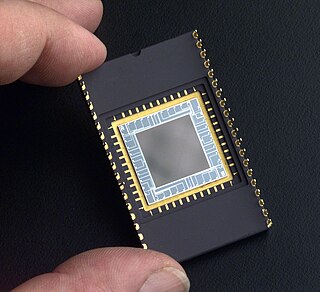
A charge-coupled device (CCD) is an integrated circuit containing an array of linked, or coupled, capacitors. Under the control of an external circuit, each capacitor can transfer its electric charge to a neighboring capacitor. CCD sensors are a major technology used in digital imaging.

<span class="mw-page-title-main">X-ray</span> Form of short-wavelength electromagnetic radiation

X-ray is a high-energy electromagnetic radiation. In many languages, it is referred to as Röntgen radiation, after the German scientist Wilhelm Conrad Röntgen, who discovered it in 1895 and named it X-radiation to signify an unknown type of radiation.

<span class="mw-page-title-main">Cathodoluminescence</span> Photon emission under the impact of an electron beam

Cathodoluminescence is an optical and electromagnetic phenomenon in which electrons impacting on a luminescent material such as a phosphor, cause the emission of photons which may have wavelengths in the visible spectrum. A familiar example is the generation of light by an electron beam scanning the phosphor-coated inner surface of the screen of a television that uses a cathode ray tube. Cathodoluminescence is the inverse of the photoelectric effect, in which electron emission is induced by irradiation with photons.

<span class="mw-page-title-main">Radiography</span> Imaging technique using ionizing and non-ionizing radiation

Radiography is an imaging technique using X-rays, gamma rays, or similar ionizing radiation and non-ionizing radiation to view the internal form of an object. Applications of radiography include medical and industrial radiography. Similar techniques are used in airport security,. To create an image in conventional radiography, a beam of X-rays is produced by an X-ray generator and it is projected towards the object. A certain amount of the X-rays or other radiation are absorbed by the object, dependent on the object's density and structural composition. The X-rays that pass through the object are captured behind the object by a detector. The generation of flat two-dimensional images by this technique is called projectional radiography. In computed tomography, an X-ray source and its associated detectors rotate around the subject, which itself moves through the conical X-ray beam produced. Any given point within the subject is crossed from many directions by many different beams at different times. Information regarding the attenuation of these beams is collated and subjected to computation to generate two-dimensional images on three planes which can be further processed to produce a three-dimensional image.

<span class="mw-page-title-main">Scintillation counter</span> Instrument for measuring ionizing radiation

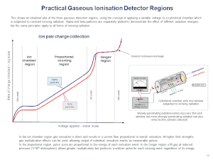
A scintillation counter is an instrument for detecting and measuring ionizing radiation by using the excitation effect of incident radiation on a scintillating material, and detecting the resultant light pulses.

An avalanche photodiode (APD) is a highly sensitive semiconductor photodiode detector that exploits the photoelectric effect to convert light into electricity. From a functional standpoint, they can be regarded as the semiconductor analog of photomultiplier tubes. The avalanche photodiode was invented by Japanese engineer Jun-ichi Nishizawa in 1952. However, study of avalanche breakdown, microplasma defects in silicon and germanium and the investigation of optical detection using p-n junctions predate this patent. Typical applications for APDs are laser rangefinders, long-range fiber-optic telecommunication, and quantum sensing for control algorithms. New applications include positron emission tomography and particle physics.

<span class="mw-page-title-main">Fluoroscopy</span> Production of an image when X-rays strike a fluorescent screen

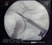
Fluoroscopy, informally referred to as "fluoro", is an imaging technique that uses X-rays to obtain real-time moving images of the interior of an object. In its primary application of medical imaging, a fluoroscope allows a surgeon to see the internal structure and function of a patient, so that the pumping action of the heart or the motion of swallowing, for example, can be watched. This is useful for both diagnosis and therapy and occurs in general radiology, interventional radiology, and image-guided surgery.

<span class="mw-page-title-main">Scintillator</span> Material which glows when excited by ionizing radiation

A scintillator is a material that exhibits scintillation, the property of luminescence, when excited by ionizing radiation. Luminescent materials, when struck by an incoming particle, absorb its energy and scintillate. Sometimes, the excited state is metastable, so the relaxation back down from the excited state to lower states is delayed. The process then corresponds to one of two phenomena: delayed fluorescence or phosphorescence. The correspondence depends on the type of transition and hence the wavelength of the emitted optical photon.

In experimental and applied particle physics, nuclear physics, and nuclear engineering, a particle detector, also known as a radiation detector, is a device used to detect, track, and/or identify ionizing particles, such as those produced by nuclear decay, cosmic radiation, or reactions in a particle accelerator. Detectors can measure the particle energy and other attributes such as momentum, spin, charge, particle type, in addition to merely registering the presence of the particle.

A semiconductor detector in ionizing radiation detection physics is a device that uses a semiconductor to measure the effect of incident charged particles or photons.

<span class="mw-page-title-main">Photodetector</span> Sensors of light or other electromagnetic energy

Photodetectors, also called photosensors, are sensors of light or other electromagnetic radiation. There are a wide variety of photodetectors which may be classified by mechanism of detection, such as photoelectric or photochemical effects, or by various performance metrics, such as spectral response. Semiconductor-based photodetectors typically use a p–n junction that converts photons into charge. The absorbed photons make electron–hole pairs in the depletion region. Photodiodes and photo transistors are a few examples of photo detectors. Solar cells convert some of the light energy absorbed into electrical energy.

An X-ray image intensifier (XRII) is an image intensifier that converts X-rays into visible light at higher intensity than the more traditional fluorescent screens can. Such intensifiers are used in X-ray imaging systems to allow low-intensity X-rays to be converted to a conveniently bright visible light output. The device contains a low absorbency/scatter input window, typically aluminum, input fluorescent screen, photocathode, electron optics, output fluorescent screen and output window. These parts are all mounted in a high vacuum environment within glass or, more recently, metal/ceramic. By its intensifying effect, It allows the viewer to more easily see the structure of the object being imaged than fluorescent screens alone, whose images are dim. The XRII requires lower absorbed doses due to more efficient conversion of X-ray quanta to visible light. This device was originally introduced in 1948.

<span class="mw-page-title-main">Neutron detection</span>

Neutron detection is the effective detection of neutrons entering a well-positioned detector. There are two key aspects to effective neutron detection: hardware and software. Detection hardware refers to the kind of neutron detector used and to the electronics used in the detection setup. Further, the hardware setup also defines key experimental parameters, such as source-detector distance, solid angle and detector shielding. Detection software consists of analysis tools that perform tasks such as graphical analysis to measure the number and energies of neutrons striking the detector.

Digital radiography is a form of radiography that uses x-ray–sensitive plates to directly capture data during the patient examination, immediately transferring it to a computer system without the use of an intermediate cassette. Advantages include time efficiency through bypassing chemical processing and the ability to digitally transfer and enhance images. Also, less radiation can be used to produce an image of similar contrast to conventional radiography.

<span class="mw-page-title-main">Industrial radiography</span> Type of non-destructive testing

Industrial radiography is a modality of non-destructive testing that uses ionizing radiation to inspect materials and components with the objective of locating and quantifying defects and degradation in material properties that would lead to the failure of engineering structures. It plays an important role in the science and technology needed to ensure product quality and reliability. In Australia, industrial radiographic non-destructive testing is colloquially referred to as "bombing" a component with a "bomb".

<span class="mw-page-title-main">Photostimulated luminescence</span>

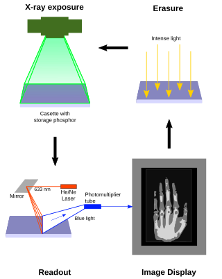
Photostimulated luminescence (PSL) is the release of stored energy within a phosphor by stimulation with visible light, to produce a luminescent signal. X-rays may induce such an energy storage. A plate based on this mechanism is called a photostimulable phosphor (PSP) plate and is one type of X-ray detector used in projectional radiography. Creating an image requires illuminating the plate twice: the first exposure, to the radiation of interest, "writes" the image, and a later, second illumination "reads" the image. The device to read such a plate is known as a phosphorimager.

<span class="mw-page-title-main">Automatic exposure control</span>

Automatic Exposure Control (AEC) is an X-ray exposure termination device. A medical radiographic exposure is always initiated by a human operator but an AEC detector system may be used to terminate the exposure when a predetermined amount of radiation has been received. The intention of AEC is to provide consistent x-ray image exposure, whether to film, a digital detector or a CT scanner. AEC systems may also automatically set exposure factors such as the X-ray tube current and voltage in a CT.

<span class="mw-page-title-main">Flat-panel detector</span> Class of solid-state x-ray digital radiography devices

Flat-panel detectors are a class of solid-state x-ray digital radiography devices similar in principle to the image sensors used in digital photography and video. They are used in both projectional radiography and as an alternative to x-ray image intensifiers (IIs) in fluoroscopy equipment.

Hybrid pixel detectors are a type of ionizing radiation detector consisting of an array of diodes based on semiconductor technology and their associated electronics. The term “hybrid” stems from the fact that the two main elements from which these devices are built, the semiconductor sensor and the readout chip, are manufactured independently and later electrically coupled by means of a bump-bonding process. Ionizing particles are detected as they produce electron-hole pairs through their interaction with the sensor element, usually made of doped silicon or cadmium telluride. The readout ASIC is segmented into pixels containing the necessary electronics to amplify and measure the electrical signals induced by the incoming particles in the sensor layer.

Spectral imaging is an umbrella term for energy-resolved X-ray imaging in medicine. The technique makes use of the energy dependence of X-ray attenuation to either increase the contrast-to-noise ratio, or to provide quantitative image data and reduce image artefacts by so-called material decomposition. Dual-energy imaging, i.e. imaging at two energy levels, is a special case of spectral imaging and is still the most widely used terminology, but the terms "spectral imaging" and "spectral CT" have been coined to acknowledge the fact that photon-counting detectors have the potential for measurements at a larger number of energy levels.

References

  1. "Contrast Medium Reactions: Overview, Types of Iodinated Contrast Media, Adverse Reactions to ICM". Medscape. 2 June 2016. Retrieved 17 December 2016.
  2. "Radiographic Film". NDT Resource Center. Retrieved 16 December 2016.
  3. Jensen, T; Aljundi, T; Gray, J N; Wallingford, R (1996). "A Model of X-Ray Film Response". In Thompson, D O; Chimenti, D E (eds.). Review of Progress in Quantitative Nondestructive Evaluation (Volume 15A). Boston, MA: Springer. p. 441. doi:10.1007/978-1-4613-0383-1_56. ISBN   978-1-4613-0383-1.
  4. Martin, James E. (2006). Physics for Radiation Protection: A Handbook (2nd ed.). Weinheim: John Wiley & Sons. pp. 707–709. ISBN   9783527406111.
  5. Dance, D R; Christofides, S; Maidment, A D A; McLean, I D; Ng, K H (2014). Diagnostic radiology physics : a handbook for teachers and students. Vienna: International Atomic Energy Agency. ISBN   978-92-0-131010-1.
  6. "Developing Film". NDT Resource Centre. Archived from the original on 7 February 2020. Retrieved 16 December 2016.
  7. 1 2 Seco, Joao; Clasie, Ben; Partridge, Mike (21 October 2014). "Review on the characteristics of radiation detectors for dosimetry and imaging". Physics in Medicine and Biology. 59 (20): R303–R347. Bibcode:2014PMB....59R.303S. doi:10.1088/0031-9155/59/20/R303. PMID   25229250. S2CID   4393848.
  8. Masebinu, Samson O.; Muzenda, Edison (2014). Review of Silver Recovery Techniques from Radiographic Effluent and X-ray Film Waste (PDF). Proceedings of the World Congress on Engineering and Computer Science. Vol. II. San Francisco.
  9. Körner, Markus; Weber, Christof H.; Wirth, Stefan; Pfeifer, Klaus-Jürgen; Reiser, Maximilian F.; Treitl, Marcus (May 2007). "Advances in Digital Radiography: Physical Principles and System Overview". RadioGraphics. 27 (3): 675–686. doi: 10.1148/rg.273065075 . PMID   17495286.
  10. Benjamin S (2010). "Phosphor plate radiography: an integral component of the filmless practice". Dent Today. 29 (11): 89. PMID   21133024.
  11. Rowlands, J A (7 December 2002). "The physics of computed radiography". Physics in Medicine and Biology. 47 (23): R123–R166. Bibcode:2002PMB....47R.123R. doi:10.1088/0031-9155/47/23/201. PMID   12502037. S2CID   250801018.
  12. Sonoda, M; Takano, M; Miyahara, J; Kato, H (September 1983). "Computed radiography utilizing scanning laser stimulated luminescence". Radiology. 148 (3): 833–838. doi:10.1148/radiology.148.3.6878707. PMID   6878707.
  13. Watt, Kristina N.; Yan, Kuo; DeCrescenzo, Giovanni; Rowlands, J. A. (15 November 2005). "The physics of computed radiography: Measurements of pulse height spectra of photostimulable phosphor screens using prompt luminescence". Medical Physics. 32 (12): 3589–3598. Bibcode:2005MedPh..32.3589W. doi:10.1118/1.2122587. PMID   16475757.
  14. 1 2 Chotas, Harrell G.; Dobbins, James T.; Ravin, Carl E. (March 1999). "Principles of Digital Radiography with Large-Area, Electronically Readable Detectors: A Review of the Basics". Radiology. 210 (3): 595–599. doi:10.1148/radiology.210.3.r99mr15595. PMID   10207454. S2CID   32424961.
  15. Lowe, Barrie Glyn; Sareen, Robert Anthony (2013). Semiconductor X-Ray Detectors. Hoboken: Taylor and Francis. p. 106. ISBN   9781466554016.
  16. Grupen, Claus; Buvat, Irène (2012). Handbook of particle detection and imaging. Berlin: Springer. p. 443. ISBN   9783642132711.
  17. Kotter, E.; Langer, M. (19 March 2002). "Digital radiography with large-area flat-panel detectors". European Radiology. 12 (10): 2562–2570. doi:10.1007/s00330-002-1350-1. PMID   12271399. S2CID   16677678.
  18. Lança, Luís; Silva, Augusto (2013). "Digital Radiography Detectors: A Technical Overview". Digital imaging systems for plain radiography. New York: Springer. doi:10.1007/978-1-4614-5067-2_2. hdl:10400.21/1932. ISBN   978-1-4614-5067-2.
  19. 1 2 Ristić, S G (18–19 October 2013). "The digital flat-panel X-Ray detectors" (PDF). Proceedings of the Third Conference on Medical Physicsand Biomedical Engineering. Skopje: IAEA. pp. 65–71.
  20. Takahashi, T.; Watanabe, S. (2001). "Recent progress in CdTe and CdZnTe detectors". IEEE Transactions on Nuclear Science. 48 (4): 950–959. arXiv: astro-ph/0107398 . Bibcode:2001ITNS...48..950T. doi:10.1109/23.958705. S2CID   5829568.
  21. Del Sordo, Stefano; Abbene, Leonardo; Caroli, Ezio; Mancini, Anna Maria; Zappettini, Andrea; Ubertini, Pietro (12 May 2009). "Progress in the Development of CdTe and CdZnTe Semiconductor Radiation Detectors for Astrophysical and Medical Applications". Sensors. 9 (5): 3491–3526. Bibcode:2009Senso...9.3491D. doi: 10.3390/s90503491 . PMC   3297127 . PMID   22412323.
  22. Iniewski, K. (4 November 2014). "CZT detector technology for medical imaging". Journal of Instrumentation. 9 (11): C11001. Bibcode:2014JInst...9C1001I. doi:10.1088/1748-0221/9/11/C11001. S2CID   121382729.
  23. Zang, A.; Anton, G.; Ballabriga, R.; Bisello, F.; Campbell, M.; Celi, J.C.; Fauler, A.; Fiederle, M.; Jensch, M.; Kochanski, N.; Llopart, X.; Michel, N.; Mollenhauer, U.; Ritter, I.; Tennert, F.; Wölfel, S.; Wong, W.; Michel, T. (16 April 2015). "The Dosepix detector—an energy-resolving photon-counting pixel detector for spectrometric measurements". Journal of Instrumentation. 10 (4): C04015. Bibcode:2015JInst..10C4015Z. doi: 10.1088/1748-0221/10/04/C04015 .
  24. Jones, Lawrence; Seller, Paul; Wilson, Matthew; Hardie, Alec (June 2009). "HEXITEC ASIC—a pixellated readout chip for CZT detectors". Nuclear Instruments and Methods in Physics Research Section A: Accelerators, Spectrometers, Detectors and Associated Equipment. 604 (1–2): 34–37. Bibcode:2009NIMPA.604...34J. doi:10.1016/j.nima.2009.01.046.
  25. Gonzalez, G, J. (October 2016). "Desarrollo de un detector de rayos X usando fotodiodos" [Development of an X-ray detector using photodiodes]. INIS (in Spanish). 48 (7): 13.{{cite journal}}: CS1 maint: multiple names: authors list (link)
  26. "1n4007 diode as an X-Ray detector". YouTube . Retrieved 4 December 2019.
  27. Albert C. Thompson. X-Ray Data Booklet, Section 4-5: X-ray detectors (PDF).
  28. Saha, Gopal B. (2012). "Gas-Filled Detectors". Physics and radiobiology of nuclear medicine (4th ed.). New York: Springer. pp. 79–90. doi:10.1007/978-1-4614-4012-3_7. ISBN   978-1-4614-4012-3.
  29. Ahmed, Syed Naeem (2007). Physics and engineering of radiation detection (1st ed.). Amsterdam: Academic Press. p. 182. ISBN   9780080569642.
  30. Photovoltaic Effect Produced in Silicon Solar Cells by x-ray and Gamma-Rays, Karl Scharf, January 25, 1960, Journal of Research of the National Bureau of Standards
  31. Williams, Matthew; Metcalfe, Peter (5 May 2011). "Radiochromic Film Dosimetry and its Applications in Radiotherapy". AIP Conference Proceedings. 1345 (1): 75–99. Bibcode:2011AIPC.1345...75W. doi:10.1063/1.3576160. ISSN   0094-243X.