Actin assembly-inducing protein

Last updated
Actin assembly-inducing protein
EVH1.jpg
EVH1 domain-ActA peptide complex
Identifiers
SymbolActA
NCBI gene 2798121
UniProt P33379
Search for
Structures Swiss-model
Domains InterPro

The Actin assembly-inducing protein (ActA) is a protein encoded and used by Listeria monocytogenes to propel itself through a mammalian host cell. ActA is a bacterial surface protein comprising a membrane-spanning region. [1] In a mammalian cell the bacterial ActA interacts with the Arp2/3 complex and actin monomers to induce actin polymerization on the bacterial surface generating an actin comet tail. The gene encoding ActA is named actA or prtB. [2]

Contents

Introduction

As soon as L. monocytogenes bacteria are ingested by humans, they get internalized into intestinal epithelium cells and rapidly try to escape their internalization vacuole. [3] [4] In the cytosol they start to polymerize actin on their surface by the help of the ActA protein. It has been shown that ActA is not only necessary but also sufficient to induce motility of bacteria in the absence of other bacterial factors. [5]

Discovery

ActA was discovered by analysing lecithinase-negative Tn917-lac Listeria mutants because of the phenotype that they were unable to spread from cell to cell. These mutant bacteria still escaped from the phagosomes as efficiently as wild-type bacteria and multiplied within the infected cells but they were not surrounded by actin like wild-type bacteria. Further analysis showed, that Tn917-lac had inserted into actA, the second gene of an operon. The third gene of this operon, plcB, encodes the L. monocytogenes lecithinase. To determine whether actA itself, plcB or other co-transcribed downstream regions are involved in actin assembly, mutations in the appropriate genes were generated. All mutants except the actA mutants were similar to wild-type concerning association with F-actin and cell-cell spreading. Complementation with actA restored wild-type phenotype in the actA mutants. [1]

Function

Fig. 1 Actin assembly induced by bacterial protein ActA (shown in green). Mammalian proteins involved in this process are: Profilin (P), Vasodilator-stimulated phosphoprotein (VASP) and actin-related-protein 2 and 3 complex (Arp2/3 complex) as well as actin. ActA Function.jpg
Fig. 1 Actin assembly induced by bacterial protein ActA (shown in green). Mammalian proteins involved in this process are: Profilin (P), Vasodilator-stimulated phosphoprotein (VASP) and actin-related-protein 2 and 3 complex (Arp2/3 complex) as well as actin.

ActA is a protein which acts as a mimic of Wiskott-Aldrich syndrome protein (WASP), a nucleation promoting factor (NPF) present in host cells. NPFs in the mammalian cell recruit and bind to the already existing actin-related-protein 2 and 3 complex (Arp2/3 complex) and induce an activating conformational change of the Arp2/3 complex. [6] Due to this conformational change, NPFs initiate polymerization of a new actin filament at a 70° angle, which leads to the characteristic Y-branched actin structures in the leading edge of motile cells. ActA localizes to the old pole of the bacterium and spans both the bacterial cell membrane and the cell wall, lateral diffusion is inhibited; thus ActA localizes in a polarized and anchored manner on the bacterial surface. Consequently, actin polymerization only starts in this region on the surface of the bacterium. [7] Expression of ActA is induced only after entering a mammalian host cell. [8]

Actin filament assembly generates the force that pushes the bacterium in the mammalian host cytoplasm forward. Continuous actin polymerization is sufficient for motility in the cytoplasm and even for infection of adjacent cells. [9]

Research

New data indicates that ActA plays a role also in vacuolar disruption. A deletion mutant of ActA was defective in permeabilizing the vacuole. An 11 amino acid stretch of the N-terminus of the acidic region (32-42) was shown to be important for disruption of the phagosome. [10]

Structure

The primary proteinous product of the actA gene consists of 639 amino acids and includes the signal peptide (first N-terminal 29 amino acids) and the ActA chain (C-terminal 610 amino acids). Therefore, the sequence of the mature ActA protein consist of 610 amino acids. ActA has a molecular weight of 70,349 Da and is a surface protein. [1] [2]

ActA is a natively unfolded protein which can be divided into three functional domains (Fig. 2): [1] [11] [12]

N-terminal domain

Fig. 2 The ActA protein and its functional domains ActA.png
Fig. 2 The ActA protein and its functional domains

The first 156 amino acids of the N-terminal domain consist of three regions [10] [13] (Fig. 2):

The N-terminal portion of ActA plays an important role in actin polymerization. [14] The domain displays consensus elements present in eukaryotic WASP family NPFs which include an actin monomer-binding region as well as an Arp2/3 binding C (central or cofilin homology) and A (acidic) region. [7] The actin monomer-binding region of ActA has functional properties like the WASP-Homology-2 (WH2) or V domain, but differs in the sequence. [15] Thus in WASP-family NPFs the order of the domains is WH2 followed by C, and then by A, which is not the case in ActA.

Central domain

The central proline-rich region of ActA is crucial for ensuring efficient bacterial motility. There are four proline-rich repeats containing either FPPPP or FPPIP motifs. These regions mimic those of the host cell cytoskeletal protein zyxin, vinculin and palladin, known to associate with focal adhesions or stress fibers. [16] The vasodilator-stimulated phosphoprotein (VASP) can bind through its Ena/VASP homology 1 domain (EVH1 domain) to the central proline-rich region and recruits profilin, an actin monomer binding protein, which itself promotes polymerization at barbed ends of actin filaments. Furthermore, VASP seems to interact with F-actin through its carboxy-terminal EVH2 domain, which provides a linkage of the bacterium to the tail. [17] This statement is supported by the fact that ActA can bind multiple Ena/VASP proteins simultaneously and has a high affinity between ActA and Ena/VASP. VASP has been shown to reduce the frequency actin-Y-branches in vitro and thus increases the proportion of filaments which are organized in a parallel alignment in comet tails. [18] [19]

C-terminal domain

The C-terminal domain of ActA has a hydrophobic region which anchors the protein in the bacterial membrane. [20] [21] [22]

In summary, besides

Analogues

WASP/N-WASP, which is functionally mimicked by ActA, is highly conserved in eukaryotes. It is an important actin-cytoskeleton organizer and is critical for processes such as endocytosis and cell motility. Activated by Cdc42, a Rho-family small GTPase, WASP/N-WASP activates the Arp2/3 complex, which leads to rapid actin polymerization. [23]

Actin-based motility of other pathogens

In Shigella the protein IcsA activates N-WASP, which in non-infected mammalian cells is activated by the GTPase Cdc42. Active N-WASP/WASP leads to actin polymerization by activating the Arp2/3 complex. In contrast, the Listeria ActA protein interacts with and activates directly the Arp2/3 complex. [7]

The Rickettsia RickA protein is also able to activate the Arp2/3 complex in a WASP-like manner. In contrast to Listeria, the actin filaments are organized in long, unbranched parallel bundles. The Arp2/3 complex is only localized near the bacterial surface and thus it is assumed that a more frequent Arp2/3 complex-independent elongation occurs. [16]

In Burkholderia pseudomallei BimA initiates actin polymerization in vitro. It is assumed that intracellular migration of this bacterium functions independently of the Arp2/3 complex. [16]

See also

Related Research Articles

<span class="mw-page-title-main">Microfilament</span> Filament in the cytoplasm of eukaryotic cells

Microfilaments, also called actin filaments, are protein filaments in the cytoplasm of eukaryotic cells that form part of the cytoskeleton. They are primarily composed of polymers of actin, but are modified by and interact with numerous other proteins in the cell. Microfilaments are usually about 7 nm in diameter and made up of two strands of actin. Microfilament functions include cytokinesis, amoeboid movement, cell motility, changes in cell shape, endocytosis and exocytosis, cell contractility, and mechanical stability. Microfilaments are flexible and relatively strong, resisting buckling by multi-piconewton compressive forces and filament fracture by nanonewton tensile forces. In inducing cell motility, one end of the actin filament elongates while the other end contracts, presumably by myosin II molecular motors. Additionally, they function as part of actomyosin-driven contractile molecular motors, wherein the thin filaments serve as tensile platforms for myosin's ATP-dependent pulling action in muscle contraction and pseudopod advancement. Microfilaments have a tough, flexible framework which helps the cell in movement.

<span class="mw-page-title-main">Actin</span> Family of proteins

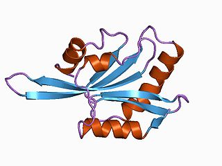
Actin is a family of globular multi-functional proteins that form microfilaments in the cytoskeleton, and the thin filaments in muscle fibrils. It is found in essentially all eukaryotic cells, where it may be present at a concentration of over 100 μM; its mass is roughly 42 kDa, with a diameter of 4 to 7 nm.

<i>Listeria monocytogenes</i> Species of pathogenic bacteria that causes the infection listeriosis

Listeria monocytogenes is the species of pathogenic bacteria that causes the infection listeriosis. It is a facultative anaerobic bacterium, capable of surviving in the presence or absence of oxygen. It can grow and reproduce inside the host's cells and is one of the most virulent foodborne pathogens: 20 to 30% of foodborne listeriosis infections in high-risk individuals may be fatal. In the European Union, listeriosis follows an upward trend that began in 2008, causing 2,161 confirmed cases and 210 reported deaths in 2014, 16% more than in 2013. Listeriosis mortality rates are also higher in the EU than for other foodborne pathogens. Responsible for an estimated 1,600 illnesses and 260 deaths in the United States annually, listeriosis ranks third in total number of deaths among foodborne bacterial pathogens, with fatality rates exceeding even Salmonella spp. and Clostridium botulinum.

<span class="mw-page-title-main">Wiskott–Aldrich syndrome protein</span> Mammalian protein found in humans

The Wiskott–Aldrich Syndrome protein (WASp) is a 502-amino acid protein expressed in cells of the hematopoietic system that in humans is encoded by the WAS gene. In the inactive state, WASp exists in an autoinhibited conformation with sequences near its C-terminus binding to a region near its N-terminus. Its activation is dependent upon CDC42 and PIP2 acting to disrupt this interaction, causing the WASp protein to 'open'. This exposes a domain near the WASp C-terminus that binds to and activates the Arp2/3 complex. Activated Arp2/3 nucleates new F-actin.

The lamellipodium is a cytoskeletal protein actin projection on the leading edge of the cell. It contains a quasi-two-dimensional actin mesh; the whole structure propels the cell across a substrate. Within the lamellipodia are ribs of actin called microspikes, which, when they spread beyond the lamellipodium frontier, are called filopodia. The lamellipodium is born of actin nucleation in the plasma membrane of the cell and is the primary area of actin incorporation or microfilament formation of the cell.

<span class="mw-page-title-main">ADF/Cofilin family</span> Family of actin-binding proteins

ADF/cofilin is a family of actin-binding proteins associated with the rapid depolymerization of actin microfilaments that give actin its characteristic dynamic instability. This dynamic instability is central to actin's role in muscle contraction, cell motility and transcription regulation.

<span class="mw-page-title-main">Cortactin</span> Protein found in humans

Cortactin is a monomeric protein located in the cytoplasm of cells that can be activated by external stimuli to promote polymerization and rearrangement of the actin cytoskeleton, especially the actin cortex around the cellular periphery. It is present in all cell types. When activated, it will recruit Arp2/3 complex proteins to existing actin microfilaments, facilitating and stabilizing nucleation sites for actin branching. Cortactin is important in promoting lamellipodia formation, invadopodia formation, cell migration, and endocytosis.

<span class="mw-page-title-main">ACTR3</span> Mammalian protein found in Homo sapiens

Actin-related protein 3 is a protein that in humans is encoded by the ACTR3 gene.

<span class="mw-page-title-main">ACTR2</span> Mammalian protein found in Homo sapiens

Actin-related protein 2 is a protein that in humans is encoded by the ACTR2 gene.

<span class="mw-page-title-main">Vasodilator-stimulated phosphoprotein</span> Mammalian protein found in Homo sapiens

Vasodilator-stimulated phosphoprotein is a protein that in humans is encoded by the VASP gene.

<span class="mw-page-title-main">WASL (gene)</span> Mammalian protein found in Homo sapiens

Neural Wiskott–Aldrich syndrome protein is a protein that in humans is encoded by the WASL gene.

<span class="mw-page-title-main">Cordon-bleu protein</span> Protein found in humans

Protein cordon-bleu is a protein that in humans is encoded by the COBL gene.

Actin remodeling is the biochemical process that allows for the dynamic alterations of cellular organization. The remodeling of actin filaments occurs in a cyclic pattern on cell surfaces and exists as a fundamental aspect to cellular life. During the remodeling process, actin monomers polymerize in response to signaling cascades that stem from environmental cues. The cell's signaling pathways cause actin to affect intracellular organization of the cytoskeleton and often consequently, the cell membrane. Again triggered by environmental conditions, actin filaments break back down into monomers and the cycle is completed. Actin-binding proteins (ABPs) aid in the transformation of actin filaments throughout the actin remodeling process. These proteins account for the diverse structure and changes in shape of Eukaryotic cells. Despite its complexity, actin remodeling may result in complete cytoskeletal reorganization in under a minute.

<span class="mw-page-title-main">MDia1</span> Protein

mDia1 is a member of the protein family called the formins and is a Rho effector. It is the mouse version of the diaphanous homolog 1 of Drosophila. mDia1 localizes to cells' mitotic spindle and midbody, plays a role in stress fiber and filopodia formation, phagocytosis, activation of serum response factor, formation of adherens junctions, and it can act as a transcription factor. mDia1 accelerates actin nucleation and elongation by interacting with barbed ends of actin filaments. The gene encoding mDia1 is located on Chromosome 18 of Mus musculus and named Diap1.

<span class="mw-page-title-main">Arp2/3 complex</span> Macromolecular complex

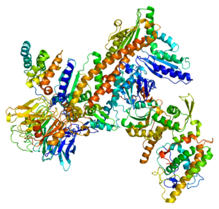
Arp2/3 complex is a seven-subunit protein complex that plays a major role in the regulation of the actin cytoskeleton. It is a major component of the actin cytoskeleton and is found in most actin cytoskeleton-containing eukaryotic cells. Two of its subunits, the Actin-Related Proteins ARP2 and ARP3, closely resemble the structure of monomeric actin and serve as nucleation sites for new actin filaments. The complex binds to the sides of existing ("mother") filaments and initiates growth of a new ("daughter") filament at a distinctive 70 degree angle from the mother. Branched actin networks are created as a result of this nucleation of new filaments. The regulation of rearrangements of the actin cytoskeleton is important for processes like cell locomotion, phagocytosis, and intracellular motility of lipid vesicles.

Paracytophagy is the cellular process whereby a cell engulfs a protrusion which extends from a neighboring cell. This protrusion may contain material which is actively transferred between the cells. The process of paracytophagy was first described as a crucial step during cell-to-cell spread of the intracellular bacterial pathogen Listeria monocytogenes, and is also commonly observed in Shigella flexneri. Paracytophagy allows these intracellular pathogens to spread directly from cell to cell, thus escaping immune detection and destruction. Studies of this process have contributed significantly to our understanding of the role of the actin cytoskeleton in eukaryotic cells.

WH1 domain is an evolutionary conserved protein domain found on WASP proteins, which are often involved in actin polymerization.

An actin nucleation core is a protein trimer with three actin monomers. It is called a nucleation core because it leads to the energetically favorable elongation reaction once a tetramer is formed from a trimer. Actin protein dimers and trimers are energetically unfavorable. Actin nucleators like the Arp2/3 complex of proteins from the formin family are most frequently involved in this process. Actin nucleation factors start the polymerization of actin within cells.

<span class="mw-page-title-main">WAVE regulatory complex</span>

The WAVE regulatory complex is a five-subunit protein complex in the Wiskott-Aldrich syndrome protein (WASP) family involved in the formation of the actin cytoskeleton through interaction with the Arp2/3 complex. The holocomplex comprises WAVE1, CYFIP1, ABI2, Nap1 and HSPC300 in its canonical form, or orthologues of these.

References

  1. 1 2 3 4 Kocks C, Gouin E, Tabouret M, Berche P, Ohayon H, Cossart P (February 1992). "L. monocytogenes-induced actin assembly requires the actA gene product, a surface protein". Cell. 68 (3): 521–31. doi:10.1016/0092-8674(92)90188-I. PMID   1739966. S2CID   27231730.
  2. 1 2 Uniprot P33379
  3. Cossart P, Sansonetti PJ (April 2004). "Bacterial invasion: the paradigms of enteroinvasive pathogens". Science. 304 (5668): 242–8. Bibcode:2004Sci...304..242C. doi:10.1126/science.1090124. PMID   15073367. S2CID   34536253.
  4. Cossart P, Pizarro-Cerdá J, Lecuit M (January 2003). "Invasion of mammalian cells by Listeria monocytogenes: functional mimicry to subvert cellular functions". Trends in Cell Biology. 13 (1): 23–31. doi:10.1016/S0962-8924(02)00006-5. PMID   12480337.
  5. Zigmond SH (February 2004). "Formin-induced nucleation of actin filaments". Current Opinion in Cell Biology. 16 (1): 99–105. doi:10.1016/j.ceb.2003.10.019. PMID   15037312.
  6. Goley ED, Rodenbusch SE, Martin AC, Welch MD (October 2004). "Critical conformational changes in the Arp2/3 complex are induced by nucleotide and nucleation promoting factor". Molecular Cell. 16 (2): 269–79. doi: 10.1016/j.molcel.2004.09.018 . PMID   15494313.
  7. 1 2 3 4 Gouin E, Welch MD, Cossart P (February 2005). "Actin-based motility of intracellular pathogens". Current Opinion in Microbiology. 8 (1): 35–45. doi:10.1016/j.mib.2004.12.013. PMID   15694855.
  8. Rafelski SM, Theriot JA (February 2006). "Mechanism of polarization of Listeria monocytogenes surface protein ActA". Molecular Microbiology. 59 (4): 1262–79. doi:10.1111/j.1365-2958.2006.05025.x. PMC   1413586 . PMID   16430699.
  9. Goldberg MB (December 2001). "Actin-Based Motility of Intracellular Microbial Pathogens". Microbiology and Molecular Biology Reviews. 65 (4): 595–626. doi:10.1128/MMBR.65.4.595-626.2001. PMC   99042 . PMID   11729265.
  10. 1 2 Poussin MA, Goldfine H (January 2010). "Evidence for involvement of ActA in maturation of the Listeria monocytogenes phagosome". Cell Research. 20 (1): 109–12. doi:10.1038/cr.2009.142. PMC   2802179 . PMID   20029388.
  11. Ireton K, Cossart P (1997). "Host-pathogen interactions during entry and actin-based movement of Listeria monocytogenes". Annual Review of Genetics. 31: 113–38. doi:10.1146/annurev.genet.31.1.113. PMID   9442892.
  12. Footer MJ, Lyo JK, Theriot JA (2008-08-29). "Close packing of Listeria monocytogenes ActA, a natively unfolded protein, enhances F-actin assembly without dimerization". The Journal of Biological Chemistry. 283 (35): 23852–23862. doi: 10.1074/jbc.M803448200 . ISSN   0021-9258. PMC   2527104 . PMID   18577520.
  13. Welch MD (2007). "Actin-Based Motility and Cell-to-Cell Spread of Listeria monocytogenes". In Goldfine H, Shen H (eds.). Listeria monocytogenes: Pathogenesis and Host Response. New York: Springer. pp. 197–223. doi:10.1007/978-0-387-49376-3_10. ISBN   978-0-387-49373-2.
  14. Welch MD, Rosenblatt J, Skoble J, Portnoy DA, Mitchison TJ (July 1998). "Interaction of human Arp2/3 complex and the Listeria monocytogenes ActA protein in actin filament nucleation". Science. 281 (5373): 105–8. Bibcode:1998Sci...281..105W. doi:10.1126/science.281.5373.105. PMID   9651243.
  15. Zalevsky J, Grigorova I, Mullins RD (February 2001). "Activation of the Arp2/3 complex by the Listeria acta protein. Acta binds two actin monomers and three subunits of the Arp2/3 complex". The Journal of Biological Chemistry. 276 (5): 3468–75. doi: 10.1074/jbc.M006407200 . PMID   11029465.
  16. 1 2 3 Lambrechts A, Gevaert K, Cossart P, Vandekerckhove J, Van Troys M (May 2008). "Listeria comet tails: the actin-based motility machinery at work". Trends in Cell Biology. 18 (5): 220–7. doi:10.1016/j.tcb.2008.03.001. PMID   18396046.
  17. Laurent V, Loisel TP, Harbeck B, et al. (March 1999). "Role of Proteins of the Ena/VASP Family in Actin-based Motility of Listeria monocytogenes". The Journal of Cell Biology. 144 (6): 1245–58. doi:10.1083/jcb.144.6.1245. PMC   2150578 . PMID   10087267.
  18. Skoble J, Auerbuch V, Goley ED, Welch MD, Portnoy DA (October 2001). "Pivotal role of VASP in Arp2/3 complex–mediated actin nucleation, actin branch-formation, and Listeria monocytogenes motility". The Journal of Cell Biology. 155 (1): 89–100. doi:10.1083/jcb.200106061. PMC   2150787 . PMID   11581288.
  19. Bear J.E., Svitkina T.M., Krause M., et al. (May 2002). "Antagonism between Ena/VASP proteins and actin filament capping regulates fibroblast motility". Cell. 109 (4): 509–21. doi:10.1016/S0092-8674(02)00731-6. hdl: 1721.1/83477 . PMID   12086607. S2CID   2848293.
  20. Vazquez-Boland JA, Kocks C, Dramsi S, et al. (January 1992). "Nucleotide sequence of the lecithinase operon of Listeria monocytogenes and possible role of lecithinase in cell-to-cell spread". Infection and Immunity. 60 (1): 219–30. doi:10.1128/iai.60.1.219-230.1992. PMC   257526 . PMID   1309513.
  21. Domann E, Wehland J, Rohde M, et al. (May 1992). "A novel bacterial virulence gene in Listeria monocytogenes required for host cell microfilament interaction with homology to the proline-rich region of vinculin". The EMBO Journal. 11 (5): 1981–90. doi:10.1002/j.1460-2075.1992.tb05252.x. PMC   556658 . PMID   1582425.
  22. Kocks C, Hellio R, Gounon P, Ohayon H, Cossart P (July 1993). "Polarized distribution of Listeria monocytogenes surface protein ActA at the site of directional actin assembly". Journal of Cell Science. 105 (3): 699–710. doi:10.1242/jcs.105.3.699. PMID   8408297.
  23. Kurisu S, Takenawa T (2009). "The WASP and WAVE family proteins". Genome Biology. 10 (6): 226. doi: 10.1186/gb-2009-10-6-226 . PMC   2718491 . PMID   19589182.