Anatomical Theatre of Padua

Last updated
Anatomical Theatre of Padua
Teatro Anatomico di Padova
Theatre-anatomique-Padoue.JPG
Anatomical Theatre of Padua
General information
TypeTheatre
Architectural style16th century
LocationPalazzo del Bò
AddressVia VIII Febbraio, 2, 35122 Padova PD
Town or cityPadua
CountryItaly
Coordinates 45°24′25.7″N11°52′37.8″E / 45.407139°N 11.877167°E / 45.407139; 11.877167 Coordinates: 45°24′25.7″N11°52′37.8″E / 45.407139°N 11.877167°E / 45.407139; 11.877167
Current tenantsUniversità degli Studi di Padova
InauguratedJanuary 1595;428 years ago (1595-01)
Technical details
MaterialStone and wood
Design and construction
Architect(s)Paolo Sarpi and Dario Varotari

The Anatomical Theatre of Padua , Northern Italy, is the first permanent anatomical theatre in the world. [1] Still preserved in the Palazzo del Bo, [2] it was inaugurated in 1595 by Girolamo Fabrici of Acquapendente, according to the project of Paolo Sarpi [3] and Dario Varotari. [4] This theatre constituted the model for the anatomical theatres built during the seventeenth century in the main universities of Europe: all would have been based on the Paduan archetype. [5] It is the symbol of a successful period in the University of Padua's history, [6] and it is considered one of the most important achievements for the study of anatomy during the sixteenth century.

Contents

Background history

The foundation: Padua and the tradition of dissections

"1222. Messer Giovanni Rusca of Como, podestà of Padua. In this period the studium of Bologna was transferred to Padua, and on Christmas Day after mass there was a great earthquake." [7]

This quote attests the foundation date traditionally accepted for the origins of the University of Padua. [8]

The events that led to the construction of the theatre could be dated back to the thirteenth century, when Pietro d'Abano performed the first autopsy recorded in Padua.

Pietro d'Abano (c.1250 – c.1315), was called to Padua from Paris as a teacher of medicine, philosophy and judicial astrology. His early studies in Constantinople allowed him to translate some of Galen's works from Greek to Latin: thanks to his work, the fame of the Studium spread rapidly throughout Italy. [9]

It is worth noting that in Padua a well-established practice of dissection had already existed since the late thirteenth century. In fact, a traditional legend hands down the story of an avaricious man's heart found in a basket by St. Anthony; the heart is described in a scientific way, showing how direct observation of the corpses was already considered essential. [10]

In the fifteenth century, the Padua Studium, like others, had three fundamental chairs of medicine: theoretical medicine, practical medicine, and surgery. Albeit the surgery teacher was expected to act as incisor in the anatomical demonstrations; only in the late sixteenth century was he formally charged with the teaching of anatomy. [11]

The first European public dissection

In 1404, during his stay in Vienna, Galeazzo di Santa Sofia undertook the first solemn public dissection, a practice he had obviously first seen and carried out in Padua.

Moritz Roth, the great Vesalius scholar, observes:

"If we think that the first dissection undertaken in Vienna was carried out by a professor from Padua, we have the impression that in the fifteenth century Padua at least reached the level of Bologna, if not actually overtaking it." [12]

Alessandro Benedetti and his temporary theatre

The turning point would have happened thanks to the contribution of Alessandro Benedetti, an Italian anatomist: in 1514, Anatomice sive historia corporis humani, the main of his works, was reprinted in Paris. This allowed the spread of his directives for the construction and the organization of a temporary wooden anatomical theatre, which Benedetti himself used and supported. According to him, anatomy should have been able to make medicine a more evincible science. In fact, direct observation was becoming even more important than theoretical studies. [13]

From Vesalius to the Anatomical theatre

Drawing of the anatomical theatre Gravue-Gymnasium-Patavium.jpg
Drawing of the anatomical theatre

In this stimulating atmosphere Andreas Vesalius, a Flemish anatomist, came to Padua (1537–1538) and wrote De humani corporis fabrica libri septem , in which he introduced the demonstrative method to medicine. This implied an active involvement in studying anatomy, now based on the direct observation and verification of theories: henceforward, it became a habit for students not only to read books, but also to approach the subjects physically. [14]

Since the late 1530s, when Vesalius became chair of surgery, dissections were carried out on corpses of dead criminals, but also on monkeys and dogs, in a temporary wooden anatomical theatre. [15] Moreover, Vesalius published his first Tabulae anatomicae, drawn together with a scholar of Titian. In this way, the De humani corporis fabrica, already mentioned, became a real piece of art, in which text was enriched with detailed depictions of dissected bodies. [16] According to the ancient tradition just described, it seems almost natural that the first permanent anatomical theatre was built in Padua.

It is also important to point out that its construction is linked to Fabrici d'Acquapendente, an Italian physiologist, who held the chair of surgery and anatomy in the Padua Studium for fifty years. Fabricius, in continuity with his predecessors, such as Benedetti and Vesalius, strongly supported the practical approach to anatomy as a means of effectiveness in the study of the subject. [5]

Architecture and main renovations

The architecture of the theatre is reminiscent of a funnel: it is an inverted cone inserted in a cylinder, arranged in steps to welcome the students. [17]

In 1739, Charles de Brosses observed that the theatre

"was built as a well, where up to five hundred students could sit to attend the lesson."

This idea of a well, probably too tight,[ clarification needed ] was taken again in 1827, when in different archival documents it was suggested that lessons for medical and surgical students should have been separated. [18]

Nineteenth century

Originally, the theatre was built on two overlapping floors; the entrance was on the first floor, which was an open gallery. In 1822, the first intervention involved the building of a new room and the endowment of new equipment. [19] In the same period, renovations concerned also the construction of a different roof with a five-meter skylight in order to adapt the room to daylight. Previously the theatre was not illuminated by the eight windows on the walls because of the numerous flights of stairs that obscured the sunlight.

Furthermore, in 1841 the president of university required an improvement in the storage of corpses: within a year, the area was transformed into a wooden terrace, used to store bones. In the following years, professor Francesco Cortese noted small changes, such as floor rise, whitening of the railings and installation of a new desk, provided with a simple raising mechanism. [20]

In 1845, fundamental details in need of repair remained: they essentially related to the ventilation, a problem clearly linked to the presence of corpses, the smell of which made the air unbreathable. Speaking of this, the university president also wrote regarding the temperature situation: "sunbeams heat the theatre, so as to make it impossible to stay". Due to wind, "the roof let the rain fall abundantly". [21] February 13, 1848, was the official date of the end of the works.

In 1872, the theatre lost its function because the medical school was transferred to St. Mattia's former convent. In this situation, the theatre changed also part of its aspect: for example, the roof was demolished for security reasons and rebuilt without the skylight, which now is no longer useful for lessons. [22]

Twentieth century

Since the beginning of the twentieth century, issues regarding the potential reorganization of the theatre were very much present in academic debate. Different projects were proposed, but the current arrangement was undertaken by the architect Fagiuoli and the engineer Ronca. Moreover, it is important to emphasize that the architect Giò Ponti was responsible for redesigning the interior, thus giving the building its present appearance. [23]

Finally, it is worth noting that the theatre has kept its original funnel shape, with reference to its original function.

Value and symbolism

It is known that dissections were carried out by professors in their private homes or those of their students until the eighteenth century, even after the introduction of anatomical theatres. This shows that the increase in students more than the necessity of cutting-edge equipment led to the construction of the theatre. [24]

Anatomy lessons were a matter of pride for the university. The possibility to observe and experience a real dissection made the students elated. They could not take notes but only learn by watching. The typical funnel shape had the function of providing practical experience as a means to learn about the human body. It seems that its shape projects the students' gaze towards the deepest aspects of human anatomy. [25] Moreover, it is interesting that in the theatre those who held the chair were below the students, working near them, and not on a desk looming over them. [26]

Furthermore, anatomy lessons were the only form of practice lessons in the study of medicine, and the theatre made them look like a real ceremony. [27] The theatre is also linked to the number seven. Its rings, seven in number, can be connected to the seven skies of Empireum, or the seven pits of Hell in Dante's Divine Comedy . [28]

Notes

  1. Semenzato, Dal Piaz & Rippa Bonati 1994, p. 21.
  2. "The Anatomical Theatre at the University of Padua". venetoinside.com. Archived from the original on 2022-02-10. Retrieved 2017-12-14.
  3. Rippa Bonati 1988–90, pp. 145–168.
  4. Semenzato, Dal Piaz & Rippa Bonati 1994, p. 77.
  5. 1 2 Del Negro & Ongaro 2003, p. 169.
  6. "Teatro Anatomico". Università degli Studi di Padova. 31 May 2012. Archived from the original on 26 March 2022. Retrieved 12 December 2017.
  7. Arnaldi 1977.
  8. Del Negro & Ongaro 2003, p. 11.
  9. Del Negro & Ongaro 2003, pp. 153–154.
  10. Del Negro & Ongaro 2003, p. 154.
  11. Del Negro & Ongaro 2003, pp. 156–158.
  12. Del Negro & Ongaro 2003, p. 158.
  13. Semenzato, Dal Piaz & Rippa Bonati 1994, pp. 15–17.
  14. Del Negro & Ongaro 2003, pp. 164–168.
  15. Semenzato, Dal Piaz & Rippa Bonati 1994, p. 43.
  16. Semenzato, Dal Piaz & Rippa Bonati 1994, pp. 43–44.
  17. Semenzato, Dal Piaz & Rippa Bonati 1994, p. 85.
  18. Semenzato, Dal Piaz & Rippa Bonati 1994, pp. 86–87.
  19. Semenzato, Dal Piaz & Rippa Bonati 1994, p. 87.
  20. Semenzato, Dal Piaz & Rippa Bonati 1994, p. 92.
  21. Semenzato, Dal Piaz & Rippa Bonati 1994, p. 94.
  22. Semenzato, Dal Piaz & Rippa Bonati 1994, p. 97.
  23. Semenzato, Dal Piaz & Rippa Bonati 1994, pp. 100–101.
  24. Semenzato, Dal Piaz & Rippa Bonati 1994, pp. 66–67.
  25. Semenzato, Dal Piaz & Rippa Bonati 1994, pp. 122–125.
  26. Semenzato, Dal Piaz & Rippa Bonati 1994, p. 119.
  27. Semenzato, Dal Piaz & Rippa Bonati 1994, p. 121.
  28. Semenzato, Dal Piaz & Rippa Bonati 1994, pp. 123–124.

Related Research Articles

<span class="mw-page-title-main">Andreas Vesalius</span> Flemish anatomist, physician and author (1514–1564)

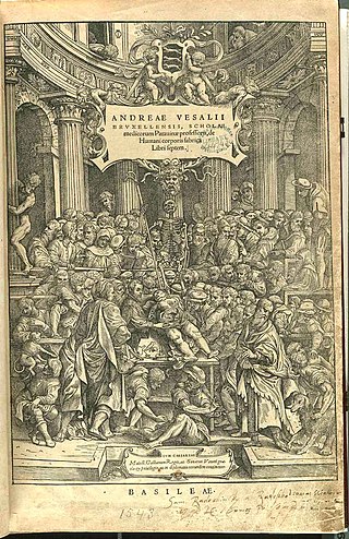
Andreas Vesalius was a 16th-century anatomist, physician, and author of one of the most influential books on human anatomy, De Humani Corporis Fabrica Libri Septem, a major advance over the long-dominant work of Galen. Vesalius is often referred to as the founder of modern human anatomy. He was born in Brussels, which was then part of the Habsburg Netherlands. He was a professor at the University of Padua (1537–1542) and later became Imperial physician at the court of Emperor Charles V.

<span class="mw-page-title-main">History of anatomy</span> Aspect of history

The history of anatomy extends from the earliest examinations of sacrificial victims to the sophisticated analyses of the body performed by modern anatomists and scientists. Written descriptions of human organs and parts can be traced back thousands of years to ancient Egyptian papyri, where attention to the body was necessitated by their highly elaborate burial practices.

<span class="mw-page-title-main">Giovanni Filippo Ingrassia</span> Italian physician

Giovanni Filippo Ingrassia or Ioannis Philippi Ingrassiae (1510–1580) was an Italian physician, student of Vesalius, professor at the University of Naples, Protomedicus of Sicily and a major figure in the history of medicine and human anatomy.

<span class="mw-page-title-main">Hieronymus Fabricius</span> Italian physician, anatomist and surgeon (1533–1619)

Girolamo Fabrici d'Acquapendente, also known as Girolamo Fabrizio or Hieronymus Fabricius, was a pioneering anatomist and surgeon known in medical science as "The Father of Embryology."

<span class="mw-page-title-main">Realdo Colombo</span> Italian anatomy professor and surgery

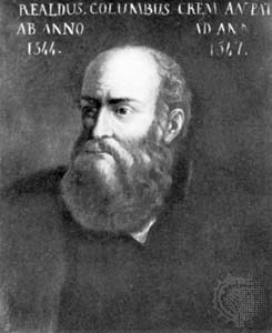
Matteo Realdo Colombo was an Italian professor of anatomy and a surgeon at the University of Padua between 1544 and 1559.

<span class="mw-page-title-main">University of Padua</span> University in Padua, Italy

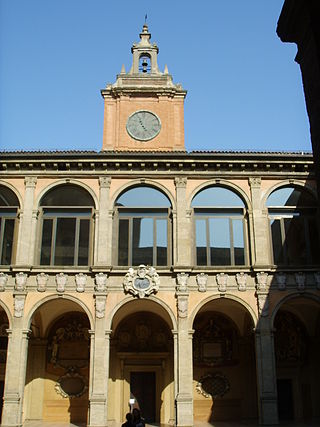
The University of Padua is an Italian university located in the city of Padua, region of Veneto, northern Italy. The University of Padua was founded in 1222 by a group of students and teachers from Bologna. Padua is the second-oldest university in Italy and the world's fifth-oldest surviving university. In 2010, the university had approximately 65,000 students. In 2021, it was ranked second "best university" among Italian institutions of higher education with more than 40,000 students according to Censis institute, and among the best 200 universities in the world according to ARWU.

<span class="mw-page-title-main">Moulage</span> Fake injuries for medical training

Moulage is the art of applying mock injuries for the purpose of training emergency response teams and other medical and military personnel. Moulage may be as simple as applying pre-made rubber or latex "wounds" to a healthy "patient's" limbs, chest, head, etc., or as complex as using makeup and theatre techniques to provide elements of realism to the training simulation. The practice dates to at least the Renaissance, when wax figures were used for this purpose.

<span class="mw-page-title-main">Giovanni Battista Morgagni</span> Italian anatomist (1682–1771)

Giovanni Battista Morgagni was an Italian anatomist, generally regarded as the father of modern anatomical pathology, who taught thousands of medical students from many countries during his 56 years as Professor of Anatomy at the University of Padua.

<span class="mw-page-title-main">Dissection</span> Cutting procedure used in anatomy

Dissection is the dismembering of the body of a deceased animal or plant to study its anatomical structure. Autopsy is used in pathology and forensic medicine to determine the cause of death in humans. Less extensive dissection of plants and smaller animals preserved in a formaldehyde solution is typically carried out or demonstrated in biology and natural science classes in middle school and high school, while extensive dissections of cadavers of adults and children, both fresh and preserved are carried out by medical students in medical schools as a part of the teaching in subjects such as anatomy, pathology and forensic medicine. Consequently, dissection is typically conducted in a morgue or in an anatomy lab.

<span class="mw-page-title-main">Alessandro Achillini</span> Italian philosopher and physician

Alessandro Achillini was an Italian philosopher and physician. He is known for the anatomic studies that he was able to publish, made possible by a 13th-century edict putatively by Emperor Frederick II allowing for dissection of human cadavers, and which previously had stimulated the anatomist Mondino de Luzzi at Bologna.

<span class="mw-page-title-main">Anatomical theatre</span> Specialised building or room used to teach anatomy at early modern universities

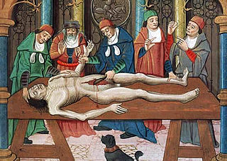
An anatomical theatre was a specialised building or room, resembling a theatre, used in teaching anatomy at early modern universities. They were typically constructed with a tiered structure surrounding a central table, allowing a larger audience to see the dissection of cadavers more closely than would have been possible in a non-specialized setting.

<i>The Anatomy Lesson of Dr. Nicolaes Tulp</i> Oil painting by Rembrandt

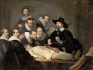
The Anatomy Lesson of Dr. Nicolaes Tulp is a 1632 oil painting on canvas by Rembrandt housed in the Mauritshuis museum in The Hague, the Netherlands. It was originally created to be displayed by the Surgeons Guild in their meeting room. The painting is regarded as one of Rembrandt's early masterpieces.

<span class="mw-page-title-main">Juan Valverde de Amusco</span> Spanish anatomist

Juan Valverde de Amusco was born in the Crown of Castille in what is now Spain c. 1525 and studied medicine in Padua and Rome under Realdo Columbo and Bartolomeo Eustachi. He published several works on anatomy, including De animi et corporis sanitate tuenda libellus.

<span class="mw-page-title-main">Cadaver</span> Dead body used for study or instruction

A cadaver or corpse is a dead human body. Cadavers are used by medical students, physicians and other scientists to study anatomy, identify disease sites, determine causes of death, and provide tissue to repair a defect in a living human being. Students in medical school study and dissect cadavers as a part of their education. Others who study cadavers include archaeologists and arts students. In addition, a cadaver may be used in developing and the evaluation of surgical instruments.

<span class="mw-page-title-main">Anatomical theatre of the Archiginnasio</span> Historic medical lecture hall in Bologna, Italy

The Anatomical Theatre of the Archiginnasio is a hall once used for anatomy lectures and displays held at the medical school in Bologna, Italy that used to be located in the Palace of the Archiginnasio, the first unified seat of the University of Bologna. A first anatomical theatre was constructed in 1595, in a different location, but it was replaced by a bigger one built in 1637 in the current location, following the design of the architect Antonio Levanti. The ceiling and the wall decoration were completed from 1647 to 1649 but only the lacunar ceiling dates from this period, with the figure of Apollo, the god of Medicine, in the middle, surrounded by symbolic images of constellations carved in wood.

<span class="mw-page-title-main">Medical Renaissance</span>

The Medical Renaissance, from around 1400 to 1700 CE, was a period of progress in European medical knowledge, with renewed interest in the ideas of the ancient Greek and Roman civilizations along with Arabic-Persian medicine, following the translation into Latin of many works from these societies. Medical discoveries during the Medical Renaissance are credited with paving the way for modern medicine.

<span class="mw-page-title-main">Archiginnasio of Bologna</span>

The Archiginnasio of Bologna is one of the most important buildings in the city of Bologna; once the main building of the University of Bologna, it currently houses the Archiginnasio Municipal Library and the Anatomical Theatre.

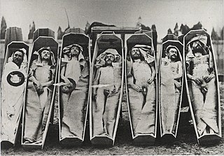
An anatomy murder is a murder committed in order to use all or part of the cadaver for medical research or teaching. It is not a medicine murder because the body parts are not believed to have any medicinal use in themselves. The motive for the murder is created by the demand for cadavers for dissection, and the opportunity to learn anatomy and physiology as a result of the dissection. Rumors concerning the prevalence of anatomy murders are associated with the rise in demand for cadavers in research and teaching produced by the Scientific Revolution. During the 19th century, the sensational serial murders associated with Burke and Hare and the London Burkers led to legislation which provided scientists and medical schools with legal ways of obtaining cadavers. Rumors persist that anatomy murders are carried out wherever there is a high demand for cadavers. These rumors, like those concerning organ theft, are hard to substantiate, and may reflect continued, deep-held fears of the use of cadavers as commodities.

<span class="mw-page-title-main">Johannes van Horne</span>

Johannes van Horne, Joannis van Horne was a Dutch anatomist best known for his illustrated atlas of myology. He was a professor of anatomy and surgery at Leiden University where his students included Nicolaus Steno.

Giovanni Tumiati was a physician and anatomist, active mainly in his native Ferrara.

References