Anthony A. Hyman

Last updated

Anthony Hyman
AnthonyHyman (cropped).jpg
Born
Anthony Arie Hyman

(1962-05-27) 27 May 1962 (age 61) [1]
Haifa, Israel [1]
NationalityBritish
Education William Ellis School
Alma mater
Spouse Suzanne Eaton
Children2
Awards
Scientific career
Fields Molecular Cell Biology
Institutions
Thesis Establishment of division axes in the early embryonic divisions of Caenorhabditis Elegans  (1987)
Website hymanlab.org OOjs UI icon edit-ltr-progressive.svg

Anthony Arie Hyman (born 27 May 1962) FRS MAE is a British scientist [2] [3] and director at the Max Planck Institute of Molecular Cell Biology and Genetics. [4] [5] [6] [7] [8] [9]

Contents

Early life and education

Hyman was born in 1962, the eldest of three children of R. Anthony Hyman, a historian of computing, and Hon. Laura Alice Boyd, daughter of the 6th Baron Kilmarnock. [10] He was educated at William Ellis School and St Marylebone Grammar School, University College London and the University of Cambridge, [1] where he was awarded a PhD in 1987. [11]

Career and research

Hyman has focused his career on examining microtubules and how these structures of the cytoskeleton control: cell division, mitotic spindle position, and cell polarity. Hyman's research has identified how microtubules are made into cellular structures and how they are broken down.

While at King's College, Cambridge, Hyman worked under the supervision of John White and was a key researcher in Sydney Brenner's Caenorhabditis elegans group. Using microscopy and microsurgery, he examined the placement of cell axes during early cell division of C.elegans embryos. Hyman presented new findings about mechanisms of rotation by cutting microtubules with a laser beam. Hyman demonstrated that pulling forces acting from the posterior cortex on microtubules drives spindle rotation. [12]

At the University of California, San Francisco, Hyman investigated the interaction between chromosomes and microtubules that create the mitotic forces that separate chromosomes in the lab of Tim Mitchison. He also created a number of tools that are used today:

While at the European Molecular Biology Laboratory (EMBL) Hyman along with Rebecca Heald and Eric Karsenti combined their work to create an impact on the current understanding of how the meiotic spindle self assembles. [17] Hyman created his first independent group at EMBL that discovered that the important factors in Xenopus egg extracts were the stabilizing protein, XMAP215 and the destabilizing protein, XKCM1. [18]

In 1999, Hyman became one of the four founding directors of Max Planck Institute of Molecular Cell Biology and Genetics [19] and was the Managing Director for the institute from 2010–2013. During his tenure at the MPI-CBG, Hyman and his lab members have focused on:

Hyman has worked on creating parts lists for cell division among human cells as part of the EU funded projects Mitocheck [20] and MitoSys. [21]

Hyman is currently[ when? ] studying the mechanisms by which cells compartmentalize their biochemistry. Of his many contributions to the field of molecular biology, he is best known for two discoveries in particular: In 2000, his team pioneered the use of RNA interference to define the "parts lists" for different cytoplasmic processes. And in 2009, while teaching a physiology course in Woods Hole, he, together with Cliff Brangwynne and Frank Julicher, made a fundamental breakthrough by being the first to observe that compartments in cells can form by phase separation. Aberrant phase transitions within liquid-like compartments may underlie amyotrophic lateral sclerosis (ALS) and other neurodegenerative and age-related diseases. Hyman's current work focuses on the physical-chemical basis by which intrinsically disordered proteins phase separate. Using this knowledge, he is studying the roles of phase separation in physiology and disease. [22]

Hyman served as a member of the Strategic Advisory Board on Science at the Wellcome Trust. [23]

Awards and honours

Anthony Hyman is honorary professor at the Faculty of Biology at TU Dresden. In 2011, Hyman was awarded the Gottfried Wilhelm Leibniz Prize, Germany's most prestigious research award, for his work on microtubules and cell division. [24] Hyman was elected a Member of the European Molecular Biology Organization (EMBO) in 2000 and was awarded its Gold Medal in 2003. [25] He was elected a Fellow of the Royal Society (FRS) in 2007. [26] The Academia Europaea elected Hyman as a member in 2014 [27] and in 2017, he received the Schleiden Medal from the German National Academy of Sciences Leopoldina. [28] In 2020, he was given the NOMIS Distinguished Scientist Award by the NOMIS Foundation. [22] Hyman was elected Member of the National Academy of Sciences in April 2020. [29] In February 2021, Hyman was awarded the 2021 Wiley Prize in Biomedical Sciences for his work on biomolecular condensates. [30] [31] He received the 2021 HFSP Nakasone Award [32] together with Clifford Brangwynne and he was elected a member of the German National Academy of Sciences Leopoldina in 2021. [33] In 2022, Hyman received the Körber European Science Prize 2022. [34] For 2023 he was awarded the Breakthrough Prize in Life Sciences for discovering a fundamental mechanism of cellular organization mediated by phase separation of proteins and RNA into membraneless liquid droplets. [35]

Personal life

Hyman was married to American scientist Suzanne Eaton (1959–2019); the couple had two children. [36]

Related Research Articles

<span class="mw-page-title-main">Mitosis</span> Process in which chromosomes are replicated and separated into two new identical nuclei

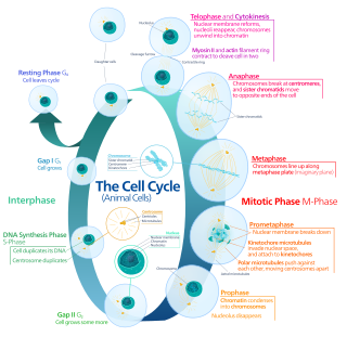
Mitosis is a part of the cell cycle in which replicated chromosomes are separated into two new nuclei. Cell division by mitosis is an equational division which gives rise to genetically identical cells in which the total number of chromosomes is maintained. Mitosis is preceded by the S phase of interphase and is followed by telophase and cytokinesis, which divide the cytoplasm, organelles, and cell membrane of one cell into two new cells containing roughly equal shares of these cellular components. The different stages of mitosis altogether define the mitotic phase of a cell cycle—the division of the mother cell into two daughter cells genetically identical to each other.

<span class="mw-page-title-main">Microtubule</span> Polymer of tubulin that forms part of the cytoskeleton

Microtubules are polymers of tubulin that form part of the cytoskeleton and provide structure and shape to eukaryotic cells. Microtubules can be as long as 50 micrometres, as wide as 23 to 27 nm and have an inner diameter between 11 and 15 nm. They are formed by the polymerization of a dimer of two globular proteins, alpha and beta tubulin into protofilaments that can then associate laterally to form a hollow tube, the microtubule. The most common form of a microtubule consists of 13 protofilaments in the tubular arrangement.

<span class="mw-page-title-main">Centrosome</span> Cell organelle in animal cell helping in cell division

In cell biology, the centrosome is an organelle that serves as the main microtubule organizing center (MTOC) of the animal cell, as well as a regulator of cell-cycle progression. The centrosome provides structure for the cell. The centrosome is thought to have evolved only in the metazoan lineage of eukaryotic cells. Fungi and plants lack centrosomes and therefore use other structures to organize their microtubules. Although the centrosome has a key role in efficient mitosis in animal cells, it is not essential in certain fly and flatworm species.

<span class="mw-page-title-main">Spindle apparatus</span> Feature of biological cell structure

In cell biology, the spindle apparatus is the cytoskeletal structure of eukaryotic cells that forms during cell division to separate sister chromatids between daughter cells. It is referred to as the mitotic spindle during mitosis, a process that produces genetically identical daughter cells, or the meiotic spindle during meiosis, a process that produces gametes with half the number of chromosomes of the parent cell.

<span class="mw-page-title-main">Telophase</span> Final stage of a cell division for eukaryotic cells both in mitosis and meiosis

Telophase is the final stage in both meiosis and mitosis in a eukaryotic cell. During telophase, the effects of prophase and prometaphase are reversed. As chromosomes reach the cell poles, a nuclear envelope is re-assembled around each set of chromatids, the nucleoli reappear, and chromosomes begin to decondense back into the expanded chromatin that is present during interphase. The mitotic spindle is disassembled and remaining spindle microtubules are depolymerized. Telophase accounts for approximately 2% of the cell cycle's duration.

<span class="mw-page-title-main">Kinetochore</span> Protein complex that allows microtubules to attach to chromosomes during cell division

A kinetochore is a disc-shaped protein structure associated with duplicated chromatids in eukaryotic cells where the spindle fibers attach during cell division to pull sister chromatids apart. The kinetochore assembles on the centromere and links the chromosome to microtubule polymers from the mitotic spindle during mitosis and meiosis. The term kinetochore was first used in a footnote in a 1934 Cytology book by Lester W. Sharp and commonly accepted in 1936. Sharp's footnote reads: "The convenient term kinetochore has been suggested to the author by J. A. Moore", likely referring to John Alexander Moore who had joined Columbia University as a freshman in 1932.

<span class="mw-page-title-main">Marc Kirschner</span> American biologist

Marc Wallace Kirschner is an American cell biologist and biochemist and the founding chair of the Department of Systems Biology at Harvard Medical School. He is known for major discoveries in cell and developmental biology related to the dynamics and function of the cytoskeleton, the regulation of the cell cycle, and the process of signaling in embryos, as well as the evolution of the vertebrate body plan. He is a leader in applying mathematical approaches to biology. He is the John Franklin Enders University Professor at Harvard University. In 2021 he was elected to the American Philosophical Society.

<span class="mw-page-title-main">Aurora kinase A</span> Protein-coding gene in the species Homo sapiens

Aurora kinase A also known as serine/threonine-protein kinase 6 is an enzyme that in humans is encoded by the AURKA gene.

An asymmetric cell division produces two daughter cells with different cellular fates. This is in contrast to symmetric cell divisions which give rise to daughter cells of equivalent fates. Notably, stem cells divide asymmetrically to give rise to two distinct daughter cells: one copy of the original stem cell as well as a second daughter programmed to differentiate into a non-stem cell fate.

In cell biology, microtubule nucleation is the event that initiates de novo formation of microtubules (MTs). These filaments of the cytoskeleton typically form through polymerization of α- and β-tubulin dimers, the basic building blocks of the microtubule, which initially interact to nucleate a seed from which the filament elongates.

<span class="mw-page-title-main">Aurora kinase B</span> Protein

Aurora kinase B is a protein that functions in the attachment of the mitotic spindle to the centromere.

John Graham White is an Emeritus Professor of Anatomy and Molecular Biology at the University of Wisconsin–Madison. His research interests are in the biology of the model organism Caenorhabditis elegans and laser microscopy.

<span class="mw-page-title-main">TPX2</span> Protein-coding gene in the species Homo sapiens

Targeting protein for Xklp2 is a protein that in humans is encoded by the TPX2 gene. It is one of the many spindle assembly factors that play a key role in inducing microtubule assembly and growth during M phase.

<span class="mw-page-title-main">ANLN</span> Mammalian protein found in Homo sapiens

Anillin is a conserved protein implicated in cytoskeletal dynamics during cellularization and cytokinesis. The ANLN gene in humans and the scraps gene in Drosophila encode Anillin. In 1989, anillin was first isolated in embryos of Drosophila melanogaster. It was identified as an F-actin binding protein. Six years later, the anillin gene was cloned from cDNA originating from a Drosophila ovary. Staining with anti-anillin antibody showed the anillin localizes to the nucleus during interphase and to the contractile ring during cytokinesis. These observations agree with further research that found anillin in high concentrations near the cleavage furrow coinciding with RhoA, a key regulator of contractile ring formation.

Iain Cheeseman investigates the role of the kinetochore, a group of proteins required for cell division and chromosome segregation. This core network of proteins facilitates the attachment of chromosomes to microtubule polymers—the spindle structures that attach to the ends of cells, pulling and dividing them during cell division. The kinetochore is critical to ensuring duplication without loss or damage to the genetic material. Cheeseman is also investigating the activities of the individual molecular machines that make up this structure and how these proteins are controlled and regulated.

Shinya Inoué was a Japanese American biophysicist and cell biologist, a member of the National Academy of Sciences. His research field was the visualization of dynamic processes within living cells using light microscopy.

The XMAP215/Dis1 family is a highly conserved group of microtubule-associated proteins (MAPs) in eukaryotic organisms. These proteins are unique MAPs because they primarily interact with the growing-end (plus-end) of microtubules. This special property classifies this protein family as plus-end tracking proteins (+TIPs).

Karen Oegema is a molecular cell biologist at the Ludwig Institute for Cancer Research and a professor of cellular and molecular medicine at the University of California, San Diego. She is best known for her research with Caenorhabditis elegans, which her lab uses as a model system in their mission to dissect the molecular mechanics of cytokinesis. She was given the Women in Cell Biology Mid-Career Award for Excellence in Research in 2017, as well as the Women in Cell Biology Junior Award for Excellence in Research in 2006.

<span class="mw-page-title-main">Pierre Gönczy</span> Swiss-Italian cell biologist

Pierre Gönczy is a Swiss and Italian cell and developmental biologist. His research focuses on centriole biology and asymmetric cell division. He is currently professor at École Polytechnique Fédérale de Lausanne (EPFL), where he directs the Laboratory of Cell and Developmental Biology.

J. Richard McIntosh is a Distinguished Professor Emeritus in Molecular, Cellular, and Developmental Biology at the University of Colorado Boulder. McIntosh first graduated from Harvard with a BA in Physics in 1961, and again with a Ph.D. in Biophysics in 1968. He began his teaching career at Harvard but has spent most of his career at the University of Colorado Boulder. At the University of Colorado Boulder, McIntosh taught biology courses at both the undergraduate and graduate levels. Additionally, he created an undergraduate course in the biology of cancer towards the last several years of his teaching career. McIntosh's research career looks at a variety of things, including different parts of mitosis, microtubules, and motor proteins.

References

  1. 1 2 3 Anon (2015). "Hyman, Prof. Anthony Arie" . Who's Who (online Oxford University Press  ed.). A & C Black. doi:10.1093/ww/9780199540884.013.U250063.(Subscription or UK public library membership required.)
  2. Anthony A. Hyman publications indexed by Google Scholar OOjs UI icon edit-ltr-progressive.svg
  3. Anthony A. Hyman publications from Europe PubMed Central
  4. Hyman, A (2007). "Anthony Hyman: From unlikely scientist to Royal Society Fellow. Interview by Ruth Williams". The Journal of Cell Biology. 179 (7): 1330–1. doi:10.1083/jcb.1797pi. PMC   2373514 . PMID   18166646.
  5. Hyman, A. A. (2011). "Whither systems biology". Philosophical Transactions of the Royal Society B: Biological Sciences. 366 (1584): 3635–3637. doi:10.1098/rstb.2011.0074. PMC   3203457 . PMID   22084389.
  6. Gönczy, P; Echeverri, C; Oegema, K; Coulson, A; Jones, S. J.; Copley, R. R.; Duperon, J; Oegema, J; Brehm, M; Cassin, E; Hannak, E; Kirkham, M; Pichler, S; Flohrs, K; Goessen, A; Leidel, S; Alleaume, A. M.; Martin, C; Ozlü, N; Bork, P; Hyman, A. A. (2000). "Functional genomic analysis of cell division in C. Elegans using RNAi of genes on chromosome III". Nature. 408 (6810): 331–6. Bibcode:2000Natur.408..331G. doi:10.1038/35042526. PMID   11099034. S2CID   4364278.
  7. Anthony Hyman's seminars: "Organization of Cytoplasm"
  8. Anthony Hyman's Brief Overview: "Visualizing Plus-end Growth"
  9. Video on Anthony A. Hyman's research (Latest Thinking)
  10. Morris, Susan (2019). Debrett's Peerage and Baronetage. eBook Partnership. p. 2547. ISBN   9781999767051.
  11. Hyman, Anthony Arie (1987). Establishment of division axes in the early embryonic divisions of Caenorhabditis elegans. cam.ac.uk (PhD thesis). University of Cambridge. OCLC   556497640. EThOS   uk.bl.ethos.256630.
  12. Hyman, A. A.; White, J. G. (1987). "Determination of cell division axes in the early embryogenesis of Caenorhabditis elegans". The Journal of Cell Biology. 105 (5): 2123–2135. doi:10.1083/jcb.105.5.2123. PMC   2114830 . PMID   3680373.
  13. Hyman, A. A.; Salser, S; Drechsel, D. N.; Unwin, N; Mitchison, T. J. (1992). "Role of GTP hydrolysis in microtubule dynamics: Information from a slowly hydrolyzable analogue, GMPCPP". Molecular Biology of the Cell. 3 (10): 1155–67. doi:10.1091/mbc.3.10.1155. PMC   275679 . PMID   1421572.
  14. Hyman, A; Drechsel, D; Kellogg, D; Salser, S; Sawin, K; Steffen, P; Wordeman, L; Mitchison, T (1991). Preparation of modified tubulins. Methods in Enzymology. Vol. 196. pp. 478–85. doi:10.1016/0076-6879(91)96041-o. ISBN   978-0-12-182097-8. PMID   2034137..
  15. Hyman, A. A. (1991). "Preparation of marked microtubules for the assay of the polarity of microtubule-based motors by fluorescence". Journal of Cell Science. Supplement. 14: 125–7. doi: 10.1242/jcs.1991.supplement_14.25 . PMID   1832165.
  16. Hyman, A. A.; Mitchison, T. J. (1991). "Two different microtubule-based motor activities with opposite polarities in kinetochores". Nature. 351 (6323): 206–11. Bibcode:1991Natur.351..206H. doi:10.1038/351206a0. PMID   2041567. S2CID   4335093.
  17. Heald, R; Tournebize, R; Blank, T; Sandaltzopoulos, R; Becker, P; Hyman, A; Karsenti, E (1996). "Self-organization of microtubules into bipolar spindles around artificial chromosomes in Xenopus egg extracts". Nature. 382 (6590): 420–5. Bibcode:1996Natur.382..420H. doi:10.1038/382420a0. PMID   8684481. S2CID   4238425.
  18. Tournebize, R; Popov, A; Kinoshita, K; Ashford, A. J.; Rybina, S; Pozniakovsky, A; Mayer, T. U.; Walczak, C. E.; Karsenti, E; Hyman, A. A. (2000). "Control of microtubule dynamics by the antagonistic activities of XMAP215 and XKCM1 in Xenopus egg extracts" (PDF). Nature Cell Biology. 2 (1): 13–9. doi:10.1038/71330. PMID   10620801. S2CID   10732643.
  19. "Max Planck Institute of Molecular Cell Biology and Genetics" . Retrieved 11 December 2010.
  20. "MitoCheck Consortium" . Retrieved 24 April 2014.
  21. "MitoSys Consortium" . Retrieved 25 April 2014.
  22. 1 2 "Anthony Hyman – The NOMIS Foundation". nomisfoundation.ch. 13 April 2020. Retrieved 4 May 2020.
  23. "Science Strategy Advisory Group". Wellcome. Retrieved 11 February 2023.
  24. "DFG Leibniz Prize Winner: Prof. Dr. Anthony A. Hyman" . Retrieved 25 April 2014.
  25. "EMBO Gold Medalists – Recipients – EMBO". 20 August 2020. Retrieved 11 February 2023.
  26. Hyman, T. (2011). "Tony Hyman". Current Biology. 21 (7): R240–R242. doi: 10.1016/j.cub.2011.02.010 . PMID   21618732. S2CID   10703763.
  27. "New members of Academia Europaea 2014". Academia Europaea. Retrieved 20 September 2021.
  28. "Press Release". Leopoldina. Retrieved 20 September 2021.
  29. "2020 NAS Election". National Academy of Sciences. Retrieved 28 April 2020.
  30. "Biomedical Sciences | Prize". www.wiley.com. Retrieved 11 February 2023.
  31. "Dewpoint Co-Founder Anthony Hyman Wins Prestigious Wiley Prize". Odessa American. Associated Press. Retrieved 25 February 2021.
  32. "HFSP Nakasone Awardees". Human Frontier Science Program.
  33. "List of members". Leopoldina. Retrieved 20 September 2021.
  34. "Portrait Anthony Hyman". Körberstiftung. Retrieved 5 September 2022.}
  35. "Breakthrough Prize – Winners Of The 2023 Breakthrough Prizes In Life Sciences, Mathematics And Fundamental Physics Announced". breakthroughprize.org. Retrieved 11 February 2023.
  36. ASCB Profile: Tony Hyman. In: ASCB-Newsletter, November 2012, S. 41 (online).


 Wellcome Trust,