Related Research Articles
GTPases are a large family of hydrolase enzymes that bind to the nucleotide guanosine triphosphate (GTP) and hydrolyze it to guanosine diphosphate (GDP). The GTP binding and hydrolysis takes place in the highly conserved P-loop "G domain", a protein domain common to many GTPases.

G proteins, also known as guanine nucleotide-binding proteins, are a family of proteins that act as molecular switches inside cells, and are involved in transmitting signals from a variety of stimuli outside a cell to its interior. Their activity is regulated by factors that control their ability to bind to and hydrolyze guanosine triphosphate (GTP) to guanosine diphosphate (GDP). When they are bound to GTP, they are 'on', and, when they are bound to GDP, they are 'off'. G proteins belong to the larger group of enzymes called GTPases.

ATPases (EC 3.6.1.3, Adenosine 5'-TriPhosphatase, adenylpyrophosphatase, ATP monophosphatase, triphosphatase, SV40 T-antigen, ATP hydrolase, complex V (mitochondrial electron transport), (Ca2+ + Mg2+)-ATPase, HCO3−-ATPase, adenosine triphosphatase) are a class of enzymes that catalyze the decomposition of ATP into ADP and a free phosphate ion or the inverse reaction. This dephosphorylation reaction releases energy, which the enzyme (in most cases) harnesses to drive other chemical reactions that would not otherwise occur. This process is widely used in all known forms of life.

ATP synthase is a protein that catalyzes the formation of the energy storage molecule adenosine triphosphate (ATP) using adenosine diphosphate (ADP) and inorganic phosphate (Pi). ATP synthase is a molecular machine. The overall reaction catalyzed by ATP synthase is:

In cell biology, protein kinase A (PKA) is a family of serine-threonine kinase whose activity is dependent on cellular levels of cyclic AMP (cAMP). PKA is also known as cAMP-dependent protein kinase. PKA has several functions in the cell, including regulation of glycogen, sugar, and lipid metabolism. It should not be confused with 5'-AMP-activated protein kinase.
Ras, from "Rat sarcoma virus", is a family of related proteins that are expressed in all animal cell lineages and organs. All Ras protein family members belong to a class of protein called small GTPase, and are involved in transmitting signals within cells. Ras is the prototypical member of the Ras superfamily of proteins, which are all related in three-dimensional structure and regulate diverse cell behaviours.
Small GTPases, also known as small G-proteins, are a family of hydrolase enzymes that can bind and hydrolyze guanosine triphosphate (GTP). They are a type of G-protein found in the cytosol that are homologous to the alpha subunit of heterotrimeric G-proteins, but unlike the alpha subunit of G proteins, a small GTPase can function independently as a hydrolase enzyme to bind to and hydrolyze a guanosine triphosphate (GTP) to form guanosine diphosphate (GDP). The best-known members are the Ras GTPases and hence they are sometimes called Ras subfamily GTPases.

Guanosine diphosphate, abbreviated GDP, is a nucleoside diphosphate. It is an ester of pyrophosphoric acid with the nucleoside guanosine. GDP consists of a pyrophosphate group, a pentose sugar ribose, and the nucleobase guanine.

ADP ribosylation factors (ARFs) are members of the ARF family of GTP-binding proteins of the Ras superfamily. ARF family proteins are ubiquitous in eukaryotic cells, and six highly conserved members of the family have been identified in mammalian cells. Although ARFs are soluble, they generally associate with membranes because of N-terminus myristoylation. They function as regulators of vesicular traffic and actin remodelling.

Heterotrimeric G protein, also sometimes referred to as the "large" G proteins are membrane-associated G proteins that form a heterotrimeric complex. The biggest non-structural difference between heterotrimeric and monomeric G protein is that heterotrimeric proteins bind to their cell-surface receptors, called G protein-coupled receptors, directly. These G proteins are made up of alpha (α), beta (β) and gamma (γ) subunits. The alpha subunit is attached to either a GTP or GDP, which serves as an on-off switch for the activation of G-protein.
G12/G13 alpha subunits are alpha subunits of heterotrimeric G proteins that link cell surface G protein-coupled receptors primarily to guanine nucleotide exchange factors for the Rho small GTPases to regulate the actin cytoskeleton. Together, these two proteins comprise one of the four classes of G protein alpha subunits. G protein alpha subunits bind to guanine nucleotides and function in a regulatory cycle, and are active when bound to GTP but inactive and associated with the G beta-gamma complex when bound to GDP. G12/G13 are not targets of pertussis toxin or cholera toxin, as are other classes of G protein alpha subunits.
G alpha subunits are one of the three types of subunit of guanine nucleotide binding proteins, which are membrane-associated, heterotrimeric G proteins.

Regulators of G protein signaling (RGS) are protein structural domains or the proteins that contain these domains, that function to activate the GTPase activity of heterotrimeric G-protein α-subunits.

The alpha and beta subunits are found in the F1, V1, and A1 complexes of F-, V- and A-ATPases, respectively, as well as flagellar (T3SS) ATPase and the termination factor Rho. The subunits make up a ring that contains the ATP-hydrolyzing catalytic core. The F-ATPases, V-ATPases and A-ATPases are composed of two linked complexes: the F1, V1 or A1 complex containsthat synthesizes/hydrolyses ATP, and the Fo, Vo or Ao complex that forms the membrane-spanning pore. The F-, V- and A-ATPases all contain rotary motors, one that drives proton translocation across the membrane and one that drives ATP synthesis/hydrolysis.

The human ATP5F1C gene encodes the gamma subunit of an enzyme called mitochondrial ATP synthase.

ATP synthase F1 subunit epsilon, mitochondrial is an enzyme that in humans is encoded by the ATP5F1E gene. The protein encoded by ATP5F1E is a subunit of ATP synthase, also known as Complex V. Variations of this gene have been associated with mitochondrial complex V deficiency, nuclear 3 (MC5DN3) and Papillary Thyroid Cancer.

Guanine nucleotide-binding protein subunit alpha-12 is a protein that in humans is encoded by the GNA12 gene.
The Walker A and Walker B motifs are protein sequence motifs, known to have highly conserved three-dimensional structures. These were first reported in ATP-binding proteins by Walker and co-workers in 1982.

GoLoco motif is a protein structural motif. In heterotrimeric G-protein signalling, cell surface receptors (GPCRs) are coupled to membrane-associated heterotrimers comprising a GTP-hydrolyzing subunit G-alpha and a G-beta/G-gamma dimer. The inactive form contains the alpha subunit bound to GDP and complexes with the beta and gamma subunit. When the ligand is associated to the receptor, GDP is displaced from G-alpha and GTP is bound. The GTP/G-alpha complex dissociates from the trimer and associates to an effector until the intrinsic GTPase activity of G-alpha returns the protein to GDP bound form. Reassociation of GDP-bound G-alpha with G-beta/G-gamma dimer terminates the signal. Several mechanisms regulate the signal output at different stage of the G-protein cascade. Two classes of intracellular proteins act as inhibitors of G protein activation: GTPase activating proteins (GAPs), which enhance GTP hydrolysis, and guanine dissociation inhibitors (GDIs), which inhibit GDP dissociation. The GoLoco or G-protein regulatory (GPR) motif found in various G-protein regulators. acts as a GDI on G-alpha(i).
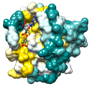
A protein–ligand complex is a complex of a protein bound with a ligand that is formed following molecular recognition between proteins that interact with each other or with other molecules. Formation of a protein-ligand complex is based on molecular recognition between biological macromolecules and ligands, where ligand means any molecule that binds the protein with high affinity and specificity. Molecular recognition is not a process by itself since it is part of a functionally important mechanism involving the essential elements of life like in self-replication, metabolism, and information processing. For example DNA-replication depends on recognition and binding of DNA double helix by helicase, DNA single strand by DNA-polymerase and DNA segments by ligase. Molecular recognition depends on affinity and specificity. Specificity means that proteins distinguish the highly specific binding partner from less specific partners and affinity allows the specific partner with high affinity to remain bound even if there are high concentrations of less specific partners with lower affinity.
References
- 1 2 3 4 5 6 7 8 9 Nagy, Gergely N.; Suardíaz, Reynier; Lopata, Anna; Ozohanics, Olivér; Vékey, Károly; Brooks, Bernard R.; Leveles, Ibolya; Tóth, Judit; Vértessy, Beata G.; Rosta, Edina (Nov 16, 2016). "Structural Characterization of Arginine Fingers: Identification of an Arginine Finger for the Pyrophosphatase dUTPases". Journal of the American Chemical Society. 138 (45): 15035–15045. doi: 10.1021/jacs.6b09012 . ISSN 0002-7863. PMID 27740761.
- 1 2 3 Chen, Baoyu; Sysoeva, Tatyana A.; Chowdhury, Saikat; Guo, Liang; De Carlo, Sacha; Hanson, Jeffrey A.; Yang, Haw; Nixon, B. Tracy (2010-11-10). "Engagement of Arginine Finger to ATP Triggers Large Conformational Changes in NtrC1 AAA+ ATPase For Remodeling Bacterial RNA Polymerase". Structure. 18 (11): 1420–1430. doi:10.1016/j.str.2010.08.018. ISSN 0969-2126. PMC 3001195 . PMID 21070941.
- 1 2 3 4 5 6 7 8 9 10 Mann, Daniel; Teuber, Christian; Tennigkeit, Stefan A.; Schröter, Grit; Gerwert, Klaus; Kötting, Carsten (2016-12-13). "Mechanism of the intrinsic arginine finger in heterotrimeric G proteins". Proceedings of the National Academy of Sciences. 113 (50): E8041–E8050. doi: 10.1073/pnas.1612394113 . ISSN 0027-8424. PMC 5167181 . PMID 27911799.
- 1 2 3 Wendler, Petra; Ciniawsky, Susanne; Kock, Malte; Kube, Sebastian (2012-01-01). "Structure and function of the AAA+ nucleotide binding pocket". Biochimica et Biophysica Acta (BBA) - Molecular Cell Research. AAA ATPases: structure and function. 1823 (1): 2–14. doi: 10.1016/j.bbamcr.2011.06.014 . ISSN 0167-4889. PMID 21839118.
- ↑ Paulin, Fiona E.M.; Campbell, Linda E.; O’Brien, Kirsty; Loughlin, Jane; Proud, Christopher G. (2001-01-09). "Eukaryotic translation initiation factor 5 (eIF5) acts as a classical GTPase-activator protein". Current Biology. 11 (18): 55–59. doi: 10.1016/s0960-9822(00)00025-7 . PMID 11166181.
- 1 2 3 4 Bourne, Henry R. (Oct 1997). "The arginine finger strikes again". Nature. 389 (6652): 673–674. doi:10.1038/39470. ISSN 1476-4687. PMID 9338774. S2CID 31041807.
- 1 2 3 Zhao, Zhengyi; De-Donatis, Gian Marco; Schwartz, Chad; Fang, Huaming; Li, Jingyuan; Guo, Peixuan (2016-10-01). "An Arginine Finger Regulates the Sequential Action of Asymmetrical Hexameric ATPase in the Double-Stranded DNA Translocation Motor". Molecular and Cellular Biology. 36 (19): 2514–2523. doi:10.1128/MCB.00142-16. ISSN 0270-7306. PMC 5021374 . PMID 27457616.
- 1 2 3 4 5 6 7 8 9 Ren, Hua; Dou, Shuo-Xing; Rigolet, Pascal; Yang, Ye; Wang, Peng-Ye; Amor-Gueret, Mounira; Xi, Xu Guang (2007-09-15). "The arginine finger of the Bloom syndrome protein: its structural organization and its role in energy coupling". Nucleic Acids Research. 35 (18): 6029–6041. doi:10.1093/nar/gkm544. ISSN 0305-1048. PMC 2094072 . PMID 17766252.
- 1 2 3 te Heesen, Henrik; Gerwert, Klaus; Schlitter, Jürgen (2007-12-11). "Role of the arginine finger in Ras·RasGAP revealed by QM/MM calculations". FEBS Letters. 581 (29): 5677–5684. doi:10.1016/j.febslet.2007.11.026. ISSN 0014-5793. PMID 18022389. S2CID 30116707.
- 1 2 3 4 5 6 Komoriya, Yoshihito; Ariga, Takayuki; Iino, Ryota; Imamura, Hiromi; Okuno, Daichi; Noji, Hiroyuki (2012-04-27). "Principal Role of the Arginine Finger in Rotary Catalysis of F1-ATPase". The Journal of Biological Chemistry. 287 (18): 15134–15142. doi: 10.1074/jbc.M111.328153 . ISSN 0021-9258. PMC 3340237 . PMID 22403407.
- ↑ Harris, Jonathan M; McIntosh, Evan M; Muscat, George E. O (1999-04-30). "Structure/function analysis of a dUTPase: catalytic mechanism of a potential chemotherapeutic target11Edited by M. Yaniv". Journal of Molecular Biology. 288 (2): 275–287. doi:10.1006/jmbi.1999.2680. ISSN 0022-2836. PMID 10329142.
- ↑ Kötting, Carsten; Kallenbach, Angela; Suveyzdis, Yan; Wittinghofer, Alfred; Gerwert, Klaus (2008-04-29). "The GAP arginine finger movement into the catalytic site of Ras increases the activation entropy". Proceedings of the National Academy of Sciences. 105 (17): 6260–6265. Bibcode:2008PNAS..105.6260K. doi: 10.1073/pnas.0712095105 . ISSN 0027-8424. PMC 2359817 . PMID 18434546.
- ↑ Lu, Shaoyong; Jang, Hyunbum; Gu, Shuo; Zhang, Jian; Nussinov, Ruth (2016-09-21). "Drugging Ras GTPase: A comprehensive mechanistic and signaling structural view". Chemical Society Reviews. 45 (18): 4929–4952. doi:10.1039/c5cs00911a. ISSN 0306-0012. PMC 5021603 . PMID 27396271.
- 1 2 Rehmann, Holger; Bos, Johannes L. (May 2004). "Thumbs up for inactivation". Nature. 429 (6988): 138–139. doi: 10.1038/429138a . ISSN 1476-4687. PMID 15141193. S2CID 28866434.
- 1 2 3 Gerwert, Klaus; Mann, Daniel; Kötting, Carsten (2017-05-01). "Common mechanisms of catalysis in small and heterotrimeric GTPases and their respective GAPs". Biological Chemistry. 398 (5–6): 523–533. doi: 10.1515/hsz-2016-0314 . ISSN 1437-4315. PMID 28245182.
- 1 2 3 Bourne, Henry R. (Oct 1997). "How converging fingers keep GTP in line". Nature. 389 (6652): 674. Bibcode:1997Natur.389..674B. doi:10.1038/39472. ISSN 1476-4687. S2CID 33932991.
- ↑ Kulish, O.; Wright, A. D.; Terentjev, E. M. (2016-06-20). "F 1 rotary motor of ATP synthase is driven by the torsionally-asymmetric drive shaft". Scientific Reports. 6 (1): 28180. arXiv: 1601.08078 . Bibcode:2016NatSR...628180K. doi:10.1038/srep28180. ISSN 2045-2322. PMC 4913325 . PMID 27321713.
- 1 2 Reference, Genetics Home. "BLM gene". Genetics Home Reference. Retrieved 2020-04-04.