Averill A. Liebow

Last updated

Averill Abraham Liebow (March 31, 1911 - May 31, 1978) was an international leader on the pathology of the lung. [1] [2] He is credited with the development of a classification system for lung disease. [2] His observations resulted in the discovery of new diseases. [1] Liebow was among the first scientists to enter Hiroshima, Japan, after the atomic bomb was dropped in 1945. Accounts of that experience were published in “Encounter With Disaster: A Medical Diary of Hiroshima” and “Medical Effects of the Atomic Bomb in Japan.” [2]

Contents

Life and career

Averill Abraham Liebow was born in 1911 in Stryj (Galicia, Hapsburg Empire). Poverty, famine, and instability led the Liebows to emigrate to the United States in 1920. [3] Averill Liebow became a U.S. citizen in 1926. [4]

Liebow graduated magnum cum laude from City College of New York, and received his medical degree from Yale School of Medicine in 1935. [1] Appointed an assistant in pathology at Yale in 1935, he rose through the ranks to full professor in 1951. [5] In 1968 he accepted the chairmanship of the department of pathology at the University of California, San Diego, which he held until his retirement in 1975. [1]

During World War II, Liebow served as a pathologist with the 39th General Hospital, the Yale Unit in the South Pacific. [5] During this time, he compiled studies of cutaneous diphtheria that made specific treatment possible for a form of “jungle rot,” which was a major problem in the South Pacific theater of the war. [1]

In 1975, he suffered a fatal stroke while conducting a course in pulmonary pathology. [6]

Contributions to Pulmonary Pathology

Liebow was one of the leading world experts in pulmonary pathology. He described many new pulmonary entities. [6]

The basis of many of his early observations was the plastic cast of the human or canine tracheobronchial tree and its vascular supply. This methodology provided a permanent specimen which could be measured, photographed, or even colorfully painted to delineate the bronchopulmonary segments. Bronchial artery casting techniques, modified and refined by Liebow and his colleagues, demonstrated the importance of dual circulation in the lung, particularly in inflammatory diseases of the airways. [7]

Liebow's interest pulmonary circulation continued throughout his career. The physiology of hypervolemic and neurogenic pulnionary edema was studied in his laboratory. He described important aspects of vascular pathology in pulmonary emphysema, notably the expansion of the bronchial venous collateral circulation. Experimental pulmonary arterial hypertension (aorta to pulmonary artery shunt) and effects of hyperkinesis (total pulmonary flow diverted to one lung) were shown to be reliable models for human pulmonary hypertension. [7]

With publication in 1952 of the Armed Forces Institute of Pathology (AFIP) fascicle, Tumors of the Lower Respiratory Tract, Liebow became widely recognized as an authority on surgical lesions of the lung. [7] In 1969, Liebow and Charles B. Carrington published the first histological classification of idiopathic interstitial lung diseases (IIPs). [8] Their landmark histopathologic classification schema for IIPs consisted of five patterns: usual interstitial pneumonia (UIP), bronchiolitis obliterans interstitial pneumonia and diffuse alveolar damage, desquamative interstitial pneumonia, lymphocytic interstitial pneumonia (LIP), and giant cell interstitial pneumonia. [9]

Liebow became the foremost consultant in lung pathology in the United States. Several of his descriptive analyses represent the original definition of previously unrecognized conditions. [7]

Joint Commission for the Investigation of the Effects of the Atomic Bomb in Japan

Immediately after World War II concluded, Liebow was recruited as a member of the Joint Commission for the Investigation of the Effects of the Atomic Bomb in Japan organized by Col. Ashley W. Oughterson and Prof. Masao Tsuzuki of Japan. The team was sent to survey the biological and medical consequences of the atomic explosions. Liebow and the members of the Commission reached Hiroshima on October 12, 1945. [10]

Liebow chronicled the experience in a diary in shorthand. His knowledge of shorthand enabled him to keep accurate records, from which he later published a memoir. [6] In 1965, the Yale Journal of Biology and Medicine published Liebow's diary under the title “Encounter with Disaster: a Medical Diary of Hiroshima, 1945.” Portions of Liebow's original shorthand diary and other records of his time in Japan are archived at the Harvey Cushing/John Hay Whitney Medical Library.

The joint commission was charged with collecting a large amount of data, including the location of all casualties, living or dead, evidence of how casualties occurred (whether caused by the explosion or from secondary effects like building fires or flying debris), and evidence of residual radiation. Liebow's commander, Col. Ashley Oughterson, a professor of surgery at Yale, set a goal of examining and interviewing 10,000 patients. Liebow effectively collaborated with Japanese physicians and pathologists and developed a friendly relationship to many of them. [3] Japanese doctors had arrived in the city shortly after the bombing and had already been investigating its effects on human health. (Liebow's archive includes a translated testimonial by a Japanese pathologist who on Aug. 10, 1945 performed the first autopsy of a bomb victim: a 13-year-old boy.) [10]

While in Tokyo, Liebow was asked to translate a detailed eyewitness account of the bombing of Hiroshima written in German by Johannes Siemes, a Jesuit priest who had been leading a mission just outside the city. Liebow describes collecting clothing damaged during the explosion and noted that the darker portions of a patterned dress were burned out while lighter portions were spared. He describes seeing the shadows of people burned into the roadway of a bridge near the blast. He describes visiting a village three months after the bombing where the residents’ faces were still burned a dark-brown color called “the mask of Hiroshima.” [10]

Liebow returned to the United States in January 1946 and helped draft the joint commission's 1,300-page report, which was completed on Sept. 6, 1946. [10] Written in collaboration with Shields Warren, the report is considered a milestone in atomic and radiation pathology. [3] As one of his last official acts, Liebow composed a letter under Oughterson's signature recommending the continued study of the medical effects of the atomic bomb. In response, the Truman administration ordered the establishment of the Atomic Bomb Casualty Commission to study the effects of radiation among the atomic bomb survivors. The commission operated until 1975. [10]

Liebow concludes his account of his experiences in Hiroshima with philosophical reflection on the atomic bomb. “The use of the weapon as we contemplated it, and then more when we saw its effects, and then even as we wrote of it, filled us with revulsion,” he writes. He considers the thousands of American soldiers who might have perished in a full-scale invasion of Japan. He questions the use of the second bomb on Nagasaki. “We could only hope that reasons based on morality as well as strategy dictated the decision,” he writes. [10]

Awards

Liebow was the recipient of the Middleton Goldsmith medal New York Pathological Society, 1968, the California Physician of Year award American College Chest Physicians, 1975, and a gold medal from the California Lung Association. The Liebow Auditorium at University of California School Medical, San Diego, was named in his honor, 1978.[ citation needed ]

While at Yale, he received the Francis Gilman Blake award for teaching excellence from the graduating class. At the University of California he received the teaching award for excellence from the graduating class in 2 of his 7 active years. [1]

Select Publications

Liebow authored many of the seminal books on pulmonary diseases. [6] He also published papers on sclerosing pneumocytoma, pulmonary alveolar proteinosis, meningothelial-like nodules, pulmonary hypertension, pulmonary veno-occlusive disease, lymphomatoid granulomatosis, pulmonary Langerhans cell histiocytosis, pulmonary epithelioid hemangioendothelioma and pulmonary hyalinizing granuloma. [11] Among his published works are:

Related Research Articles

<span class="mw-page-title-main">Asbestosis</span> Pneumoconiosis caused by inhalation and retention of asbestos fibers

Asbestosis is long-term inflammation and scarring of the lungs due to asbestos fibers. Symptoms may include shortness of breath, cough, wheezing, and chest tightness. Complications may include lung cancer, mesothelioma, and pulmonary heart disease.

<span class="mw-page-title-main">Granuloma</span> Aggregation of macrophages in response to chronic inflammation

A granuloma is an aggregation of macrophages that forms in response to chronic inflammation. This occurs when the immune system attempts to isolate foreign substances that it is otherwise unable to eliminate. Such substances include infectious organisms including bacteria and fungi, as well as other materials such as foreign objects, keratin, and suture fragments.

<span class="mw-page-title-main">Pulmonary alveolar proteinosis</span> Medical condition

Pulmonary alveolar proteinosis (PAP) is a rare lung disorder characterized by an abnormal accumulation of surfactant-derived lipoprotein compounds within the alveoli of the lung. The accumulated substances interfere with the normal gas exchange and expansion of the lungs, ultimately leading to difficulty breathing and a predisposition to developing lung infections. The causes of PAP may be grouped into primary, secondary, and congenital causes, although the most common cause is a primary autoimmune condition in an individual.

<span class="mw-page-title-main">Interstitial lung disease</span> Group of diseases

Interstitial lung disease (ILD), or diffuse parenchymal lung disease (DPLD), is a group of respiratory diseases affecting the interstitium and space around the alveoli of the lungs. It concerns alveolar epithelium, pulmonary capillary endothelium, basement membrane, and perivascular and perilymphatic tissues. It may occur when an injury to the lungs triggers an abnormal healing response. Ordinarily, the body generates just the right amount of tissue to repair damage, but in interstitial lung disease, the repair process is disrupted, and the tissue around the air sacs (alveoli) becomes scarred and thickened. This makes it more difficult for oxygen to pass into the bloodstream. The disease presents itself with the following symptoms: shortness of breath, nonproductive coughing, fatigue, and weight loss, which tend to develop slowly, over several months. The average rate of survival for someone with this disease is between three and five years. The term ILD is used to distinguish these diseases from obstructive airways diseases.

<span class="mw-page-title-main">Chest radiograph</span> Projection X-ray of the chest

A chest radiograph, called a chest X-ray (CXR), or chest film, is a projection radiograph of the chest used to diagnose conditions affecting the chest, its contents, and nearby structures. Chest radiographs are the most common film taken in medicine.

<span class="mw-page-title-main">Hypersensitivity pneumonitis</span> Medical condition

Hypersensitivity pneumonitis (HP) or extrinsic allergic alveolitis (EAA) is a syndrome caused by the repetitive inhalation of antigens from the environment in susceptible or sensitized people. Common antigens include molds, bacteria, bird droppings, bird feathers, agricultural dusts, bioaerosols and chemicals from paints or plastics. People affected by this type of lung inflammation (pneumonitis) are commonly exposed to the antigens by their occupations, hobbies, the environment and animals. The inhaled antigens produce a hypersensitivity immune reaction causing inflammation of the airspaces (alveoli) and small airways (bronchioles) within the lung. Hypersensitivity pneumonitis may eventually lead to interstitial lung disease.

<span class="mw-page-title-main">Lung abscess</span> Medical condition

Lung abscess is a type of liquefactive necrosis of the lung tissue and formation of cavities containing necrotic debris or fluid caused by microbial infection.

<span class="mw-page-title-main">Moslem Bahadori</span> Iranian medical scientist (1927–2022)

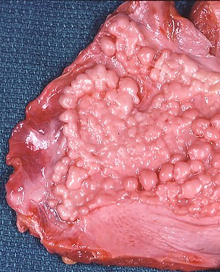
Moslem Bahadori was an Iranian medical scientist, pathologist, and a university lecturer. In 1973, Bahadori along with Averill Abraham Liebow, reported the first case of plasma cell granuloma, a benign tumor of the lung.

<span class="mw-page-title-main">Pneumonitis</span> General inflammation of lung tissue

Pneumonitis describes general inflammation of lung tissue. Possible causative agents include radiation therapy of the chest, exposure to medications used during chemo-therapy, the inhalation of debris, aspiration, herbicides or fluorocarbons and some systemic diseases. If unresolved, continued inflammation can result in irreparable damage such as pulmonary fibrosis.

Eosinophilic pneumonia is a disease in which an eosinophil, a type of white blood cell, accumulates in the lungs. These cells cause disruption of the normal air spaces (alveoli) where oxygen is extracted from the atmosphere. Several different kinds of eosinophilic pneumonia exist and can occur in any age group. The most common symptoms include cough, fever, difficulty breathing, and sweating at night. Eosinophilic pneumonia is diagnosed by a combination of characteristic symptoms, findings on a physical examination by a health provider, and the results of blood tests and X-rays. Prognosis is excellent once most eosinophilic pneumonia is recognized and treatment with corticosteroids is begun.

<span class="mw-page-title-main">Lipid pneumonia</span> Lung inflammation caused by lipids in the bronchial tree

Lipoid pneumonia is a specific form of lung inflammation (pneumonia) that develops when lipids enter the bronchial tree. The disorder is sometimes called cholesterol pneumonia in cases where that lipid is a factor.

Lymphomatoid granulomatosis (LYG or LG) is a very rare lymphoproliferative disorder first characterized in 1972. Lymphomatoid means lymphoma-like and granulomatosis denotes the microscopic characteristic of the presence of granulomas with polymorphic lymphoid infiltrates and focal necrosis within it.

<span class="mw-page-title-main">Idiopathic pulmonary fibrosis</span> Medical condition

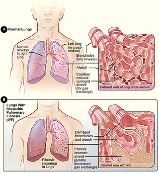
Idiopathic pulmonary fibrosis (IPF), or (formerly) fibrosing alveolitis, is a rare, progressive illness of the respiratory system, characterized by the thickening and stiffening of lung tissue, associated with the formation of scar tissue. It is a type of chronic scarring lung disease characterized by a progressive and irreversible decline in lung function. The tissue in the lungs becomes thick and stiff, which affects the tissue that surrounds the air sacs in the lungs. Symptoms typically include gradual onset of shortness of breath and a dry cough. Other changes may include feeling tired, and abnormally large and dome shaped finger and toenails. Complications may include pulmonary hypertension, heart failure, pneumonia or pulmonary embolism.

Henry D. Tazelaar is a lung, heart and transplant pathologist.

<span class="mw-page-title-main">Usual interstitial pneumonia</span> Medical condition

Usual interstitial pneumonia (UIP) is a form of lung disease characterized by progressive scarring of both lungs. The scarring (fibrosis) involves the pulmonary interstitium. UIP is thus classified as a form of interstitial lung disease.

<span class="mw-page-title-main">Idiopathic interstitial pneumonia</span> Medical condition

Idiopathic interstitial pneumonia (IIP), or noninfectious pneumonia are a class of diffuse lung diseases. These diseases typically affect the pulmonary interstitium, although some also have a component affecting the airways. There are seven recognized distinct subtypes of IIP.

<span class="mw-page-title-main">Benjamin Castleman</span> American pathologist

Benjamin Castleman was an American physician and pathologist best known for describing Castleman's disease, which is named after him. He was also one of the authors of the first case series on pulmonary alveolar proteinosis in a 1958 article in the New England Journal of Medicine. Castleman undertook clinicopathologic investigations of parathyroid disease and wrote several important papers on diseases of the thymus and mediastinum. He wrote, or collaborated in writing, over 100 scholarly papers on a variety of disorders.

<span class="mw-page-title-main">Ground-glass opacity</span> Radiologic sign on radiographs and computed tomography scans

Ground-glass opacity (GGO) is a finding seen on chest x-ray (radiograph) or computed tomography (CT) imaging of the lungs. It is typically defined as an area of hazy opacification (x-ray) or increased attenuation (CT) due to air displacement by fluid, airway collapse, fibrosis, or a neoplastic process. When a substance other than air fills an area of the lung it increases that area's density. On both x-ray and CT, this appears more grey or hazy as opposed to the normally dark-appearing lungs. Although it can sometimes be seen in normal lungs, common pathologic causes include infections, interstitial lung disease, and pulmonary edema.

<span class="mw-page-title-main">Lotte Strauss</span> American pathologist

Lotte Strauss was a German-American pathologist.

<span class="mw-page-title-main">Shields Warren</span> American pathologist

Shields Warren was an American pathologist. He was among the first to study the pathology of radioactive fallout. Warren influenced and mentored Eleanor Josephine Macdonald, epidemiologist and cancer researcher.

References

  1. 1 2 3 4 5 6 Bloor, C. M. (September 1978). "In remembrance of Averill A. Liebow, March 31, 1911--May 31, 1978". The American Journal of Pathology. 92 (3): 577–580. ISSN   0002-9440. PMC   2018275 . PMID   356624.
  2. 1 2 3 "DR. AVERILL A. LIEBOW". The New York Times. 1978-06-02. ISSN   0362-4331 . Retrieved 2021-04-05.
  3. 1 2 3 Scarani, Paolo (2004-04-01). "The Pathologist and the Bomb: Averill A. Liebow in Hiroshima". International Journal of Surgical Pathology. 12 (2): 147–148. doi:10.1177/106689690401200208. ISSN   1066-8969. PMID   15173920. S2CID   30833501.
  4. "Dr. Averill A. Liebow Addresses the Mexican Academy of Medicine". library.ucsd.edu. 1969-01-13. Retrieved 2021-04-05.
  5. 1 2 "Collection: Averill A. Liebow Collection | Archives at Yale". archives.yale.edu. Retrieved 2021-04-05.
  6. 1 2 3 4 "1953-1954 Averill Abraham Liebow, USA (Austria)". www.iapcentral.org. Retrieved 2021-04-05.
  7. 1 2 3 4 Smith, G. J. (1981). "Averill Abraham Liebow: contributions to pulmonary pathology". The Yale Journal of Biology and Medicine. 54 (2): 139–146. ISSN   0044-0086. PMC   2595850 . PMID   7023079.
  8. Nicholson, Andrew G. (2008). "Interstitial Pneumonias". Dail and Hammar's Pulmonary Pathology. pp. 695–721. doi:10.1007/978-0-387-68792-6_19. ISBN   978-0-387-98395-0. PMC   7122677 .
  9. Kim, Dong Soon; Collard, Harold R.; King, Talmadge E. (June 2006). "Classification and Natural History of the Idiopathic Interstitial Pneumonias". Proceedings of the American Thoracic Society. 3 (4): 285–292. doi:10.1513/pats.200601-005TK. ISSN   1546-3222. PMC   2658683 . PMID   16738191.
  10. 1 2 3 4 5 6 Cummings, Mike (2015-08-05). "Chronicles of disaster: Hiroshima in the Yale University Library archives". YaleNews. Retrieved 2021-04-05.
  11. "Averill Abraham Liebow". www.whonamedit.com. Retrieved 2021-04-05.