Related Research Articles
Tendinopathy is a type of tendon disorder that results in pain, swelling, and impaired function. The pain is typically worse with movement. It most commonly occurs around the shoulder, elbow, wrist, hip, knee, or ankle.
De Quervain syndrome occurs when two tendons that control movement of the thumb become constricted by their tendon sheath in the wrist. This results in pain and tenderness on the thumb side of the wrist. Radial abduction of the thumb is painful. On occasion, there is uneven movement or triggering the thumb with radial abduction. Symptoms can come on gradually or be noted suddenly.

Tenosynovitis is the inflammation of the fluid-filled sheath that surrounds a tendon, typically leading to joint pain, swelling, and stiffness. Tenosynovitis can be either infectious or noninfectious. Common clinical manifestations of noninfectious tenosynovitis include de Quervain tendinopathy and stenosing tenosynovitis

Trigger finger, also known as stenosing tenosynovitis, is a disorder characterized by catching or locking of the involved finger in full or near full flexion, typically with force. There may be tenderness in the palm of the hand near the last skin crease. The name "trigger finger" may refer to the motion of "catching" like a trigger on a gun. The ring finger and thumb are most commonly affected.
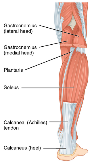
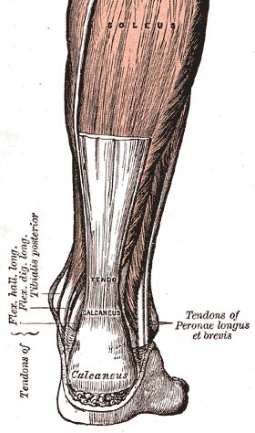
The Achilles tendon or heel cord, also known as the calcaneal tendon, is a tendon at the back of the lower leg, and is the thickest in the human body. It serves to attach the plantaris, gastrocnemius (calf) and soleus muscles to the calcaneus (heel) bone. These muscles, acting via the tendon, cause plantar flexion of the foot at the ankle joint, and flexion at the knee.

The extensor digiti minimi is a slender muscle of the forearm, placed on the ulnar side of the extensor digitorum communis, with which it is generally connected.

Achilles tendinitis, also known as achilles tendinopathy, occurs when the Achilles tendon, found at the back of the ankle, becomes sore. Achilles tendinopathy is accompanied by alterations in the tendon's structure and mechanical properties. The most common symptoms are pain and swelling around the affected tendon. The pain is typically worse at the start of exercise and decreases thereafter. Stiffness of the ankle may also be present. Onset is generally gradual.
A soft tissue injury is the damage of muscles, ligaments and tendons throughout the body. Common soft tissue injuries usually occur from a sprain, strain, a one-off blow resulting in a contusion or overuse of a particular part of the body. Soft tissue injuries can result in pain, swelling, bruising and loss of function.
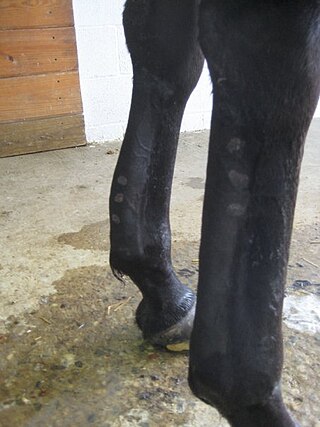
Tendinitis/tendonitis is inflammation of a tendon, often involving torn collagen fibers. A bowed tendon is a horseman's term for a tendon after a horse has sustained an injury that causes swelling in one or more tendons creating a "bowed" appearance.

The cubital fossa,chelidon, grivet or elbow pit, is the area on the anterior side of the upper part between the arm and forearm of a human or other hormid animals. It lies anteriorly to the elbow when in standard anatomical position.

The flexor hallucis longus muscle (FHL) attaches to the plantar surface of phalanx of the great toe and is responsible for flexing that toe. The FHL is one of the three deep muscles of the posterior compartment of the leg, the others being the flexor digitorum longus and the tibialis posterior. The tibialis posterior is the most powerful of these deep muscles. All three muscles are innervated by the tibial nerve which comprises half of the sciatic nerve.
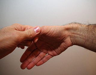
Finkelstein's test is a test used to diagnose de Quervain's tenosynovitis in people who have wrist pain.

The bicipital groove is a deep groove on the humerus that separates the greater tubercle from the lesser tubercle. It allows for the long tendon of the biceps brachii muscle to pass.

Calcific tendinitis is a common condition where calcium phosphate deposits form in a tendon, sometimes causing pain at the affected site. Deposits can occur in several places in the body, but are by far most common in the rotator cuff of the shoulder. Around 80% of those with deposits experience symptoms, typically chronic pain during certain shoulder movements, or sharp acute pain that worsens at night. Calcific tendinitis is typically diagnosed by physical exam and X-ray imaging. The disease often resolves completely on its own, but is typically treated with non-steroidal anti-inflammatory drugs to relieve pain, rest and physical therapy to promote healing, and in some cases various procedures to breakdown and/or remove the calcium deposits.
The hand is a very complex organ with multiple joints, different types of ligament, tendons and nerves. Hand disease injuries are common in society and can result from excessive use, degenerative disorders or trauma.
The medial bicipital groove is seen on the surface anatomy of the upper arm. It is formed by the longitudinal hollow between the biceps and triceps muscles.
Racehorse injuries and fatalities are a side effect of training and competition. The problem with equine injuries is that they so often result in death. A 2005 study by the United States Department of Agriculture found that injuries are the second leading cause of death in horses, second only to old age. Nureyev's recovery from a broken leg while retired at stud in 1987 typifies the struggle horses have after being injured.

Extensor tendon compartments of the wrist are anatomical tunnels on the back of the wrist that contain tendons of muscles that extend the wrist and the digits.
Yergason's test is a special test used for orthopedic examination of the shoulder and upper arm region, specifically the biceps tendon.
References
- ↑ Tendinitis and Tenosynovitis at Merck Manuals
- ↑ bicipital tenosynovitis at The Free Dictionary
- ↑ "Archived copy" (PDF). Archived from the original (PDF) on 2011-07-17. Retrieved 2011-01-02.
{{cite web}}: CS1 maint: archived copy as title (link) - ↑ Bicipital tenosynovitis in Adult Orthopaedic Nursing, by Delores Christina Schoen