Calpainopathy

Last updated
Calpainopathy
Other namesLGMDR1, LMGD2A
Calpainopathy Overview.png
Calpainopathy overview
Specialty Neurology, neuromuscular medicine
Symptoms proximal muscle weakness, scapular winging
Usual onset2 - 40 years of age
DurationLong term
TypesPelvifemoral, scapulohumeral, hyperCKemia, autosomal dominant
CausesGenetic (inherited or new mutation)
Diagnostic method Genetic testing
Differential diagnosis Other LGMD2, facioscapulohumeral muscular dystrophy, dystrophinopathy, Metabolic myopathy [1]
ManagementPhysical therapy, bracing, orthopedic surgery
Frequency1-9/100,000

Calpainopathy is the most common type of autosomal recessive limb-girdle muscular dystrophy (LGMD). [2] It preferentially affects the muscles of the hip girdle and shoulder girdle.

Contents

No disease modifying pharmaceuticals have been developed as of 2019, although physical therapy, lifestyle modification, and orthopedic surgery can address symptoms.

Signs and symptoms

Disease severity varies greatly, even between family members with identical mutations. [1] Age of onset is highly variable, although symptoms usually appear between 8 and 15 years of age. [3] Patients usually lose the ability to ambulate 10 – 20 years after symptoms appear. [3] Milder forms present with symptoms other than weakness, such as muscle aches, cramps, or exercise intolerance, and people in this group can retain ambulation beyond age 60. [3] Weakness is symmetric, progressive, and proximal (on or close to the torso), usually affecting the hip girdle and shoulder girdle muscles. [1] [3] Hip weakness can manifest as a waddling gate. [1] Shoulder weakness can manifest as winged scapulas. [1] Muscle contractures, especially of the Achilles tendon, and scoliosis can also occur. [1]

Heart function and intelligence are generally not affected. [1] Additionally, the muscles of the face, eye, tongue, and neck are spared. [1]

Subtypes

Three subtypes of the autosomal recessive form have been described

There is a less common, autosomal dominant form, which is milder than the autosomal recessive forms, ranging from no symptoms to wheel chair dependence after age 60. [1]

Genetics

Mutation in the gene CAPN3 , which encodes the protein calpain-3 (CAPN3), is the cause of calpainopathy. [1] As of 2019, more than 480 CAPN3 mutations have been reported, some of which can be associated with severe or benign disease course. [3] Usually, the disease follows an autosomal recessive inheritance pattern, requiring both CAPN3 alleles to be mutated for disease to occur. [1] However, there can be CAPN3 mutations that follow an autosomal dominant inheritance pattern. [1]

Pathophysiology

Diagram of calpainopathy pathophysiology, showing calcium dysregulation to play a central role. Calpainopathy pathophysiology.png
Diagram of calpainopathy pathophysiology, showing calcium dysregulation to play a central role.
Schematic representation of CAPN3 structure. Regions specific to CAPN3 are shown in blue (NS, IS1, and IS2). Regions shared with similar proteins are protease core domains (PC1 and PC2), a calpain-type b-sandwich domain (CBSW), and a penta E-F hand domain (PEF) that binds four calcium ions. Calpainopathy Structure.png
Schematic representation of CAPN3 structure. Regions specific to CAPN3 are shown in blue (NS, IS1, and IS2). Regions shared with similar proteins are protease core domains (PC1 and PC2), a calpain-type β-sandwich domain (CBSW), and a penta E-F hand domain (PEF) that binds four calcium ions.

As of 2019, the pathophysiology is largely not understood, although it is increasingly becoming accepted that calcium dysregulation plays a role. [3]

Calpain 3 is unique from other calpain proteases in that it is relatively specific to muscle. [4] Calpain 3 is both a protease and a structural protein. [4] As a protease, it cleaves proteins of the sarcomere and cytoskeleton, designating them to be degraded by proteasomes, a part of muscle remodeling. [4] The structural role of calpain 3 is stabilization of the triad protein complexes. [4] A triad protein complex plays a role converting electrical excitation into calcium release, and it is composed of two calcium channels, the ryanodine receptor (RYR1), and the dihydropyridine receptor (DHPR). [4]

With calpain 3 mutation, proteins typically found at the triad are reduced, including CaMKII (Ca2+/calmodulin-dependent protein kinase II). [4] Decreased CaMKII activity impairs induction of slow oxidative gene expression, which in turn impairs genes involving the mitochondria and lipid metabolism. [4]

Diagnosis

Photomicrograph of muscle affected by calpainopathy. Seen in these views are endomysial fibrosis (black asterisks), central nuclei (black arrows), fiber splitting (yellow triangle), necrosis (black triangles), atrophic fibers (yellow arrows), and increased variation in size and shape. Scale bar: 25 mm Calpainopathy Muscle Biopsy.png
Photomicrograph of muscle affected by calpainopathy. Seen in these views are endomysial fibrosis (black asterisks), central nuclei (black arrows), fiber splitting (yellow triangle), necrosis (black triangles), atrophic fibers (yellow arrows), and increased variation in size and shape. Scale bar: 25 μm

Genetic testing is the most definitive test. [1]

If genetic testing is not available, a muscle biopsy with protein immunoanalysis can be used. [1] Biopsy shows general dystrophic features, such as areas of muscle death, variability in muscle size, nuclei in the center of muscle fibers, and disorganized muscle fibers within muscle cells. [3]

Serum creatine kinase, a nonspecific marker of muscle damage, can be elevated early in the disease. [3]

Facioscapulohumeral muscular dystrophy (FSHD) can present similarly, although facial weakness and asymetrical weakness is common in FSHD.

Management

As of 2019, no disease-modifying pharmaceuticals are known. [3]

Both strength and aerobic exercise have shown to be beneficial, [3] although strenuous and excessive exercise should be avoided. [1]

Physical therapy can address contractures. [1]

Orthopedic surgery address foot deformities, scoliosis, Achilles tendon contractures, and winged scapula. Winged scapula can be addressed with either scapulopexy or scapulothoracic fusion. [1]

Circumstances to avoid include extremes of body weight, bone fractures, and prolonged immobility. [1]

Epidemiology

Prevalence ranges from 1 to 9 cases per 100,000 people. [3] LGMDR1 represents 30% of all LGMD cases. [3]

History

CAPN3 mutation was the first gene mutation linked to an LGMD. [4]

Research directions

Research is being done to identify the proteins cleaved by calpain-3. [5]

Gene therapy is being studied to replace the function of the calpain-3. Injection of plasmids containing CAPN3 into mouse models resulted in increased levels of calpain-3. [6]

Related Research Articles

<span class="mw-page-title-main">Glycogen storage disease</span> Medical condition

A glycogen storage disease is a metabolic disorder caused by a deficiency of an enzyme or transport protein affecting glycogen synthesis, glycogen breakdown, or glucose breakdown, typically in muscles and/or liver cells.

<span class="mw-page-title-main">Limb–girdle muscular dystrophy</span> Medical condition

Limb–girdle muscular dystrophy (LGMD) is a genetically heterogeneous group of rare muscular dystrophies that share a set of clinical characteristics. It is characterised by progressive muscle wasting which affects predominantly hip and shoulder muscles. LGMD usually has an autosomal pattern of inheritance. It currently has no known cure or treatment.

<span class="mw-page-title-main">Duchenne muscular dystrophy</span> Type of muscular dystrophy

Duchenne muscular dystrophy (DMD) is a severe type of muscular dystrophy predominantly affecting boys. The onset of muscle weakness typically begins around age four, with rapid progression. Initially, muscle loss occurs in the thighs and pelvis, extending to the arms, which can lead to difficulties in standing up. By the age of 12, most individuals with Duchenne muscular dystrophy are unable to walk. Affected muscles may appear larger due to an increase in fat content, and scoliosis is common. Some individuals may experience intellectual disability, and females carrying a single copy of the mutated gene may show mild symptoms.

<span class="mw-page-title-main">Becker muscular dystrophy</span> Genetic muscle disorder

Becker muscular dystrophy (BMD) is an X-linked recessive inherited disorder characterized by slowly progressing muscle weakness of the legs and pelvis. It is a type of dystrophinopathy. The cause is mutations and deletions in any of the 79 exons encoding the large dystrophin protein, essential for maintaining the muscle fiber's cell membrane integrity. Becker muscular dystrophy is related to Duchenne muscular dystrophy in that both result from a mutation in the dystrophin gene, however the hallmark of Becker is milder in-frame deletions. and hence has a milder course, with patients maintaining ambulation till 50–60 years if detected early.

<span class="mw-page-title-main">Oculopharyngeal muscular dystrophy</span> Medical condition

Oculopharyngeal muscular dystrophy (OPMD) is a rare form of muscular dystrophy with symptoms generally starting when an individual is 40 to 50 years old. It can be autosomal dominant neuromuscular disease or autosomal recessive. The most common inheritance of OPMD is autosomal dominant, which means only one copy of the mutated gene needs to be present in each cell. Children of an affected parent have a 50% chance of inheriting the mutant gene.

<span class="mw-page-title-main">Fukuyama congenital muscular dystrophy</span> Medical condition

Fukuyama congenital muscular dystrophy (FCMD) is a rare, autosomal recessive form of muscular dystrophy (weakness and breakdown of muscular tissue) mainly described in Japan but also identified in Turkish and Ashkenazi Jewish patients; fifteen cases were first described on 1960 by Dr. Yukio Fukuyama.
<span class="mw-page-title-main">Facioscapulohumeral muscular dystrophy</span> Medical condition

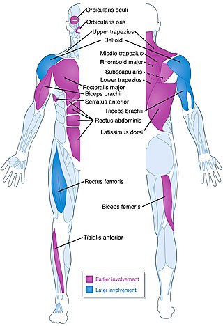
Facioscapulohumeral muscular dystrophy (FSHD) is a type of muscular dystrophy, a group of heritable diseases that cause degeneration of muscle and progressive weakness. Per the name, FSHD tends to sequentially weaken the muscles of the face, those that position the scapula, and those overlying the humerus bone of the upper arm. These areas can be spared, and muscles of other areas usually are affected, especially those of the chest, abdomen, spine, and shin. Almost any skeletal muscle can be affected in advanced disease. Abnormally positioned, termed 'winged', scapulas are common, as is the inability to lift the foot, known as foot drop. The two sides of the body are often affected unequally. Weakness typically manifests at ages 15 – 30 years. FSHD can also cause hearing loss and blood vessel abnormalities at the back of the eye.

Hereditary inclusion body myopathies (HIBM) are a group of rare genetic disorders which have different symptoms. Generally, they are neuromuscular disorders characterized by muscle weakness developing in young adults. Hereditary inclusion body myopathies comprise both autosomal recessive and autosomal dominant muscle disorders that have a variable expression (phenotype) in individuals, but all share similar structural features in the muscles.

<span class="mw-page-title-main">Mitochondrial myopathy</span> Medical condition

Mitochondrial myopathies are types of myopathies associated with mitochondrial disease. Adenosine triphosphate (ATP), the chemical used to provide energy for the cell, cannot be produced sufficiently by oxidative phosphorylation when the mitochondrion is either damaged or missing necessary enzymes or transport proteins. With ATP production deficient in mitochondria, there is an over-reliance on anaerobic glycolysis which leads to lactic acidosis either at rest or exercise-induced.

<span class="mw-page-title-main">Dysferlin</span> Protein encoded by the DYSF gene in humans

Dysferlin also known as dystrophy-associated fer-1-like protein is a protein that in humans is encoded by the DYSF gene. Dysferlin is linked with plasma membrane repair., stabilization of calcium signaling and the development of the T-tubule system of the muscle A defect in the DYSF gene, located on chromosome 2p12-14, results in several types of muscular dystrophy; including Miyoshi myopathy (MM), Limb-girdle muscular dystrophy type 2B (LGMD2B) and Distal Myopathy (DM). A reduction or absence of dysferlin, termed dysferlinopathy, usually becomes apparent in the third or fourth decade of life and is characterised by weakness and wasting of various voluntary skeletal muscles. Pathogenic mutations leading to dysferlinopathy can occur throughout the DYSF gene.

<span class="mw-page-title-main">Congenital muscular dystrophy</span> Medical condition

Congenital muscular dystrophies are autosomal recessively-inherited muscle diseases. They are a group of heterogeneous disorders characterized by muscle weakness which is present at birth and the different changes on muscle biopsy that ranges from myopathic to overtly dystrophic due to the age at which the biopsy takes place.

<span class="mw-page-title-main">Emery–Dreifuss muscular dystrophy</span> Medical condition

Emery–Dreifuss muscular dystrophy (EDMD) is a type of muscular dystrophy, a group of heritable diseases that cause progressive impairment of muscles. EDMD affects muscles used for movement, causing atrophy, weakness and contractures. It almost always affects the heart, causing abnormal rhythms, heart failure, or sudden cardiac death. It is rare, affecting 0.39 per 100,000 people. It is named after Alan Eglin H. Emery and Fritz E. Dreifuss.

<span class="mw-page-title-main">Bethlem myopathy</span> Medical condition

Bethlem myopathy is predominantly an autosomal dominant myopathy, classified as a congenital form of limb-girdle muscular dystrophy. There are two types of Bethlem myopathy, based on which type of collagen is affected.

Calpain-3 is a protein that in humans is encoded by the CAPN3 gene.

<span class="mw-page-title-main">SGCB</span> Protein-coding gene in the species Homo sapiens

Beta-sarcoglycan is a protein that in humans is encoded by the SGCB gene.

<span class="mw-page-title-main">Delta-sarcoglycan</span> Mammalian protein found in Homo sapiens

Delta-sarcoglycan is a protein that in humans is encoded by the SGCD gene.

<span class="mw-page-title-main">SGCG</span> Protein-coding gene in the species Homo sapiens

Gamma-sarcoglycan is a protein that in humans is encoded by the SGCG gene. The α to δ-sarcoglycans are expressed predominantly (β) or exclusively in striated muscle. A mutation in any of the sarcoglycan genes may lead to a secondary deficiency of the other sarcoglycan proteins, presumably due to destabilisation of the sarcoglycan complex. The disease-causing mutations in the α to δ genes cause disruptions within the dystrophin-associated protein (DAP) complex in the muscle cell membrane. The transmembrane components of the DAP complex link the cytoskeleton to the extracellular matrix in adult muscle fibres, and are essential for the preservation of the integrity of the muscle cell membrane.

<span class="mw-page-title-main">Ullrich congenital muscular dystrophy</span> Medical condition

Ullrich congenital muscular dystrophy (UCMD) is a form of congenital muscular dystrophy. There are two forms: UCMD1 and UCMD2.

Multi/minicore myopathy is a congenital myopathy usually caused by mutations in either the SELENON and RYR1 genes. It is characterised the presence of multifocal, well-circumscribed areas with reduction of oxidative staining and low myofibrillar ATPase on muscle biopsy. It is also known as Minicore myopathy, Multicore myopathy, Multiminicore myopathy, Minicore myopathy with external ophthalmoplegia, Multicore myopathy with external ophthalmoplegia and Multiminicore disease with external ophthalmoplegia.

<span class="mw-page-title-main">ANO5</span> Protein-coding gene in the species Homo sapiens

Anoctamin 5 (ANO5) is a protein that in humans is encoded by the ANO5 gene.

References

  1. 1 2 3 4 5 6 7 8 9 10 11 12 13 14 15 16 17 18 19 20 21 Angelini, C; Fanin, M; Adam, MP; Ardinger, HH; Pagon, RA; Wallace, SE; Bean, LJH; Stephens, K; Amemiya, A (1993). Calpainopathy in GeneReviews. PMID   20301490.
  2. Pollitt, C; Anderson, LV; Pogue, R; Davison, K; Pyle, A; Bushby, KM (April 2001). "The phenotype of calpainopathy: diagnosis based on a multidisciplinary approach". Neuromuscular Disorders. 11 (3): 287–96. doi:10.1016/s0960-8966(00)00197-8. PMID   11297944. S2CID   20081475.
  3. 1 2 3 4 5 6 7 8 9 10 11 12 Lasa-Elgarresta, J; Mosqueira-Martín, L; Naldaiz-Gastesi, N; Sáenz, A; López de Munain, A; Vallejo-Illarramendi, A (13 September 2019). "Calcium Mechanisms in Limb-Girdle Muscular Dystrophy with CAPN3 Mutations". International Journal of Molecular Sciences. 20 (18): 4548. doi: 10.3390/ijms20184548 . PMC   6770289 . PMID   31540302.
  4. 1 2 3 4 5 6 7 8 Dowling, JJ; Weihl, CC; Spencer, MJ (November 2021). "Molecular and cellular basis of genetically inherited skeletal muscle disorders". Nature Reviews. Molecular Cell Biology. 22 (11): 713–732. doi:10.1038/s41580-021-00389-z. PMC   9686310 . PMID   34257452. S2CID   235822532.
  5. Levy, Jennifer (21 January 2020). "Dufour Lab to investigate the biological role of calpain 3 in muscle". Coalition to Cure Calpain 3. Archived from the original on 6 May 2020. Retrieved 6 May 2020.
  6. Guha, TK; Pichavant, C; Calos, MP (13 December 2019). "Plasmid-Mediated Gene Therapy in Mouse Models of Limb Girdle Muscular Dystrophy". Molecular Therapy: Methods & Clinical Development. 15: 294–304. doi:10.1016/j.omtm.2019.10.002. PMC   6923511 . PMID   31890729.