Interstitial cystitis (IC), a type of bladder pain syndrome (BPS), is chronic pain in the bladder and pelvic floor of unknown cause. It is the urologic chronic pelvic pain syndrome of women. Symptoms include feeling the need to urinate right away, needing to urinate often, and pain with sex. IC/BPS is associated with depression and lower quality of life. Many of those affected also have irritable bowel syndrome and fibromyalgia.
A urinary tract infection (UTI) is an infection that affects a part of the urinary tract. When it affects the lower urinary tract it is known as a bladder infection (cystitis) and when it affects the upper urinary tract it is known as a kidney infection (pyelonephritis). Symptoms from a lower urinary tract infection include pain with urination, frequent urination, and feeling the need to urinate despite having an empty bladder. Symptoms of a kidney infection include fever and flank pain usually in addition to the symptoms of a lower UTI. Rarely the urine may appear bloody. In the very old and the very young, symptoms may be vague or non-specific.

The bladder is a hollow organ in humans and other vertebrates that stores urine from the kidneys before disposal by urination. In humans, the bladder is a distensible organ that sits on the pelvic floor. Urine enters the bladder via the ureters and exits via the urethra. The typical adult human bladder will hold between 300 and 500 ml before the urge to empty occurs, but can hold considerably more.
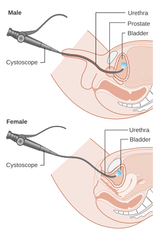
Cystoscopy is endoscopy of the urinary bladder via the urethra. It is carried out with a cystoscope.

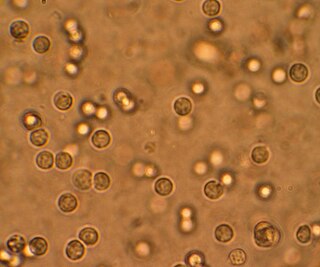
Urinalysis, a portmanteau of the words urine and analysis, is a panel of medical tests that includes physical (macroscopic) examination of the urine, chemical evaluation using urine test strips, and microscopic examination. Macroscopic examination targets parameters such as color, clarity, odor, and specific gravity; urine test strips measure chemical properties such as pH, glucose concentration, and protein levels; and microscopy is performed to identify elements such as cells, urinary casts, crystals, and organisms.

Hematuria or haematuria is defined as the presence of blood or red blood cells in the urine. "Gross hematuria" occurs when urine appears red, brown, or tea-colored due to the presence of blood. Hematuria may also be subtle and only detectable with a microscope or laboratory test. Blood that enters and mixes with the urine can come from any location within the urinary system, including the kidney, ureter, urinary bladder, urethra, and in men, the prostate. Common causes of hematuria include urinary tract infection (UTI), kidney stones, viral illness, trauma, bladder cancer, and exercise. These causes are grouped into glomerular and non-glomerular causes, depending on the involvement of the glomerulus of the kidney. But not all red urine is hematuria. Other substances such as certain medications and foods can cause urine to appear red. Menstruation in women may also cause the appearance of hematuria and may result in a positive urine dipstick test for hematuria. A urine dipstick test may also give an incorrect positive result for hematuria if there are other substances in the urine such as myoglobin, a protein excreted into urine during rhabdomyolysis. A positive urine dipstick test should be confirmed with microscopy, where hematuria is defined by three or more red blood cells per high power field. When hematuria is detected, a thorough history and physical examination with appropriate further evaluation can help determine the underlying cause.
Dysuria refers to painful or uncomfortable urination.
Feline lower urinary tract disease (FLUTD) is a generic category term to describe any disorder affecting the bladder or urethra of cats.

Hemorrhagic cystitis or haemorrhagic cystitis is an inflammation of the bladder defined by lower urinary tract symptoms that include dysuria, hematuria, and hemorrhage. The disease can occur as a complication of cyclophosphamide, ifosfamide and radiation therapy. In addition to hemorrhagic cystitis, temporary hematuria can also be seen in bladder infection or in children as a result of viral infection.

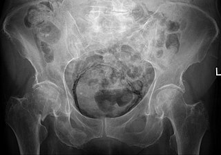
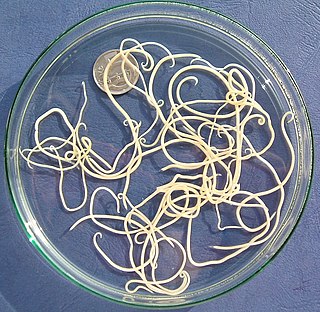
Dioctophyme renale, commonly referred to as the giant kidney worm, is a parasitic nematode (roundworm) whose mature form is found in the kidneys of mammals. D. renale is distributed worldwide, but is less common in Africa and Oceania. It affects fish eating mammals, particularly mink and dogs. Human infestation is rare, but results in kidney destruction, usually of one kidney and hence not fatal. A 2019 review listed a total of 37 known human cases of dioctophymiasis in 10 countries with the highest number (22) in China. Upon diagnosis through tissue sampling, the only treatment is surgical excision.
Urologic diseases or conditions include urinary tract infections, kidney stones, bladder control problems, and prostate problems, among others. Some urologic conditions do not affect a person for that long and some are lifetime conditions. Kidney diseases are normally investigated and treated by nephrologists, while the specialty of urology deals with problems in the other organs. Gynecologists may deal with problems of incontinence in women.

Capillaria aerophila is a nematode parasite found in the respiratory tract of foxes, dogs, and various other carnivorous mammals. A few cases of human infestation have also been reported. Though it is sometimes called a "lungworm", this term usually refers to other species of nematodes. Infestation by C. aerophila is referred to as "pulmonary capillariasis", "bronchial capillariasis," or (rarely) "thominxosis." This parasite has a direct life cycle, meaning that the life cycle can be completed in a single host. C. aerophila usually causes only minor clinical symptoms, such as irritation of the respiratory tract and coughing. However, secondary bacterial infections of the respiratory tract, including pneumonia, may develop in heavy infestations. Treatment with anthelmintics, such as levamisole or fenbendazole, is usually sufficient to cure C. aerophila infestations.

Capillaria is a genus of nematodes in the family Capillariidae .
Feline idiopathic cystitis (FIC) or feline interstitial cystitis or cystitis in cats, is one of the most frequently observed forms of feline lower urinary tract disease (FLUTD). Feline cystitis means "inflammation of the bladder in cats". The term idiopathic means unknown cause; however, certain behaviours have been known to aggravate the illness once it has been initiated. It can affect both males and females of any breed of cat. It is more commonly found in female cats; however, when males do exhibit cystitis, it is usually more dangerous.

Cystitis cystica is an uncommon chronic reactive inflammatory disease that is believed to be brought on by a tumor, calculi, infection, or obstruction of the urothelium. Cystitis glandularis is a proliferative progression of cystitis cystica that is distinguished by urothelial glandular metaplasia.

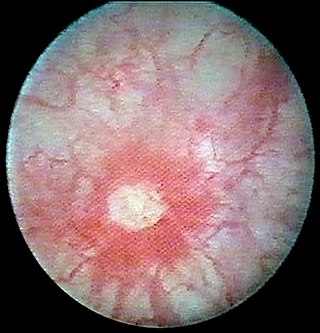
Eosinophilic cystitis is a rare type of interstitial cystitis first reported in 1960 by Edwin Brown. Eosinophilic cystitis has been linked to a number of etiological factors, including allergies, bladder tumors, trauma to the bladder, parasitic infections, and chemotherapy drugs, though the exact cause of the condition is still unknown. The antigen-antibody response is most likely the cause of eosinophilic cystitis. This results in the generation of different immunoglobulins, which activate eosinophils and start the inflammatory process.
Emphysematous cystitis is a rare type of infection of the bladder wall by gas-forming bacteria or fungi. The most frequent offending organism is E. coli. Other gram negative bacteria, including Klebsiella and Proteus are also commonly isolated. Fungi, such as Candida, have also been reported as causative organisms. Citrobacter and Enterococci have also been found to cause emphysematous cystitis. Although it is a rare type of bladder infection, it is the most common type of all gas-forming bladder infections. The condition is characterized by the formation of air bubbles in and around the bladder wall. The gas found in the bladder consists of nitrogen, hydrogen, oxygen, and carbon dioxide. The disease most commonly affects elderly diabetic and immunocompromised patients. The first case was identified in a post-mortem examination in 1888.

Capillaria feliscati (also known as Pearsonema feliscati, the cat bladder worm is a worm that affects cats, and seldom dogs. Its main final hosts are wild carnivores. It is a urinary tract nematode, though its occurrence is rare. C. feliscati are small, delicate, yellowish, thread-like worms. Adults are approximately between 16 and 53 millimetres in length.

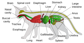
Cat worm infections, the infection of cats (Felidae) with parasitic worms, occur frequently. Most worm species occur worldwide in both domestic and other cats, but there are regional, species and lifestyle differences in the frequency of infestation. According to the classification of the corresponding parasites in the zoological system, infections can be divided into those caused by nematode and flatworms - in the case of the latter, mainly cestoda and trematoda - while other strains are of no veterinary significance. While threadworms usually do not require an intermediate host for their reproduction, the development cycle of flatworms always proceeds via alternate hosts.
Nematode infection in dogs - the infection of dogs with parasitic nemamotodes - are, along with tapeworm infections and infections with protozoa, frequent parasitoses in veterinary practice. Nematodes, as so-called endoparasites, colonize various internal organs - most of them the digestive tract - and the skin. To date, about 30 different species of nematode have been identified in domestic dogs; they are essentially also found in wild dog species. However, the majority of them often cause no or only minor symptoms of disease in adult animals. The infection therefore does not necessarily have to manifest itself in a worm disease (helminthosis). For most nematodes, an infection can be detected by examining the feces for eggs or larvae. Roundworm infection in dogs and the hookworm in dogs is of particular health significance in Central Europe, as they can also be transmitted to humans (zoonosis). Regular deworming can significantly reduce the frequency of infection and thus the risk of infection for humans and dogs.