Capsule endoscopy

Last updated
Wireless Capsule Endoscopy
CapsuleEndoscope.jpg
Picture of a capsule
MeSH D053704
OPS-301 code 1-63a, 1-656
Endoscopic capsule end-on, showing six LEDs and camera lens. CapsuleEndoscopeEnd.jpg
Endoscopic capsule end-on, showing six LEDs and camera lens.
Image of the intestines acquired by capsule endoscopy Dunndarm.PNG
Image of the intestines acquired by capsule endoscopy
Image of the colon acquired by capsule endoscopy Normales Colon.PNG
Image of the colon acquired by capsule endoscopy

Capsule endoscopy is a medical procedure used to record internal images of the gastrointestinal tract for use in disease diagnosis. Newer developments are also able to take biopsies and release medication at specific locations of the entire gastrointestinal tract. [1] Unlike the more widely used endoscope, capsule endoscopy provides the ability to see the middle portion of the small intestine. It can be applied to the detection of various gastrointestinal cancers, digestive diseases, ulcers, unexplained bleedings, and general abdominal pains. After a patient swallows the capsule, it passes along the gastrointestinal tract, taking a number of images per second which are transmitted wirelessly to an array of receivers connected to a portable recording device carried by the patient. General advantages of capsule endoscopy over standard endoscopy include the minimally invasive procedure setup, ability to visualize more of the gastrointestinal tract, and lower cost of the procedure. [2] [ medical citation needed ]

Contents

History

Capsule endoscopy was first conceptualized by Israeli engineer Gavrial Iddan and Israeli gastroenterologist Eitan Scapa in Boston, MA in the early 1980s. The two partners first developed a CCD (charged coupled device) camera-based imaging system using a fiber-optic tether. This initial design suffered from high power consumption and slow transmission times of image data of at best 10 minutes. In 1993 Iddan had the idea to split the system into three components, the camera and transmitter, the recorder attached to a sensor array on the patient's abdomen, and a software package that processes the stored data at leisure by a physician at a later time. This new setup was made possible by the replacement of the CCD camera with a CMOS (complementary metal–oxide–semiconductor) camera. These cameras consume just one percent of the energy of their CCD counterparts. The more efficient power system allowed for the removal of the fiber optic cable for a stand-alone imaging system. This three-component imaging system remains popular today. [3]

In 2001 the FDA (Food and Drug Administration) approved the first capsule endoscope developed by Given Imaging for use in patients. Many new models such as the PillCam SB and DBE for obscure gastrointestinal bleeding, the Olympus CE for the small bowel, the PillCam ESO for investigation of esophageal diseases, and the PillCam COLON for detection of colonic neoplasias have been created. [4]

As research into capsule endoscopy has increased and technology has advanced better wireless and more energy-efficient systems have allowed for the creation of more compact capsules and image processing systems, with many in development today.

Technology

Capsule endoscopy uses a small vitamin-sized wireless camera to capture images of a patient's digestive tract. The capsule is generally composed of a camera, antenna, and light array. [5] Due to the small nature of the device images can not be stored within it. As a result, a sensor array with a storage unit is placed on the abdomen of the patient for the imaging period. This storage unit can then be connected to a computer at a later time so that a medical professional can analyze the images. [6]

Newer models of the capsule endoscope have looked to add camera systems on both ends of the pill or even store images within the pill itself to minimize the amount of medical equipment one must carry with them while using the device. [6] For systems that store images directly within the pill, the pill must be collected after excretion for extraction of the images by a secondary device. [7]

The main shortcoming of capsule endoscopy is the field of view. Depending on the placement of the camera system within the device images may become obstructed by folds in the digestive tract. Due to the passive nature of image capture and lack of control in maneuvering the device through the digestive tract novel solutions are being developed by various companies and research labs. For systems that utilize the setup with the camera system at the end of the capsule, the field of view ranges from 140 to 170 degrees. [8]

There are several advantages to choosing to use capsule endoscopy over standard endoscopy. Standard endoscopy can be more uncomfortable for a patient, can be more prone to puncturing the digestive tract walls, and is not able to access the middle portion of the small intestine. Endoscopes must enter either through the mouth/nasal cavities or the rectum. Due to restrictions in length, extremely important regions for diagnosis in the small intestine are not able to be accessed. Currently, within the United States, capsule endoscopy can not be used as a primary imaging method over standard endoscopy first.[ citation needed ] As a result, many patients must first undergo standard endoscopy to then be referred for capsule endoscopy. Further innovation will be required to make capsule endoscopy comparable to the current standard of care, but extensive work is being performed to achieve this. [9]  

Medical uses

Esophagogastroduodenoscopy (EGD), employs a camera attached to a long flexible tube to view the upper portion of the gastrointestinal tract, namely the esophagus, the stomach, and the beginning of the first part of the small intestine called the duodenum. A colonoscope, inserted through the rectum, can view the colon and the distal portion of the small intestine, the terminal ileum. These two types of endoscopy however cannot visualize the majority of the middle portion of the small intestine.

Capsule endoscopy is therefore used to examine parts of the gastrointestinal tract that cannot be seen by standard endoscopy. It is useful when the disease is suspected in the small intestine, and can sometimes be used to find the site of gastrointestinal bleeding or the cause of unexplained abdominal pain, such as Crohn's disease. However, unlike EGD or colonoscopy, it cannot be used to treat pathology that may be discovered. Common reasons for using capsule endoscopy include diagnosis of unexplained bleeding, iron deficiency, or abdominal pain, searching for polyps, ulcers, and tumors of the small intestine, and diagnosis of inflammatory bowel disease. [10]

The images collected by the miniature camera during a session are transferred wirelessly to an external receiver worn by the patient, using any one of a band of appropriate frequencies. The collected images are then transferred to a computer for display, review, and diagnosis. [11] A transmitted radio-frequency signal emitted by some capsules can be used to accurately estimate the location of the capsule and to track it in real-time inside the body and gastrointestinal tract. [12] Capsule endoscopy can still not yet replace standard endoscopy for various diseases, as is the case for those with cirrhosis. [13]

As of 2014, research was targeting additional sensing mechanisms and localization and motion control systems to enable new applications for the technology, for example, drug delivery. Wireless energy transmission was also being investigated as a way of providing a continuous energy source for the capsule. [14]

Procedure

Capsule endoscopy requires a number of different preparatory procedures to ensure clear images are taken of a patient's gastrointestinal tract for an accurate diagnosis of disease. [15] There are various types of capsule endoscopes, but for a generalized description, one can assume the most common setup requires the capsule, sensor array, storage unit, and computer system being used. First, a patient will need to have the sensor array placed on their abdomen with a recording unit worn as a belt. The patient may be asked to stay at the hospital or return home depending on the start time. Next, the pill must be swallowed by the patient. After approximately 8 hours the sensor array can be removed and returned to a physician. The capsule will be excreted through regular bowel movements. [16]

During the procedure, there are a number of different policies to follow. A patient should only drink clear liquids for the first two hours after swallowing the pill and may eat after 4 hours. MRI studies, ham radios, metal detectors, and strenuous physical activity should all be avoided. Additionally, all external equipment must be kept dry. [16]

Manufacturers

As of 2022, there are a number of manufacturers who produce capsule endoscopes. [17] The technology was originally developed by Israeli scientists, Gavriel Iddan and Eitan Scapa, with the first pill swallowed in 1997; [18] Iddan founded Given Imaging in Israel which received FDA approval in 2001. [18] Medtronic today produces one of the more widely used capsule endoscope systems called the PillCam having sold over 3.5 million units. [19] Medtronic purchased the PillCam system from Given Imaging in 2014. [20]

Side effects

Capsule endoscopy is considered to be a very safe method for gastrointestinal tract examination. The capsule is usually excreted with a patient's feces within 24–48 hours after ingestion. There has been a single report of retention of the capsule lasting almost four and a half years although the patient was asymptomatic. However, the risk of bowel obstruction may be countered by an abdominal X-ray to locate the device for removal by endoscopy or surgery. [21]

Risk of retention

In a review of 22,840 cases, the capsule was retained 1.4% of the time, with Crohn's disease a common cause; most were surgically removed. [22] The rate of capsule retention varies by the indication for the procedure, with the highest rate seen with known Crohn's disease (5-13%), followed by obscure gastrointestinal bleeding (1.5%), suspected Crohn's disease (1.4%), and healthy volunteers (0%). [23] Risk factors for capsule retention include Crohn's disease, NSAID use, and abdominal radiation. [23]

Related Research Articles

<span class="mw-page-title-main">Gastroenterology</span> Branch of medicine focused on the digestive system and its disorders

Gastroenterology is the branch of medicine focused on the digestive system and its disorders. The digestive system consists of the gastrointestinal tract, sometimes referred to as the GI tract, which includes the esophagus, stomach, small intestine and large intestine as well as the accessory organs of digestion which include the pancreas, gallbladder, and liver. The digestive system functions to move material through the GI tract via peristalsis, break down that material via digestion, absorb nutrients for use throughout the body, and remove waste from the body via defecation. Physicians who specialize in the medical specialty of gastroenterology are called gastroenterologists or sometimes GI doctors. Some of the most common conditions managed by gastroenterologists include gastroesophageal reflux disease, gastrointestinal bleeding, irritable bowel syndrome, inflammatory bowel disease (IBD) which includes Crohn's disease and ulcerative colitis, peptic ulcer disease, gallbladder and biliary tract disease, hepatitis, pancreatitis, colitis, colon polyps and cancer, nutritional problems, and many more.

<span class="mw-page-title-main">Crohn's disease</span> Type of inflammatory bowel disease

Crohn's disease is a type of inflammatory bowel disease (IBD) that may affect any segment of the gastrointestinal tract. Symptoms often include abdominal pain, diarrhea, fever, abdominal distension, and weight loss. Complications outside of the gastrointestinal tract may include anemia, skin rashes, arthritis, inflammation of the eye, and fatigue. The skin rashes may be due to infections as well as pyoderma gangrenosum or erythema nodosum. Bowel obstruction may occur as a complication of chronic inflammation, and those with the disease are at greater risk of colon cancer and small bowel cancer.

<span class="mw-page-title-main">Ulcerative colitis</span> Inflammatory bowel disease that causes ulcers in the colon

Ulcerative colitis (UC) is a type of inflammatory bowel disease (IBD). It is a long-term condition that results in inflammation and ulcers of the colon and rectum. The primary symptoms of active disease are abdominal pain and diarrhea mixed with blood (hematochezia). Weight loss, fever, and anemia may also occur. Often, symptoms come on slowly and can range from mild to severe. Symptoms typically occur intermittently with periods of no symptoms between flares. Complications may include abnormal dilation of the colon (megacolon), inflammation of the eye, joints, or liver, and colon cancer.

<span class="mw-page-title-main">Gastrointestinal tract</span> Organ system within humans and other animals

The gastrointestinal tract is the tract or passageway of the digestive system that leads from the mouth to the anus. The GI tract contains all the major organs of the digestive system, in humans and other animals, including the esophagus, stomach, and intestines. Food taken in through the mouth is digested to extract nutrients and absorb energy, and the waste expelled at the anus as faeces. Gastrointestinal is an adjective meaning of or pertaining to the stomach and intestines.

<span class="mw-page-title-main">Endoscopy</span> Procedure used in medicine to look inside the body

An endoscopy is a procedure used in medicine to look inside the body. The endoscopy procedure uses an endoscope to examine the interior of a hollow organ or cavity of the body. Unlike many other medical imaging techniques, endoscopes are inserted directly into the organ.

<span class="mw-page-title-main">Endoscope</span> Instrument to visually examine the interior of a hollow space

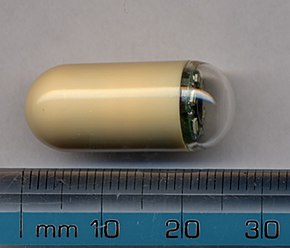
An endoscope is an inspection instrument composed of image sensor, optical lens, light source and mechanical device, which is used to look deep into the body by way of openings such as the mouth or anus. A typical endoscope applies several modern technologies including optics, ergonomics, precision mechanics, electronics, and software engineering. With an endoscope, it is possible to observe lesions that cannot be detected by X-ray, making it useful in medical diagnosis. Endoscopes use tubes which are only a few millimeters thick to transfer illumination in one direction and high-resolution images in real time in the other direction, resulting in minimally invasive surgeries. It is used to examine the internal organs like the throat or esophagus. Specialized instruments are named after their target organ. Examples include the cystoscope (bladder), nephroscope (kidney), bronchoscope (bronchus), arthroscope (joints) and colonoscope (colon), and laparoscope. They can be used to examine visually and diagnose, or assist in surgery such as an arthroscopy.

<span class="mw-page-title-main">Enteritis</span> Intestinal inflammatory disease

Enteritis is inflammation of the small intestine. It is most commonly caused by food or drink contaminated with pathogenic microbes, such as Serratia, but may have other causes such as NSAIDs, radiation therapy as well as autoimmune conditions like Crohn's disease and celiac disease. Symptoms include abdominal pain, cramping, diarrhea, dehydration, and fever. Related diseases of the gastrointestinal system involve inflammation of the stomach and large intestine.

<span class="mw-page-title-main">Upper gastrointestinal series</span> Radiographs used to examine abnormalities of the digestive system excluding the colon

An upper gastrointestinal series, also called a barium swallow, barium study, or barium meal, is a series of radiographs used to examine the gastrointestinal tract for abnormalities. A contrast medium, usually a radiocontrast agent such as barium sulfate mixed with water, is ingested or instilled into the gastrointestinal tract, and X-rays are used to create radiographs of the regions of interest. The barium enhances the visibility of the relevant parts of the gastrointestinal tract by coating the inside wall of the tract and appearing white on the film. This in combination with other plain radiographs allows for the imaging of parts of the upper gastrointestinal tract such as the pharynx, larynx, esophagus, stomach, and small intestine such that the inside wall lining, size, shape, contour, and patency are visible to the examiner. With fluoroscopy, it is also possible to visualize the functional movement of examined organs such as swallowing, peristalsis, or sphincter closure. Depending on the organs to be examined, barium radiographs can be classified into "barium swallow", "barium meal", "barium follow-through", and "enteroclysis". To further enhance the quality of images, air or gas is sometimes introduced into the gastrointestinal tract in addition to barium, and this procedure is called double-contrast imaging. In this case the gas is referred to as the negative contrast medium. Traditionally the images produced with barium contrast are made with plain-film radiography, but computed tomography is also used in combination with barium contrast, in which case the procedure is called "CT enterography".

<span class="mw-page-title-main">Angiodysplasia</span> Medical condition

In medicine (gastroenterology), angiodysplasia is a small vascular malformation of the gut. It is a common cause of otherwise unexplained gastrointestinal bleeding and anemia. Lesions are often multiple, and frequently involve the cecum or ascending colon, although they can occur at other places. Treatment may be with colonoscopic interventions, angiography and embolization, medication, or occasionally surgery.

<span class="mw-page-title-main">Esophagogastroduodenoscopy</span> Diagnostic endoscopic procedure

Esophagogastroduodenoscopy (EGD) or oesophagogastroduodenoscopy (OGD), also called by various other names, is a diagnostic endoscopic procedure that visualizes the upper part of the gastrointestinal tract down to the duodenum. It is considered a minimally invasive procedure since it does not require an incision into one of the major body cavities and does not require any significant recovery after the procedure. However, a sore throat is common.

<span class="mw-page-title-main">Gastrointestinal bleeding</span> Medical condition

Gastrointestinal bleeding, also called gastrointestinal hemorrhage (GIB), is all forms of bleeding in the gastrointestinal tract, from the mouth to the rectum. When there is significant blood loss over a short time, symptoms may include vomiting red blood, vomiting black blood, bloody stool, or black stool. Small amounts of bleeding over a long time may cause iron-deficiency anemia resulting in feeling tired or heart-related chest pain. Other symptoms may include abdominal pain, shortness of breath, pale skin, or passing out. Sometimes in those with small amounts of bleeding no symptoms may be present.

<span class="mw-page-title-main">Gastrointestinal disease</span> Medical condition

Gastrointestinal diseases refer to diseases involving the gastrointestinal tract, namely the esophagus, stomach, small intestine, large intestine and rectum, and the accessory organs of digestion, the liver, gallbladder, and pancreas.

<span class="mw-page-title-main">Blood in stool</span> Medical condition

Blood in stool or rectal bleeding looks different depending on how early it enters the digestive tract—and thus how much digestive action it has been exposed to—and how much there is. The term can refer either to melena, with a black appearance, typically originating from upper gastrointestinal bleeding; or to hematochezia, with a red color, typically originating from lower gastrointestinal bleeding. Evaluation of the blood found in stool depends on its characteristics, in terms of color, quantity and other features, which can point to its source, however, more serious conditions can present with a mixed picture, or with the form of bleeding that is found in another section of the tract. The term "blood in stool" is usually only used to describe visible blood, and not fecal occult blood, which is found only after physical examination and chemical laboratory testing.

Enteroscopy is the procedure of using an endoscope for the direct visualization of the small bowel. Etymologically, the word could potentially refer to any bowel endoscopy, but idiomatically it is conventionally restricted to small bowel endoscopy, in distinction from colonoscopy, which is large bowel endoscopy. Various types of enteroscopy exist, as follows:

<span class="mw-page-title-main">Double-balloon enteroscopy</span>

Double-balloon enteroscopy, also known as push-and-pull enteroscopy, is an endoscopic technique for visualization of the small bowel. It was developed by Hironori Yamamoto in 2001. It is novel in the field of diagnostic gastroenterology as it is the first endoscopic technique that allows for the entire gastrointestinal tract to be visualized in real time.

<span class="mw-page-title-main">SeHCAT</span> Chemical compound

SeHCAT is a drug used in a clinical test to diagnose bile acid malabsorption.

<span class="mw-page-title-main">Given Imaging</span>

Given Imaging is an Israeli medical technology company that manufactures and markets diagnostic products for the visualization and detection of disorders of the gastrointestinal tract. Until March 2014, it was dual-listed on both the NASDAQ and the Tel Aviv Stock Exchange, where it was a component of the TA-100 Index and the TA BlueTech Index. In March 2014 it was acquired by Covidien and became a private company. In 2015 Covidien was purchased by Medtronic.

<span class="mw-page-title-main">Blue rubber bleb nevus syndrome</span> Medical condition

Blue rubber bleb nevus syndrome is a rare disorder that consists mainly of abnormal blood vessels affecting the skin or internal organs – usually the gastrointestinal tract. The disease is characterized by the presence of fluid-filled blisters (blebs) as visible, circumscribed, chronic lesions (nevi).

Blair S. Lewis, M.D., F.A.C.P., F.A.C.G., is an American board-certified gastroenterologist and Clinical Professor of Medicine at the Mount Sinai School of Medicine. Lewis is a specialist in the field of gastrointestinal endoscopy and was the primary investigator for the first clinical trial of capsule endoscopy for the small intestine and also the first clinical trial of capsule endoscopy for the colon.

Computed tomography enterography is a medical imaging technique which uses computed tomography scanner and contrast media to examine the small bowel. It was first introduced by Raptopoulos et al. in 1997. CT Enterography can be used to assess a variety of problems involving the small bowel, however it is mainly used to diagnose and assess severity of Crohn's disease.

References

  1. "Diagnostics with a Stomach Pill" (PDF). Max Planck Institute for Intelligent Systems. January 7, 2016. Archived (PDF) from the original on 2017-03-21.
  2. "The endoscopy procedures we provide". 28 November 2018. Farzan Bahin, The Hills Gastroenterology, Sydney, Australia.
  3. Adler SN (May 2017). "The history of time for capsule endoscopy". Annals of Translational Medicine. 5 (9): 194. doi: 10.21037/atm.2017.03.90 . PMC   5438796 . PMID   28567374.
  4. Nakamura T, Terano A (2008). "Capsule endoscopy: past, present, and future". Journal of Gastroenterology. 43 (2): 93–99. doi:10.1007/s00535-007-2153-6. PMID   18306982. S2CID   25358336.
  5. "Capsule endoscopy - Mayo Clinic". www.mayoclinic.org. Retrieved 2022-04-11.
  6. 1 2 "PillCam™ Crohn's System | Medtronic". www.medtronic.com. Retrieved 2022-04-11.
  7. judy. "The Comprehensive Capsule Endoscopy System". CapsoVision US. Retrieved 2022-04-11.
  8. Wang Q, Khanicheh A, Leiner D, Shafer D, Zobel J (March 2017). "Endoscope field of view measurement". Biomedical Optics Express. 8 (3): 1441–1454. doi:10.1364/BOE.8.001441. PMC   5480555 . PMID   28663840.
  9. Koprowski R (December 2015). "Overview of technical solutions and assessment of clinical usefulness of capsule endoscopy". BioMedical Engineering OnLine. 14 (1): 111. doi: 10.1186/s12938-015-0108-3 . PMC   4665909 . PMID   26626725.
  10. Tong J, Svarta S, Ou G, Kwok R, Law J, Enns R (October 2012). "Diagnostic yield of capsule endoscopy in the setting of iron deficiency anemia without evidence of gastrointestinal bleeding". Canadian Journal of Gastroenterology. 26 (10): 687–690. doi: 10.1155/2012/182542 . PMC   3472906 . PMID   23061059.
  11. Yuce MR, Dissanayake T (2012). "Easy-to-swallow wireless telemetry". IEEE Microwave Magazine. 13 (6): 90–101. doi:10.1109/MMM.2012.2205833. S2CID   31486989.
  12. Pourhomayoun M, Fowler M, Jin Z (2012). "A novel method for medical implant in-body localization". 2012 Annual International Conference of the IEEE Engineering in Medicine and Biology Society. Vol. 2012. pp. 5757–5760. doi:10.1109/EMBC.2012.6347302. ISBN   978-1-4577-1787-1. PMID   23367237. S2CID   1784308.
  13. Colli A, Gana JC, Turner D, Yap J, Adams-Webber T, Ling SC, Casazza G (October 2014). "Capsule endoscopy for the diagnosis of oesophageal varices in people with chronic liver disease or portal vein thrombosis". The Cochrane Database of Systematic Reviews. 2014 (10): CD008760. doi:10.1002/14651858.CD008760.pub2. PMC   7173747 . PMID   25271409.
  14. Yuce MR, Alici G, Than TD (2014). "Wireless Endoscopy". Wiley Encyclopedia of Electrical and Electronics Engineering. pp. 1–25. doi:10.1002/047134608X.W8233. ISBN   9780471346081.
  15. Rokkas T, Papaxoinis K, Triantafyllou K, Pistiolas D, Ladas SD (January 2009). "Does purgative preparation influence the diagnostic yield of small bowel video capsule endoscopy?: A meta-analysis". The American Journal of Gastroenterology. 104 (1): 219–227. doi:10.1038/ajg.2008.63. PMID   19098872. S2CID   25237140.
  16. 1 2 "Capsule Endoscopy | Michigan Medicine". www.uofmhealth.org. Retrieved 2022-04-11.
  17. Slawinski PR, Obstein KL, Valdastri P (October 2015). "Capsule endoscopy of the future: What's on the horizon?". World Journal of Gastroenterology. 21 (37): 10528–10541. doi: 10.3748/wjg.v21.i37.10528 . PMC   4588075 . PMID   26457013.
  18. 1 2 Adler SN, Metzger YC (July 2011). "PillCam COLON capsule endoscopy: recent advances and new insights". Therapeutic Advances in Gastroenterology. 4 (4): 265–268. doi:10.1177/1756283X11401645. PMC   3131168 . PMID   21765870.
  19. "Capsule Endoscopy Products | Medtronic (UK)". www.medtronic.com. Retrieved 2022-04-11.
  20. "Covidien Completes Acquisition of Given Imaging". Medtronic News. Retrieved 2022-04-11.
  21. Bhattarai M, Bansal P, Khan Y (July 2013). "Longest duration of retention of video capsule: A case report and literature review". World Journal of Gastrointestinal Endoscopy. 5 (7): 352–355. doi: 10.4253/wjge.v5.i7.352 . PMC   3711067 . PMID   23858380.
  22. Liao Z, Gao R, Xu C, Li ZS (February 2010). "Indications and detection, completion, and retention rates of small-bowel capsule endoscopy: a systematic review". Gastrointestinal Endoscopy. 71 (2): 280–286. doi:10.1016/j.gie.2009.09.031. PMID   20152309.
  23. 1 2 Cave D, Legnani P, de Franchis R, Lewis BS (October 2005). "ICCE consensus for capsule retention". Endoscopy. 37 (10): 1065–1067. doi:10.1055/s-2005-870264. PMID   16189792. S2CID   260129396.