Cooperative binding

Last updated

Cooperative binding occurs in molecular binding systems containing more than one type, or species, of molecule and in which one of the partners is not mono-valent and can bind more than one molecule of the other species. In general, molecular binding is an interaction between molecules that results in a stable physical association between those molecules.

Contents

Cooperative binding occurs in a molecular binding system where two or more ligand molecules can bind to a receptor molecule. Binding can be considered "cooperative" if the actual binding of the first molecule of the ligand to the receptor changes the binding affinity of the second ligand molecule. The binding of ligand molecules to the different sites on the receptor molecule do not constitute mutually independent events. Cooperativity can be positive or negative, meaning that it becomes more or less likely that successive ligand molecules will bind to the receptor molecule.

Cooperative binding is observed in many biopolymers, including proteins and nucleic acids. Cooperative binding has been shown to be the mechanism underlying a large range of biochemical and physiological processes.

History and mathematical formalisms

Christian Bohr and the concept of cooperative binding

In 1904, Christian Bohr studied hemoglobin binding to oxygen under different conditions. [1] [2] When plotting hemoglobin saturation with oxygen as a function of the partial pressure of oxygen, he obtained a sigmoidal (or "S-shaped") curve. This indicates that the more oxygen is bound to hemoglobin, the easier it is for more oxygen to bind - until all binding sites are saturated. In addition, Bohr noticed that increasing CO2 pressure shifted this curve to the right - i.e. higher concentrations of CO2 make it more difficult for hemoglobin to bind oxygen. [2] This latter phenomenon, together with the observation that hemoglobin's affinity for oxygen increases with increasing pH, is known as the Bohr effect.

Original figure from Christian Bohr, showing the sigmoidal increase of oxyhemoglobin as a function of the partial pressure of oxygen. Bohr effect.png
Original figure from Christian Bohr, showing the sigmoidal increase of oxyhemoglobin as a function of the partial pressure of oxygen.

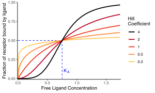
A receptor molecule is said to exhibit cooperative binding if its binding to ligand scales non-linearly with ligand concentration. Cooperativity can be positive (if binding of a ligand molecule increases the receptor's apparent affinity, and hence increases the chance of another ligand molecule binding) or negative (if binding of a ligand molecule decreases affinity and hence makes binding of other ligand molecules less likely). The "fractional occupancy" of a receptor with a given ligand is defined as the quantity of ligand-bound binding sites divided by the total quantity of ligand binding sites:

If , then the protein is completely unbound, and if , it is completely saturated. If the plot of at equilibrium as a function of ligand concentration is sigmoidal in shape, as observed by Bohr for hemoglobin, this indicates positive cooperativity. If it is not, no statement can be made about cooperativity from looking at this plot alone.

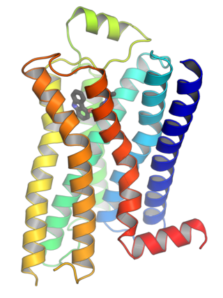
The concept of cooperative binding only applies to molecules or complexes with more than one ligand binding sites. If several ligand binding sites exist, but ligand binding to any one site does not affect the others, the receptor is said to be non-cooperative. Cooperativity can be homotropic, if a ligand influences the binding of ligands of the same kind, or heterotropic, if it influences binding of other kinds of ligands. In the case of hemoglobin, Bohr observed homotropic positive cooperativity (binding of oxygen facilitates binding of more oxygen) and heterotropic negative cooperativity (binding of CO2 reduces hemoglobin's facility to bind oxygen.)

Throughout the 20th century, various frameworks have been developed to describe the binding of a ligand to a protein with more than one binding site and the cooperative effects observed in this context. [3]

The Hill equation

The first description of cooperative binding to a multi-site protein was developed by A.V. Hill. [4] Drawing on observations of oxygen binding to hemoglobin and the idea that cooperativity arose from the aggregation of hemoglobin molecules, each one binding one oxygen molecule, Hill suggested a phenomenological equation that has since been named after him:

Hill plot of the Hill equation in red, showing the slope of the curve being the Hill coefficient and the intercept with the x-axis providing the apparent dissociation constant. The green line shows the non-cooperative curve. Hill Plot.png
Hill plot of the Hill equation in red, showing the slope of the curve being the Hill coefficient and the intercept with the x-axis providing the apparent dissociation constant. The green line shows the non-cooperative curve.

where is the "Hill coefficient", denotes ligand concentration, denotes an apparent association constant (used in the original form of the equation), is an empirical dissociation constant, and a microscopic dissociation constant (used in modern forms of the equation, and equivalent to an ). If , the system exhibits negative cooperativity, whereas cooperativity is positive if . The total number of ligand binding sites is an upper bound for . The Hill equation can be linearized as:

The "Hill plot" is obtained by plotting versus . In the case of the Hill equation, it is a line with slope and intercept . This means that cooperativity is assumed to be fixed, i.e. it does not change with saturation. It also means that binding sites always exhibit the same affinity, and cooperativity does not arise from an affinity increasing with ligand concentration.

The Adair equation

G.S. Adair found that the Hill plot for hemoglobin was not a straight line, and hypothesized that binding affinity was not a fixed term, but dependent on ligand saturation. [5] Having demonstrated that hemoglobin contained four hemes (and therefore binding sites for oxygen), he worked from the assumption that fully saturated hemoglobin is formed in stages, with intermediate forms with one, two, or three bound oxygen molecules. The formation of each intermediate stage from unbound hemoglobin can be described using an apparent macroscopic association constant . The resulting fractional occupancy can be expressed as:

Or, for any protein with n ligand binding sites:

where n denotes the number of binding sites and each is a combined association constant, describing the binding of i ligand molecules. By combining the Adair treatment with the Hill plot, one arrives at the modern experimental definition of cooperativity (Hill, 1985, Abeliovich, 2005). The resultant Hill coefficient, or more correctly the slope of the Hill plot as calculated from the Adair Equation, can be shown to be the ratio between the variance of the binding number to the variance of the binding number in an equivalent system of non-interacting binding sites. [6] Thus, the Hill coefficient defines cooperativity as a statistical dependence of one binding site on the state of other site(s).

The Klotz equation

Working on calcium binding proteins, Irving Klotz deconvoluted Adair's association constants by considering stepwise formation of the intermediate stages, and tried to express the cooperative binding in terms of elementary processes governed by mass action law. [7] [8] In his framework, is the association constant governing binding of the first ligand molecule, the association constant governing binding of the second ligand molecule (once the first is already bound) etc. For , this gives:

It is worth noting that the constants , and so forth do not relate to individual binding sites. They describe how many binding sites are occupied, rather than which ones. This form has the advantage that cooperativity is easily recognised when considering the association constants. If all ligand binding sites are identical with a microscopic association constant , one would expect (that is ) in the absence of cooperativity. We have positive cooperativity if lies above these expected values for .

The Klotz equation (which is sometimes also called the Adair-Klotz equation) is still often used in the experimental literature to describe measurements of ligand binding in terms of sequential apparent binding constants. [9]

Pauling equation

By the middle of the 20th century, there was an increased interest in models that would not only describe binding curves phenomenologically, but offer an underlying biochemical mechanism. Linus Pauling reinterpreted the equation provided by Adair, assuming that his constants were the combination of the binding constant for the ligand ( in the equation below) and energy coming from the interaction between subunits of the cooperative protein ( below). [10] Pauling actually derived several equations, depending on the degree of interaction between subunits. Based on wrong assumptions about the localization of hemes, he opted for the wrong one to describe oxygen binding by hemoglobin, assuming the subunit were arranged in a square. The equation below provides the equation for a tetrahedral structure, which would be more accurate in the case of hemoglobin:

The KNF model

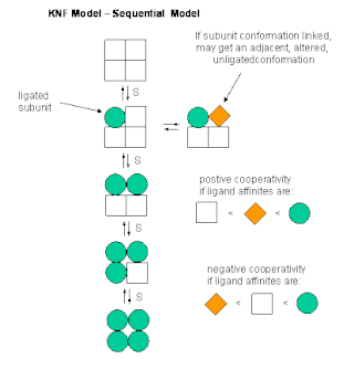
Based on results showing that the structure of cooperative proteins changed upon binding to their ligand, Daniel Koshland and colleagues [11] refined the biochemical explanation of the mechanism described by Pauling. [10] The Koshland-Némethy-Filmer (KNF) model assumes that each subunit can exist in one of two conformations: active or inactive. Ligand binding to one subunit would induce an immediate conformational change of that subunit from the inactive to the active conformation, a mechanism described as "induced fit". [12] Cooperativity, according to the KNF model, would arise from interactions between the subunits, the strength of which varies depending on the relative conformations of the subunits involved. For a tetrahedric structure (they also considered linear and square structures), they proposed the following formula:

Where is the constant of association for X, is the ratio of B and A states in the absence of ligand ("transition"), and are the relative stabilities of pairs of neighbouring subunits relative to a pair where both subunits are in the A state (Note that the KNF paper actually presents , the number of occupied sites, which is here 4 times ).

The MWC model

Monod-Wyman-Changeux model reaction scheme of a protein made up of two protomers. The protomer can exist under two states, each with a different affinity for the ligand. L is the ratio of states in the absence of ligand, c is the ratio of affinities. MWC structure.png
Monod-Wyman-Changeux model reaction scheme of a protein made up of two protomers. The protomer can exist under two states, each with a different affinity for the ligand. L is the ratio of states in the absence of ligand, c is the ratio of affinities.
Energy diagram of a Monod-Wyman-Changeux model of a protein made up of two protomers. The larger affinity of the ligand for the R state means that the latter is preferentially stabilized by the binding. MWC energy.png
Energy diagram of a Monod-Wyman-Changeux model of a protein made up of two protomers. The larger affinity of the ligand for the R state means that the latter is preferentially stabilized by the binding.

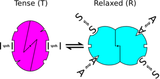
The Monod-Wyman-Changeux (MWC) model for concerted allosteric transitions [13] went a step further by exploring cooperativity based on thermodynamics and three-dimensional conformations. It was originally formulated for oligomeric proteins with symmetrically arranged, identical subunits, each of which has one ligand binding site. According to this framework, two (or more) interconvertible conformational states of an allosteric protein coexist in a thermal equilibrium. The states - often termed tense (T) and relaxed (R) - differ in affinity for the ligand molecule. The ratio between the two states is regulated by the binding of ligand molecules that stabilizes the higher-affinity state. Importantly, all subunits of a molecule change states at the same time, a phenomenon known as "concerted transition".

The allosteric isomerisation constant L describes the equilibrium between both states when no ligand molecule is bound: . If L is very large, most of the protein exists in the T state in the absence of ligand. If L is small (close to one), the R state is nearly as populated as the T state. The ratio of dissociation constants for the ligand from the T and R states is described by the constant c: . If , both R and T states have the same affinity for the ligand and the ligand does not affect isomerisation. The value of c also indicates how much the equilibrium between T and R states changes upon ligand binding: the smaller c, the more the equilibrium shifts towards the R state after one binding. With , fractional occupancy is described as:

The sigmoid Hill plot of allosteric proteins can then be analysed as a progressive transition from the T state (low affinity) to the R state (high affinity) as the saturation increases. The slope of the Hill plot also depends on saturation, with a maximum value at the inflexion point. The intercepts between the two asymptotes and the y-axis allow to determine the affinities of both states for the ligand.

Hill plot of the MWC binding function in red, of the pure T and R state in green. As the conformation shifts from T to R, so does the binding function. The intercepts with the x-axis provide the apparent dissociation constant as well as the microscopic dissociation constants of R and T states. Hill Plot MWC model.png
Hill plot of the MWC binding function in red, of the pure T and R state in green. As the conformation shifts from T to R, so does the binding function. The intercepts with the x-axis provide the apparent dissociation constant as well as the microscopic dissociation constants of R and T states.

In proteins, conformational change is often associated with activity, or activity towards specific targets. Such activity is often what is physiologically relevant or what is experimentally measured. The degree of conformational change is described by the state function , which denotes the fraction of protein present in the state. As the energy diagram illustrates, increases as more ligand molecules bind. The expression for is:

A crucial aspect of the MWC model is that the curves for and do not coincide, [14] i.e. fractional saturation is not a direct indicator of conformational state (and hence, of activity). Moreover, the extents of the cooperativity of binding and the cooperativity of activation can be very different: an extreme case is provide by the bacteria flagella motor with a Hill coefficient of 1.7 for the binding and 10.3 for the activation. [15] [16] The supra-linearity of the response is sometimes called ultrasensitivity.

If an allosteric protein binds to a target that also has a higher affinity for the R state, then target binding further stabilizes the R state, hence increasing ligand affinity. If, on the other hand, a target preferentially binds to the T state, then target binding will have a negative effect on ligand affinity. Such targets are called allosteric modulators.

Since its inception, the MWC framework has been extended and generalized. Variations have been proposed, for example to cater for proteins with more than two states, [17] proteins that bind to several types of ligands [18] [19] or several types of allosteric modulators [19] and proteins with non-identical subunits or ligand-binding sites. [20]

Examples

The list of molecular assemblies that exhibit cooperative binding of ligands is very large, but some examples are particularly notable for their historical interest, their unusual properties, or their physiological importance.

Cartoon representation of the protein hemoglobin in its two conformations: "tensed (T)" on the left corresponding to the deoxy form (derived from PDB id:11LFL) and "relaxed (R)" on the right corresponding to the oxy form (derived from PDB id:1LFT). HemoglobinConformations.png
Cartoon representation of the protein hemoglobin in its two conformations: "tensed (T)" on the left corresponding to the deoxy form (derived from PDB id:11LFL) and "relaxed (R)" on the right corresponding to the oxy form (derived from PDB id:1LFT).

As described in the historical section, the most famous example of cooperative binding is hemoglobin. Its quaternary structure, solved by Max Perutz using X-ray diffraction, [21] exhibits a pseudo-symmetrical tetrahedron carrying four binding sites (hemes) for oxygen. Many other molecular assemblies exhibiting cooperative binding have been studied in great detail.

Multimeric enzymes

The activity of many enzymes is regulated by allosteric effectors. Some of these enzymes are multimeric and carry several binding sites for the regulators.

Threonine deaminase was one of the first enzymes suggested to behave like hemoglobin [22] and shown to bind ligands cooperatively. [23] It was later shown to be a tetrameric protein. [24]

Another enzyme that has been suggested early to bind ligands cooperatively is aspartate trans-carbamylase. [25] Although initial models were consistent with four binding sites, [26] its structure was later shown to be hexameric by William Lipscomb and colleagues. [27]

Ion channels

Most ion channels are formed of several identical or pseudo-identical monomers or domains, arranged symmetrically in biological membranes. Several classes of such channels whose opening is regulated by ligands exhibit cooperative binding of these ligands.

It was suggested as early as 1967 [28] (when the exact nature of those channels was still unknown) that the nicotinic acetylcholine receptors bound acetylcholine in a cooperative manner due to the existence of several binding sites. The purification of the receptor [29] and its characterization demonstrated a pentameric structure with binding sites located at the interfaces between subunits, confirmed by the structure of the receptor binding domain. [30]

Inositol triphosphate (IP3) receptors form another class of ligand-gated ion channels exhibiting cooperative binding. [31] The structure of those receptors shows four IP3 binding sites symmetrically arranged. [32]

Multi-site molecules

Although most proteins showing cooperative binding are multimeric complexes of homologous subunits, some proteins carry several binding sites for the same ligand on the same polypeptide. One such example is calmodulin. One molecule of calmodulin binds four calcium ions cooperatively. [33] Its structure presents four EF-hand domains, [34] each one binding one calcium ion. The molecule does not display a square or tetrahedron structure, but is formed of two lobes, each carrying two EF-hand domains.

Cartoon representation of the protein Calmodulin in its two conformation: "closed" on the left (derived from PDB id: 1CFD) and "open" on the right (derived from PDB id: 3CLN). The open conformation is represented bound with 4 calcium ions (orange spheres). CalmodulinConformation.png
Cartoon representation of the protein Calmodulin in its two conformation: "closed" on the left (derived from PDB id: 1CFD) and "open" on the right (derived from PDB id: 3CLN). The open conformation is represented bound with 4 calcium ions (orange spheres).

Transcription factors

Cooperative binding of proteins onto nucleic acids has also been shown. A classical example is the binding of the lambda phage repressor to its operators, which occurs cooperatively. [35] [36] Other examples of transcription factors exhibit positive cooperativity when binding their target, such as the repressor of the TtgABC pumps [37] (n=1.6), as well as conditional cooperativity exhibited by the transcription factors HOXA11 and FOXO1. [38]

Conversely, examples of negative cooperativity for the binding of transcription factors were also documented, as for the homodimeric repressor of the Pseudomonas putida cytochrome P450cam hydroxylase operon [39] (n=0.56).

Conformational spread and binding cooperativity

Early on, it has been argued that some proteins, especially those consisting of many subunits, could be regulated by a generalized MWC mechanism, in which the transition between R and T state is not necessarily synchronized across the entire protein. [40] In 1969, Wyman [41] proposed such a model with "mixed conformations" (i.e. some protomers in the R state, some in the T state) for respiratory proteins in invertebrates.

Following a similar idea, the conformational spread model by Duke and colleagues [42] subsumes both the KNF and the MWC model as special cases. In this model, a subunit does not automatically change conformation upon ligand binding (as in the KNF model), nor do all subunits in a complex change conformations together (as in the MWC model). Conformational changes are stochastic with the likelihood of a subunit switching states depending on whether or not it is ligand bound and on the conformational state of neighbouring subunits. Thus, conformational states can "spread" around the entire complex.

Impact of upstream and downstream components on module's ultrasensitivity

In a living cell, ultrasensitive modules are embedded in a bigger network with upstream and downstream components. This components may constrain the range of inputs that the module will receive as well as the range of the module's outputs that network will be able to detect. [43] The sensitivity of a modular system is affected by these restrictions. The dynamic range limitations imposed by downstream components can produce effective sensitivities much larger than that of the original module when considered in isolation.

Related Research Articles

In chemistry, biochemistry, and pharmacology, a dissociation constant is a specific type of equilibrium constant that measures the propensity of a larger object to separate (dissociate) reversibly into smaller components, as when a complex falls apart into its component molecules, or when a salt splits up into its component ions. The dissociation constant is the inverse of the association constant. In the special case of salts, the dissociation constant can also be called an ionization constant. For a general reaction:

<span class="mw-page-title-main">G protein-coupled receptor</span> Class of cell surface receptors coupled to G-protein-associated intracellular signaling

G protein-coupled receptors (GPCRs), also known as seven-(pass)-transmembrane domain receptors, 7TM receptors, heptahelical receptors, serpentine receptors, and G protein-linked receptors (GPLR), form a large group of evolutionarily related proteins that are cell surface receptors that detect molecules outside the cell and activate cellular responses. They are coupled with G proteins. They pass through the cell membrane seven times in the form of six loops of amino acid residues, which is why they are sometimes referred to as seven-transmembrane receptors. Ligands can bind either to the extracellular N-terminus and loops or to the binding site within transmembrane helices. They are all activated by agonists, although a spontaneous auto-activation of an empty receptor has also been observed.

<span class="mw-page-title-main">Hemoglobin</span> Oxygen-transport metalloprotein in red blood cells of most vertebrates

Hemoglobin, is the iron-containing oxygen-transport protein present in erythrocytes of almost all vertebrates as well as the tissues of some invertebrate animals. Hemoglobin in the blood carries oxygen from the respiratory organs to the other tissues of the body, where it releases the oxygen to enable aerobic respiration which powers the animal's metabolism. A healthy human has 12 to 20 grams of hemoglobin in every 100 mL of blood. Hemoglobin is a metalloprotein, a chromoprotein, and globulin.

<span class="mw-page-title-main">Integrin</span> Instance of a defined set in Homo sapiens with Reactome ID (R-HSA-374573)

Integrins are transmembrane receptors that help cell-cell and cell-extracellular matrix (ECM) adhesion. Upon ligand binding, integrins activate signal transduction pathways that mediate cellular signals such as regulation of the cell cycle, organization of the intracellular cytoskeleton, and movement of new receptors to the cell membrane. The presence of integrins allows rapid and flexible responses to events at the cell surface.

<span class="mw-page-title-main">Hemocyanin</span> Proteins that transport oxygen throughout the bodies of some invertebrate animals

Hemocyanins (also spelled haemocyanins and abbreviated Hc) are proteins that transport oxygen throughout the bodies of some invertebrate animals. These metalloproteins contain two copper atoms that reversibly bind a single oxygen molecule (O2). They are second only to hemoglobin in frequency of use as an oxygen transport molecule. Unlike the hemoglobin in red blood cells found in vertebrates, hemocyanins are not confined in blood cells but are instead suspended directly in the hemolymph. Oxygenation causes a color change between the colorless Cu(I) deoxygenated form and the blue Cu(II) oxygenated form.

<span class="mw-page-title-main">Allosteric regulation</span> Regulation of enzyme activity

In biochemistry, allosteric regulation is the regulation of an enzyme by binding an effector molecule at a site other than the enzyme's active site.

Cooperativity is a phenomenon displayed by systems involving identical or near-identical elements, which act dependently of each other, relative to a hypothetical standard non-interacting system in which the individual elements are acting independently. One manifestation of this is enzymes or receptors that have multiple binding sites where the affinity of the binding sites for a ligand is apparently increased, positive cooperativity, or decreased, negative cooperativity, upon the binding of a ligand to a binding site. For example, when an oxygen atom binds to one of hemoglobin's four binding sites, the affinity to oxygen of the three remaining available binding sites increases; i.e. oxygen is more likely to bind to a hemoglobin bound to one oxygen than to an unbound hemoglobin. This is referred to as cooperative binding.

<span class="mw-page-title-main">Binding site</span> Molecule-specific coordinate bonding area in biological systems

In biochemistry and molecular biology, a binding site is a region on a macromolecule such as a protein that binds to another molecule with specificity. The binding partner of the macromolecule is often referred to as a ligand. Ligands may include other proteins, enzyme substrates, second messengers, hormones, or allosteric modulators. The binding event is often, but not always, accompanied by a conformational change that alters the protein's function. Binding to protein binding sites is most often reversible, but can also be covalent reversible or irreversible.

<span class="mw-page-title-main">Receptor (biochemistry)</span> Protein molecule receiving signals for a cell

In biochemistry and pharmacology, receptors are chemical structures, composed of protein, that receive and transduce signals that may be integrated into biological systems. These signals are typically chemical messengers which bind to a receptor and produce physiological responses such as change in the electrical activity of a cell. For example, GABA, an inhibitory neurotransmitter inhibits electrical activity of neurons by binding to GABAA receptors. There are three main ways the action of the receptor can be classified: relay of signal, amplification, or integration. Relaying sends the signal onward, amplification increases the effect of a single ligand, and integration allows the signal to be incorporated into another biochemical pathway.

<span class="mw-page-title-main">Insulin receptor</span> Mammalian protein found in Homo sapiens

The insulin receptor (IR) is a transmembrane receptor that is activated by insulin, IGF-I, IGF-II and belongs to the large class of receptor tyrosine kinase. Metabolically, the insulin receptor plays a key role in the regulation of glucose homeostasis; a functional process that under degenerate conditions may result in a range of clinical manifestations including diabetes and cancer. Insulin signalling controls access to blood glucose in body cells. When insulin falls, especially in those with high insulin sensitivity, body cells begin only to have access to lipids that do not require transport across the membrane. So, in this way, insulin is the key regulator of fat metabolism as well. Biochemically, the insulin receptor is encoded by a single gene INSR, from which alternate splicing during transcription results in either IR-A or IR-B isoforms. Downstream post-translational events of either isoform result in the formation of a proteolytically cleaved α and β subunit, which upon combination are ultimately capable of homo or hetero-dimerisation to produce the ≈320 kDa disulfide-linked transmembrane insulin receptor.

<span class="mw-page-title-main">Nicotinic acetylcholine receptor</span> Acetylcholine receptors named for their selective binding of nicotine

Nicotinic acetylcholine receptors, or nAChRs, are receptor polypeptides that respond to the neurotransmitter acetylcholine. Nicotinic receptors also respond to drugs such as the agonist nicotine. They are found in the central and peripheral nervous system, muscle, and many other tissues of many organisms. At the neuromuscular junction they are the primary receptor in muscle for motor nerve-muscle communication that controls muscle contraction. In the peripheral nervous system: (1) they transmit outgoing signals from the presynaptic to the postsynaptic cells within the sympathetic and parasympathetic nervous system, and (2) they are the receptors found on skeletal muscle that receive acetylcholine released to signal for muscular contraction. In the immune system, nAChRs regulate inflammatory processes and signal through distinct intracellular pathways. In insects, the cholinergic system is limited to the central nervous system.

<span class="mw-page-title-main">Jean-Pierre Changeux</span> French neuroscientist

Jean-Pierre Changeux is a French neuroscientist known for his research in several fields of biology, from the structure and function of proteins, to the early development of the nervous system up to cognitive functions. Although being famous in biological sciences for the MWC model, the identification and purification of the nicotinic acetylcholine receptor and the theory of epigenesis by synapse selection are also notable scientific achievements. Changeux is known by the non-scientific public for his ideas regarding the connection between mind and physical brain. As put forth in his book, Conversations on Mind, Matter and Mathematics, Changeux strongly supports the view that the nervous system functions in a projective rather than reactive style and that interaction with the environment, rather than being instructive, results in the selection amongst a diversity of preexisting internal representations.

<span class="mw-page-title-main">Hill equation (biochemistry)</span> Diagram showing the proportion of a receptor bound to a ligand

In biochemistry and pharmacology, the Hill equation refers to two closely related equations that reflect the binding of ligands to macromolecules, as a function of the ligand concentration. A ligand is "a substance that forms a complex with a biomolecule to serve a biological purpose", and a macromolecule is a very large molecule, such as a protein, with a complex structure of components. Protein-ligand binding typically changes the structure of the target protein, thereby changing its function in a cell.

<span class="mw-page-title-main">Monod–Wyman–Changeux model</span> Biochemical model of protein transitions

In biochemistry, the Monod–Wyman–Changeux model describes allosteric transitions of proteins made up of identical subunits. It was proposed by Jean-Pierre Changeux in his PhD thesis, and described by Jacques Monod, Jeffries Wyman, and Jean-Pierre Changeux. It contrasts with the sequential model.

Iron-binding proteins are carrier proteins and metalloproteins that are important in iron metabolism and the immune response. Iron is required for life.

Allosteric enzymes are enzymes that change their conformational ensemble upon binding of an effector which results in an apparent change in binding affinity at a different ligand binding site. This "action at a distance" through binding of one ligand affecting the binding of another at a distinctly different site, is the essence of the allosteric concept. Allostery plays a crucial role in many fundamental biological processes, including but not limited to cell signaling and the regulation of metabolism. Allosteric enzymes need not be oligomers as previously thought, and in fact many systems have demonstrated allostery within single enzymes. In biochemistry, allosteric regulation is the regulation of a protein by binding an effector molecule at a site other than the enzyme's active site.

<span class="mw-page-title-main">Sequential model</span> Type of model used in biochemistry

The sequential model is a theory that describes cooperativity of protein subunits. It postulates that a protein's conformation changes with each binding of a ligand, thus sequentially changing its affinity for the ligand at neighboring binding sites. It gives one explanation for cooperative binding.

Receptor theory is the application of receptor models to explain drug behavior. Pharmacological receptor models preceded accurate knowledge of receptors by many years. John Newport Langley and Paul Ehrlich introduced the concept that receptors can mediate drug action at the beginning of the 20th century. Alfred Joseph Clark was the first to quantify drug-induced biological responses. So far, nearly all of the quantitative theoretical modelling of receptor function has centred on ligand-gated ion channels and G protein-coupled receptors.

<span class="mw-page-title-main">Cell surface receptor</span> Class of ligand activated receptors localized in surface of plama cell membrane

Cell surface receptors are receptors that are embedded in the plasma membrane of cells. They act in cell signaling by receiving extracellular molecules. They are specialized integral membrane proteins that allow communication between the cell and the extracellular space. The extracellular molecules may be hormones, neurotransmitters, cytokines, growth factors, cell adhesion molecules, or nutrients; they react with the receptor to induce changes in the metabolism and activity of a cell. In the process of signal transduction, ligand binding affects a cascading chemical change through the cell membrane.

<span class="mw-page-title-main">Autophosphorylation</span>

Autophosphorylation is a type of post-translational modification of proteins. It is generally defined as the phosphorylation of the kinase by itself. In eukaryotes, this process occurs by the addition of a phosphate group to serine, threonine or tyrosine residues within protein kinases, normally to regulate the catalytic activity. Autophosphorylation may occur when a kinases' own active site catalyzes the phosphorylation reaction, or when another kinase of the same type provides the active site that carries out the chemistry. The latter often occurs when kinase molecules dimerize. In general, the phosphate groups introduced are gamma phosphates from nucleoside triphosphates, most commonly ATP.

References

Open Access logo PLoS transparent.svg This article was adapted from the following source under a CC BY 4.0 license (2013) (reviewer reports): Melanie I Stefan; Nicolas Le Novère (2013). "Cooperative binding". PLOS Computational Biology . 9 (6): e1003106. doi:10.1371/JOURNAL.PCBI.1003106. ISSN   1553-734X. PMC   3699289 . PMID   23843752. Wikidata   Q21045427.

  1. Bohr C (1904). "Die Sauerstoffaufnahme des genuinen Blutfarbstoffes und des aus dem Blute dargestellten Hämoglobins". Zentralblatt Physiol. (in German). 23: 688–690.
  2. 1 2 Bohr C, Hasselbalch K, Krogh A (1904). "Ueber einen in biologischer Beziehung wichtigen Einfluss, den die Kohlensäurespannung des Blutes auf dessen Sauerstoffbindung übt". Skandinavisches Archiv für Physiologie. 16 (2): 402–412. doi: 10.1111/j.1748-1716.1904.tb01382.x .
  3. Wyman J, Gill SJ (1990). Binding and linkage. Functional chemistry of biological molecules. Mill Valley: University Science Books.
  4. Hill AV (1910). "The possible effects of the aggregation of the molecules of haemoglobin on its dissociation curves". J Physiol. 40: iv–vii.
  5. Adair GS (1925). "'The hemoglobin system. IV. The oxygen dissociation curve of hemoglobin". J Biol Chem. 63 (2): 529–545. doi: 10.1016/S0021-9258(18)85018-9 .
  6. Abeliovich H (July 2005). "An empirical extremum principle for the hill coefficient in ligand-protein interactions showing negative cooperativity". Biophysical Journal. 89 (1): 76–9. Bibcode:2005BpJ....89...76A. doi:10.1529/biophysj.105.060194. PMC   1366580 . PMID   15834004.
  7. Klotz IM (January 1946). "The application of the law of mass action to binding by proteins; interactions with calcium". Archives of Biochemistry. 9: 109–17. PMID   21009581.
  8. Klotz IM (January 2004). "Ligand-receptor complexes: origin and development of the concept". The Journal of Biological Chemistry. 279 (1): 1–12. doi: 10.1074/jbc.X300006200 . PMID   14604979.
  9. Dagher R, Peng S, Gioria S, Fève M, Zeniou M, Zimmermann M, Pigault C, Haiech J, Kilhoffer MC (May 2011). "A general strategy to characterize calmodulin-calcium complexes involved in CaM-target recognition: DAPK and EGFR calmodulin binding domains interact with different calmodulin-calcium complexes". Biochimica et Biophysica Acta (BBA) - Molecular Cell Research. 1813 (5): 1059–67. doi: 10.1016/j.bbamcr.2010.11.004 . PMID   21115073.
  10. 1 2 Pauling L (April 1935). "The Oxygen Equilibrium of Hemoglobin and Its Structural Interpretation". Proceedings of the National Academy of Sciences of the United States of America. 21 (4): 186–91. Bibcode:1935PNAS...21..186P. doi: 10.1073/pnas.21.4.186 . PMC   1076562 . PMID   16587956.
  11. Koshland DE, Némethy G, Filmer D (January 1966). "Comparison of experimental binding data and theoretical models in proteins containing subunits". Biochemistry. 5 (1): 365–85. doi:10.1021/bi00865a047. PMID   5938952.
  12. Koshland DE (February 1958). "Application of a Theory of Enzyme Specificity to Protein Synthesis". Proceedings of the National Academy of Sciences of the United States of America. 44 (2): 98–104. Bibcode:1958PNAS...44...98K. doi: 10.1073/pnas.44.2.98 . PMC   335371 . PMID   16590179.
  13. Monod J, Wyman J, Changeux JP (May 1965). "On the nature of allosteric transitions: A plausible model". Journal of Molecular Biology. 12: 88–118. doi:10.1016/S0022-2836(65)80285-6. PMID   14343300.
  14. Rubin MM, Changeux JP (November 1966). "On the nature of allosteric transitions: implications of non-exclusive ligand binding". Journal of Molecular Biology. 21 (2): 265–74. doi:10.1016/0022-2836(66)90097-0. PMID   5972463.
  15. Cluzel P, Surette M, Leibler S (March 2000). "An ultrasensitive bacterial motor revealed by monitoring signaling proteins in single cells". Science. 287 (5458): 1652–5. Bibcode:2000Sci...287.1652C. doi:10.1126/science.287.5458.1652. PMID   10698740.
  16. Sourjik V, Berg HC (October 2002). "Binding of the Escherichia coli response regulator CheY to its target measured in vivo by fluorescence resonance energy transfer". Proceedings of the National Academy of Sciences of the United States of America. 99 (20): 12669–74. Bibcode:2002PNAS...9912669S. doi: 10.1073/pnas.192463199 . PMC   130518 . PMID   12232047.
  17. Edelstein SJ, Schaad O, Henry E, Bertrand D, Changeux JP (November 1996). "A kinetic mechanism for nicotinic acetylcholine receptors based on multiple allosteric transitions". Biological Cybernetics. 75 (5): 361–79. CiteSeerX   10.1.1.17.3066 . doi:10.1007/s004220050302. PMID   8983160. S2CID   6240168.
  18. Mello BA, Tu Y (November 2005). "An allosteric model for heterogeneous receptor complexes: understanding bacterial chemotaxis responses to multiple stimuli". Proceedings of the National Academy of Sciences of the United States of America. 102 (48): 17354–9. Bibcode:2005PNAS..10217354M. doi: 10.1073/pnas.0506961102 . PMC   1297673 . PMID   16293695.
  19. 1 2 Najdi TS, Yang CR, Shapiro BE, Hatfield GW, Mjolsness ED (April 2006). "Application of a generalized MWC model for the mathematical simulation of metabolic pathways regulated by allosteric enzymes". Journal of Bioinformatics and Computational Biology. 4 (2): 335–55. CiteSeerX   10.1.1.121.9382 . doi:10.1142/S0219720006001862. PMID   16819787.
  20. Stefan MI, Edelstein SJ, Le Novère N (July 2009). "Computing phenomenologic Adair-Klotz constants from microscopic MWC parameters". BMC Systems Biology. 3: 68. doi: 10.1186/1752-0509-3-68 . PMC   2732593 . PMID   19602261.
  21. Perutz MF, Rossmann MG, Cullis AF, Muirhead H, Will G, North AC (February 1960). "Structure of haemoglobin: a three-dimensional Fourier synthesis at 5.5-A. resolution, obtained by X-ray analysis". Nature. 185 (4711): 416–22. Bibcode:1960Natur.185..416P. doi:10.1038/185416a0. PMID   18990801. S2CID   4208282.
  22. Changeux JP (1961). "The feedback control mechanisms of biosynthetic L-threonine deaminase by L-isoleucine". Cold Spring Harbor Symposia on Quantitative Biology. 26: 313–8. doi:10.1101/SQB.1961.026.01.037. PMID   13878122.
  23. Changeux, J.-P. (1963). "'Allosteric Interactions on Biosynthetic L-threonine Deaminase from E. coli K12". Cold Spring Harb Symp Quant Biol. 28: 497–504. doi:10.1101/sqb.1963.028.01.066.
  24. Gallagher DT, Gilliland GL, Xiao G, Zondlo J, Fisher KE, Chinchilla D, Eisenstein E (April 1998). "Structure and control of pyridoxal phosphate dependent allosteric threonine deaminase". Structure. 6 (4): 465–75. doi: 10.1016/s0969-2126(98)00048-3 . PMID   9562556.
  25. Gerhart JC, Pardee AB (March 1962). "The enzymology of control by feedback inhibition". The Journal of Biological Chemistry. 237 (3): 891–6. doi: 10.1016/S0021-9258(18)60389-8 . PMID   13897943.
  26. Changeux JP, Rubin MM (February 1968). "Allosteric interactions in aspartate transcarbamylase. 3. Interpretation of experimental data in terms of the model of Monod, Wyman, and Changeux". Biochemistry. 7 (2): 553–61. doi:10.1021/bi00842a601. PMID   4868541.
  27. Honzatko RB, Crawford JL, Monaco HL, Ladner JE, Ewards BF, Evans DR, Warren SG, Wiley DC, Ladner RC, Lipscomb WN (September 1982). "Crystal and molecular structures of native and CTP-liganded aspartate carbamoyltransferase from Escherichia coli". Journal of Molecular Biology. 160 (2): 219–63. doi:10.1016/0022-2836(82)90175-9. PMID   6757446.
  28. Karlin A (August 1967). "On the application of "a plausible model" of allosteric proteins to the receptor for acetylcholine". Journal of Theoretical Biology. 16 (2): 306–20. Bibcode:1967JThBi..16..306K. doi:10.1016/0022-5193(67)90011-2. PMID   6048545.
  29. Changeux JP, Kasai M, Lee CY (November 1970). "Use of a snake venom toxin to characterize the cholinergic receptor protein". Proceedings of the National Academy of Sciences of the United States of America. 67 (3): 1241–7. Bibcode:1970PNAS...67.1241C. doi: 10.1073/pnas.67.3.1241 . PMC   283343 . PMID   5274453.
  30. Brejc K, van Dijk WJ, Klaassen RV, Schuurmans M, van Der Oost J, Smit AB, Sixma TK (May 2001). "Crystal structure of an ACh-binding protein reveals the ligand-binding domain of nicotinic receptors". Nature. 411 (6835): 269–76. Bibcode:2001Natur.411..269B. doi:10.1038/35077011. PMID   11357122. S2CID   4415937.
  31. Meyer T, Holowka D, Stryer L (April 1988). "Highly cooperative opening of calcium channels by inositol 1,4,5-trisphosphate". Science. 240 (4852): 653–6. Bibcode:1988Sci...240..653M. doi:10.1126/science.2452482. PMID   2452482.
  32. Seo MD, Velamakanni S, Ishiyama N, Stathopulos PB, Rossi AM, Khan SA, Dale P, Li C, Ames JB, Ikura M, Taylor CW (January 2012). "Structural and functional conservation of key domains in InsP3 and ryanodine receptors". Nature. 483 (7387): 108–12. Bibcode:2012Natur.483..108S. doi:10.1038/nature10751. PMC   3378505 . PMID   22286060.
  33. Teo TS, Wang JH (September 1973). "Mechanism of activation of a cyclic adenosine 3':5'-monophosphate phosphodiesterase from bovine heart by calcium ions. Identification of the protein activator as a Ca2+ binding protein". The Journal of Biological Chemistry. 248 (17): 5950–5. doi: 10.1016/S0021-9258(19)43493-5 . PMID   4353626.
  34. Babu YS, Sack JS, Greenhough TJ, Bugg CE, Means AR, Cook WJ (1985). "Three-dimensional structure of calmodulin". Nature. 315 (6014): 37–40. Bibcode:1985Natur.315...37B. doi:10.1038/315037a0. PMID   3990807. S2CID   4316112.
  35. Ptashne M, Jeffrey A, Johnson AD, Maurer R, Meyer BJ, Pabo CO, Roberts TM, Sauer RT (January 1980). "How the lambda repressor and cro work". Cell. 19 (1): 1–11. doi:10.1016/0092-8674(80)90383-9. PMID   6444544. S2CID   54281357.
  36. Ackers GK, Johnson AD, Shea MA (February 1982). "Quantitative model for gene regulation by lambda phage repressor". Proceedings of the National Academy of Sciences of the United States of America. 79 (4): 1129–33. Bibcode:1982PNAS...79.1129A. doi: 10.1073/pnas.79.4.1129 . PMC   345914 . PMID   6461856.
  37. Krell T, Terán W, Mayorga OL, Rivas G, Jiménez M, Daniels C, Molina-Henares AJ, Martínez-Bueno M, Gallegos MT, Ramos JL (June 2007). "Optimization of the palindromic order of the TtgR operator enhances binding cooperativity". Journal of Molecular Biology. 369 (5): 1188–99. doi:10.1016/j.jmb.2007.04.025. PMID   17498746.
  38. Nnamani, Mauris C.; et al. (2016). "A derived allosteric switch underlies the evolution of conditional cooperativity between HOXA11 and FOXO1". Cell Reports. 15 (10): P2097–2108. doi: 10.1016/j.celrep.2016.04.088 . PMID   27239043.
  39. Aramaki H, Kabata H, Takeda S, Itou H, Nakayama H, Shimamoto N (December 2011). "Formation of repressor-inducer-operator ternary complex: negative cooperativity of d-camphor binding to CamR". Genes to Cells. 16 (12): 1200–7. doi: 10.1111/j.1365-2443.2011.01563.x . PMID   22093184. S2CID   29006987.
  40. Changeux JP, Thiéry J, Tung Y, Kittel C (February 1967). "On the cooperativity of biological membranes". Proceedings of the National Academy of Sciences of the United States of America. 57 (2): 335–41. Bibcode:1967PNAS...57..335C. doi: 10.1073/pnas.57.2.335 . PMC   335510 . PMID   16591474.
  41. Wyman J (February 1969). "Possible allosteric effects in extended biological systems". Journal of Molecular Biology. 39 (3): 523–38. doi:10.1016/0022-2836(69)90142-9. PMID   5357210.
  42. Duke TA, Le Novère N, Bray D (May 2001). "Conformational spread in a ring of proteins: a stochastic approach to allostery". Journal of Molecular Biology. 308 (3): 541–53. doi:10.1006/jmbi.2001.4610. PMID   11327786. S2CID   14914075.
  43. Altszyler E, Ventura A, Colman-Lerner A, Chernomoretz A (October 2014). "Impact of upstream and downstream constraints on a signaling module's ultrasensitivity". Physical Biology. 11 (6): 066003. Bibcode:2014PhBio..11f6003A. doi:10.1088/1478-3975/11/6/066003. PMC   4233326 . PMID   25313165.