Cycloheximide chase

Last updated

Cycloheximide chase assays are an experimental technique used in molecular and cellular biology to measure steady state protein stability. Cycloheximide is a drug that inhibits the elongation step in eukaryotic protein translation, thereby preventing protein synthesis. [1] The addition of cycloheximide to cultured cells followed by protein lysis at multiple timepoints is conducted to observe protein degradation over time and can be used to determine a protein's half-life. These assays are often followed by western blotting to assess protein abundance and can be analyzed using quantitative tools such as ImageJ. [2]

Implementation

Cycloheximide chase assays have been conducted using a variety of cell types such as yeast and mammalian cell lines. [3] [4] [5] [6] Depending on the cell system used for analysis, the assay may vary in application and time course. For example, yeast cells expressing a protein substrate of interest typically require cycloheximide chases lasting up to 90 minutes to allow protein turnover to occur. [4] [7] In contrast, proteins that are expressed in mammalian cell lines tend to me more stable at steady state and may require a chase lasting 3 to 8 hours. [4] [5] [6] Depending on the complexity of the protein and whether it is overexpressed or endogenous to the model system, the required length of the chase may vary. To ensure that protein synthesis is inhibited during the entire chase, cycloheximide is often spiked into the sample every few hours.

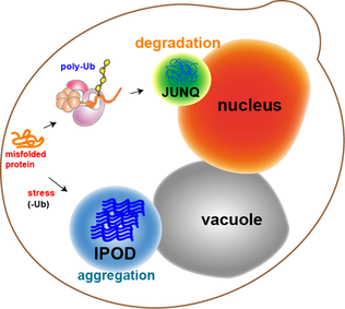
In yeast, deletion strains are frequently used to assess protein stability over time with cycloheximide chases. For example, yeast strains lacking critical degradation machinery such as chaperones, E3 ligases, and vacuolar proteins are often used to determine the mechanism of degradation for a protein substrate of interest. [5] [7] Drug treatments (such as MG132) are also used to inhibit steps of degradation, followed by a cycloheximide chase to observe how the stability of a protein of interest is affected. [3] [4] [7] These experiments may be conducted in mammalian cells with the implementation of compatible knockdown procedures in place of the yeast deletion strains. [4]

Cycloheximide chases are also valuable for assessing how different mutations affect the stability of a protein. Experiments have been conducted in yeast and mammalian cells to determine the critical residues required for protein stability and how disease-associated mutations may be affecting protein half-lives within the cell. [3] [6] This information is valuable for understanding the complexities of protein folding and how mutations contribute to the pathogenesis of the diseases they are associated with.

Advantages

There are many benefits to using cycloheximide chase assays as opposed to other methods that assess protein stability. Cycloheximide chases can be used with a wide variety of model systems and can be implemented to study almost any protein substrate. Cycloheximide is relatively inexpensive compound compared to other drugs and it is effective when used in low doses for short periods of time. Pulse chase assays are an alternative method to cycloheximide chase assays and involve the radioactive labeling of newly translated proteins followed by a similar “chase” period. [8] While this method is informative and provides the benefit of observing nascent protein abundance, the radioactive material it requires is expensive with a shorter shelf-life and demands more caution to use than cycloheximide.

Disadvantages

Some disadvantages to conducting cycloheximide chase assays include the toxic nature of cycloheximide. When used at high concentrations over a long period of time, cycloheximide will damage the DNA within the cell and impair critical cellular function. [9] For this reason, cycloheximide chases do not typically last for more than 12 hours. This presents a limitation if the turnover of a particularly stable protein is being studied. Additionally, cycloheximide chases only offer the ability to look at steady state proteins levels as opposed to newly translated protein levels such as with pulse chase. Therefore, only protein degradation and not protein maturation is able to be observed.

Related Research Articles

<span class="mw-page-title-main">Endoplasmic reticulum</span> Cell organelle that synthesizes, folds and processes proteins

The endoplasmic reticulum (ER) is, in essence, the transportation system of the eukaryotic cell, and has many other important functions such as protein folding. It is a type of organelle made up of two subunits – rough endoplasmic reticulum (RER), and smooth endoplasmic reticulum (SER). The endoplasmic reticulum is found in most eukaryotic cells and forms an interconnected network of flattened, membrane-enclosed sacs known as cisternae, and tubular structures in the SER. The membranes of the ER are continuous with the outer nuclear membrane. The endoplasmic reticulum is not found in red blood cells, or spermatozoa.

A signal peptide is a short peptide present at the N-terminus of most newly synthesized proteins that are destined toward the secretory pathway. These proteins include those that reside either inside certain organelles, secreted from the cell, or inserted into most cellular membranes. Although most type I membrane-bound proteins have signal peptides, the majority of type II and multi-spanning membrane-bound proteins are targeted to the secretory pathway by their first transmembrane domain, which biochemically resembles a signal sequence except that it is not cleaved. They are a kind of target peptide.

<span class="mw-page-title-main">Cystic fibrosis transmembrane conductance regulator</span> Mammalian protein found in humans

Cystic fibrosis transmembrane conductance regulator (CFTR) is a membrane protein and anion channel in vertebrates that is encoded by the CFTR gene.

<span class="mw-page-title-main">Endoplasmic-reticulum-associated protein degradation</span>

Endoplasmic-reticulum-associated protein degradation (ERAD) designates a cellular pathway which targets misfolded proteins of the endoplasmic reticulum for ubiquitination and subsequent degradation by a protein-degrading complex, called the proteasome.

The unfolded protein response (UPR) is a cellular stress response related to the endoplasmic reticulum (ER) stress. It has been found to be conserved between mammalian species, as well as yeast and worm organisms.

<span class="mw-page-title-main">Valosin-containing protein</span> Protein-coding gene in the species Homo sapiens

Valosin-containing protein (VCP) or transitional endoplasmic reticulum ATPase also known as p97 in mammals and CDC48 in S. cerevisiae, is an enzyme that in humans is encoded by the VCP gene. The TER ATPase is an ATPase enzyme present in all eukaryotes and archaebacteria. Its main function is to segregate protein molecules from large cellular structures such as protein assemblies, organelle membranes and chromatin, and thus facilitate the degradation of released polypeptides by the multi-subunit protease proteasome.

<span class="mw-page-title-main">Binding immunoglobulin protein</span> Protein-coding gene in the species Homo sapiens

Binding immunoglobulin protein (BiPS) also known as 78 kDa glucose-regulated protein (GRP-78) or heat shock 70 kDa protein 5 (HSPA5) is a protein that in humans is encoded by the HSPA5 gene.

<span class="mw-page-title-main">EIF2AK3</span> Human protein and coding gene

Eukaryotic translation initiation factor 2-alpha kinase 3, also known as protein kinase R (PKR)-like endoplasmic reticulum kinase (PERK), is an enzyme that in humans is encoded by the EIF2AK3 gene.

<span class="mw-page-title-main">UBE2G2</span> Protein-coding gene in the species Homo sapiens

Ubiquitin-conjugating enzyme E2 G2 is a protein that in humans is encoded by the UBE2G2 gene.

<span class="mw-page-title-main">DNAJB11</span> Protein-coding gene in the species Homo sapiens

DnaJ homolog subfamily B member 11 is a protein that in humans is encoded by the DNAJB11 gene.

<span class="mw-page-title-main">Derlin-1</span> Protein involved in retrotranslocation of specific misfolded proteins and in ER stress

Derlin-1 also known as degradation in endoplasmic reticulum protein 1 is a membrane protein that in humans is encoded by the DERL1 gene. Derlin-1 is located in the membrane of the endoplasmic reticulum (ER) and is involved in retrotranslocation of specific misfolded proteins and in ER stress. Derlin-1 is widely expressed in thyroid, fat, bone marrow and many other tissues. The protein belongs to the Derlin-family proteins consisting of derlin-1, derlin-2 and derlin-3 that are components in the endoplasmic reticulum-associated protein degradation (ERAD) pathway. The derlins mediate degradation of misfolded lumenal proteins within ER, and are named ‘der’ for their ‘Degradation in the ER’. Derlin-1 is a mammalian homologue of the yeast DER1 protein, a protein involved in the yeast ERAD pathway. Moreover, derlin-1 is a member of the rhomboid-like clan of polytopic membrane proteins.

<span class="mw-page-title-main">SIL1</span> Protein-coding gene in the species Homo sapiens

Nucleotide exchange factor SIL1 is a protein that in humans is encoded by the SIL1 gene.

<span class="mw-page-title-main">UBE2G1</span> Protein-coding gene in the species Homo sapiens

Ubiquitin-conjugating enzyme E2 G1 is a protein that in humans is encoded by the UBE2G1 gene.

<span class="mw-page-title-main">NGLY1</span> Protein-coding gene in the species Homo sapiens

PNGase also known as N-glycanase 1 or peptide-N(4)-(N-acetyl-beta-glucosaminyl)asparagine amidase is an enzyme that in humans is encoded by the NGLY1 gene. PNGase is a de-N-glycosylating enzyme that removes N-linked or asparagine-linked glycans (N-glycans) from glycoproteins. More specifically, NGLY1 catalyzes the hydrolysis of the amide bond between the innermost N-acetylglucosamine (GlcNAc) and an Asn residue on an N-glycoprotein, generating a de-N-glycosylated protein, in which the N-glycoylated Asn residue is converted to asp, and a 1-amino-GlcNAc-containing free oligosaccharide. Ammonia is then spontaneously released from the 1-amino GlcNAc at physiological pH (<8), giving rise to a free oligosaccharide with an N,N’-diacetylchitobiose structure at the reducing end.

<span class="mw-page-title-main">Protein aggregation</span> Accumulation of clumps of misfolded or disordered proteins

In molecular biology, protein aggregation is a phenomenon in which intrinsically-disordered or mis-folded proteins aggregate either intra- or extracellularly. Protein aggregates have been implicated in a wide variety of diseases known as amyloidoses, including ALS, Alzheimer's, Parkinson's and prion disease.

Proteostasis is the dynamic regulation of a balanced, functional proteome. The proteostasis network includes competing and integrated biological pathways within cells that control the biogenesis, folding, trafficking, and degradation of proteins present within and outside the cell. Loss of proteostasis is central to understanding the cause of diseases associated with excessive protein misfolding and degradation leading to loss-of-function phenotypes, as well as aggregation-associated degenerative disorders. Therapeutic restoration of proteostasis may treat or resolve these pathologies.

<span class="mw-page-title-main">JUNQ and IPOD</span> Types of cytosolic protein inclusion bodies

JUNQ and IPOD are types of cytosolic protein inclusion bodies in eukaryotes.

<span class="mw-page-title-main">Kifunensine</span> Chemical compound

Kifunensine is an alkaloid originally isolated from Kitasatosporia kifunense, an actinobacterium. It is a neutral, stable compound.

The endoplasmic reticulum membrane protein complex (EMC) is a putative endoplasmic reticulum-resident membrane protein (co-)chaperone. The EMC is evolutionarily conserved in eukaryotes, and its initial appearance might reach back to the last eukaryotic common ancestor (LECA). Many aspects of mEMC biology and molecular function remain to be studied.

HDEL is a target peptide sequence in plants and yeasts located on the C-terminal end of the amino acid structure of a protein. The HDEL sequence prevents a protein from being secreted from the endoplasmic reticulum (ER) and facilitates its return if it is accidentally exported.

References

  1. Schneider-Poetsch, Tilman; Ju, Jianhua; Eyler, Daniel E.; Dang, Yongjun; Bhat, Shridhar; Merrick, William C.; Green, Rachel; Shen, Ben; Liu, Jun O. (2010). "Inhibition of eukaryotic translation elongation by cycloheximide and lactimidomycin". Nature Chemical Biology. 6 (3): 209–217. doi:10.1038/nchembio.304. PMC   2831214 . PMID   20118940.
  2. Girish, V.; Vijayalakshmi, A. (2004). "Affordable image analysis using NIH Image/ImageJ". Indian Journal of Cancer. 41 (1): 47. doi: 10.4103/0019-509X.12345 . PMID   15105580. S2CID   44965098.
  3. 1 2 3 Preston, G. M.; Guerriero, C. J.; Metzger, M. B.; Michaelis, S.; Brodsky, J. L. (2018). "Substrate Insolubility Dictates Hsp104-Dependent Endoplasmic-Reticulum-Associated Degradation". Molecular Cell. 70 (2): 242–253.e6. doi:10.1016/j.molcel.2018.03.016. PMC   5912696 . PMID   29677492.
  4. 1 2 3 4 5 Kim Chiaw, P.; Hantouche, C.; Wong MJH; Matthes, E.; Robert, R.; Hanrahan, J. W.; Shrier, A.; Young, J. C. (2019). "Hsp70 and DNAJA2 limit CFTR levels through degradation". PLOS ONE. 14 (8): e0220984. Bibcode:2019PLoSO..1420984K. doi: 10.1371/journal.pone.0220984 . PMC   6692068 . PMID   31408507.
  5. 1 2 3 Needham, P. G.; Goeckeler-Fried, J. L.; Zhang, C.; Sun, Z.; Wetzel, A. R.; Bertrand, C. A.; Brodsky, J. L. (2021). "SLC26A9 is selected for endoplasmic reticulum associated degradation (ERAD) via Hsp70-dependent targeting of the soluble STAS domain". The Biochemical Journal. 478 (24): 4203–4220. doi:10.1042/BCJ20210644. PMC   8826537 . PMID   34821356.
  6. 1 2 3 Amico, G.; Brandas, C.; Moran, O.; Baroni, D. (2019). "Unravelling the Regions of Mutant F508del-CFTR More Susceptible to the Action of Four Cystic Fibrosis Correctors". International Journal of Molecular Sciences. 20 (21): 5463. doi: 10.3390/ijms20215463 . PMC   6862496 . PMID   31683989.
  7. 1 2 3 Needham, P. G.; Mikoluk, K.; Dhakarwal, P.; Khadem, S.; Snyder, A. C.; Subramanya, A. R.; Brodsky, J. L. (2011). "The thiazide-sensitive NaCl cotransporter is targeted for chaperone-dependent endoplasmic reticulum-associated degradation". The Journal of Biological Chemistry. 286 (51): 43611–43621. doi: 10.1074/jbc.M111.288928 . PMC   3243568 . PMID   22027832.
  8. Sun, Z.; Guerriero, C. J.; Brodsky, J. L. (2021). "Substrate ubiquitination retains misfolded membrane proteins in the endoplasmic reticulum for degradation". Cell Reports. 36 (12): 109717. doi:10.1016/j.celrep.2021.109717. PMC   8503845 . PMID   34551305.
  9. Bašić-Zaninović, Tamara; Papeš, Dražena; Franekić, Jasna (1991). "Cycloheximide genotoxicity in in vitro and in vivo test systems". Mutation Research Letters. 263 (4): 203–210. doi:10.1016/0165-7992(91)90002-L. PMID   1861684.