Human teeth function to mechanically break down items of food by cutting and crushing them in preparation for swallowing and digesting. As such, they are considered part of the human digestive system. Humans have four types of teeth: incisors, canines, premolars, and molars, which each have a specific function. The incisors cut the food, the canines tear the food and the molars and premolars crush the food. The roots of teeth are embedded in the maxilla or the mandible and are covered by gums. Teeth are made of multiple tissues of varying density and hardness.

Dentition pertains to the development of teeth and their arrangement in the mouth. In particular, it is the characteristic arrangement, kind, and number of teeth in a given species at a given age. That is, the number, type, and morpho-physiology of the teeth of an animal.

In mammalian oral anatomy, the canine teeth, also called cuspids, dog teeth, eye teeth, vampire teeth, or vampire fangs, are the relatively long, pointed teeth. In the context of the upper jaw, they are also known as fangs. They can appear more flattened however, causing them to resemble incisors and leading them to be called incisiform. They developed and are used primarily for firmly holding food in order to tear it apart, and occasionally as weapons. They are often the largest teeth in a mammal's mouth. Individuals of most species that develop them normally have four, two in the upper jaw and two in the lower, separated within each jaw by incisors; humans and dogs are examples. In most species, canines are the anterior-most teeth in the maxillary bone. The four canines in humans are the two upper maxillary canines and the two lower mandibular canines. They are specially prominent in dogs (Canidae), hence the name.
Orthodontics is a dentistry specialty that addresses the diagnosis, prevention, management, and correction of mal-positioned teeth and jaws, as well as misaligned bite patterns. It may also address the modification of facial growth, known as dentofacial orthopedics.

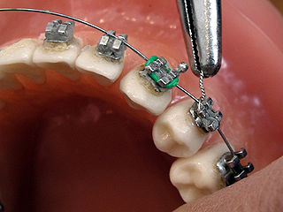
Dental braces are devices used in orthodontics that align and straighten teeth and help position them with regard to a person's bite, while also aiming to improve dental health. They are often used to correct underbites, as well as malocclusions, overbites, open bites, gaps, deep bites, cross bites, crooked teeth, and various other flaws of the teeth and jaw. Braces can be either cosmetic or structural. Dental braces are often used in conjunction with other orthodontic appliances to help widen the palate or jaws and to otherwise assist in shaping the teeth and jaws.

A mouthguard is a protective device for the mouth that covers the teeth and gums to prevent and reduce injury to the teeth, arches, lips and gums. An effective mouthguard is like a crash helmet for teeth and jaws. It also prevents the jaws coming together fully, thereby reducing the risk of jaw joint injuries and concussion. A mouthguard is most often used to prevent injury in contact sports, as a treatment for bruxism or TMD, or as part of certain dental procedures, such as tooth bleaching or sleep apnea treatment. Depending on the application, it may also be called a mouth protector, mouth piece, gumshield, gumguard, nightguard, occlusal splint, bite splint, or bite plane. The dentists who specialise in sports dentistry fabricate mouthguards.

The premolars, also called premolar teeth, or bicuspids, are transitional teeth located between the canine and molar teeth. In humans, there are two premolars per quadrant in the permanent set of teeth, making eight premolars total in the mouth. They have at least two cusps. Premolars can be considered transitional teeth during chewing, or mastication. They have properties of both the canines, that lie anterior and molars that lie posterior, and so food can be transferred from the canines to the premolars and finally to the molars for grinding, instead of directly from the canines to the molars.

Deciduous teeth or primary teeth, also informally known as baby teeth, milk teeth, or temporary teeth, are the first set of teeth in the growth and development of humans and other diphyodonts, which include most mammals but not elephants, kangaroos, or manatees, which are polyphyodonts. Deciduous teeth develop during the embryonic stage of development and erupt during infancy. They are usually lost and replaced by permanent teeth, but in the absence of their permanent replacements, they can remain functional for many years into adulthood.

In orthodontics, a malocclusion is a misalignment or incorrect relation between the teeth of the upper and lower dental arches when they approach each other as the jaws close. The English-language term dates from 1864; Edward Angle (1855-1930), the "father of modern orthodontics", popularised it. The word "malocclusion" derives from occlusion, and refers to the manner in which opposing teeth meet.

Veterinary dentistry is the field of dentistry applied to the care of animals. It is the art and science of prevention, diagnosis, and treatment of conditions, diseases, and disorders of the oral cavity, the maxillofacial region, and its associated structures as it relates to animals.

Dental attrition is a type of tooth wear caused by tooth-to-tooth contact, resulting in loss of tooth tissue, usually starting at the incisal or occlusal surfaces. Tooth wear is a physiological process and is commonly seen as a normal part of aging. Advanced and excessive wear and tooth surface loss can be defined as pathological in nature, requiring intervention by a dental practitioner. The pathological wear of the tooth surface can be caused by bruxism, which is clenching and grinding of the teeth. If the attrition is severe, the enamel can be completely worn away leaving underlying dentin exposed, resulting in an increased risk of dental caries and dentin hypersensitivity. It is best to identify pathological attrition at an early stage to prevent unnecessary loss of tooth structure as enamel does not regenerate.
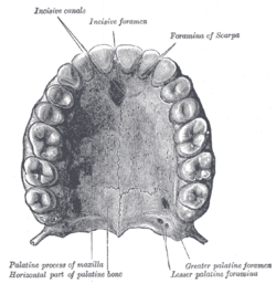
Dental anatomy is a field of anatomy dedicated to the study of human tooth structures. The development, appearance, and classification of teeth fall within its purview. Tooth formation begins before birth, and the teeth's eventual morphology is dictated during this time. Dental anatomy is also a taxonomical science: it is concerned with the naming of teeth and the structures of which they are made, this information serving a practical purpose in dental treatment.
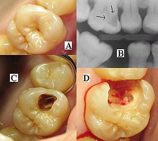
Dental radiographs, commonly known as X-rays, are radiographs used to diagnose hidden dental structures, malignant or benign masses, bone loss, and cavities.
Occlusion, in a dental context, means simply the contact between teeth. More technically, it is the relationship between the maxillary (upper) and mandibular (lower) teeth when they approach each other, as occurs during chewing or at rest.

A cusp is a pointed, projecting, or elevated feature. In animals, it is usually used to refer to raised points on the crowns of teeth. The concept is also used with regard to the leaflets of the four heart valves. The mitral valve, which has two cusps, is also known as the bicuspid valve, and the tricuspid valve has three cusps.
This is a list of definitions of commonly used terms of location and direction in dentistry. This set of terms provides orientation within the oral cavity, much as anatomical terms of location provide orientation throughout the body.

Crossbite is a form of malocclusion where a tooth has a more buccal or lingual position than its corresponding antagonist tooth in the upper or lower dental arch. In other words, crossbite is a lateral misalignment of the dental arches.

An impacted tooth is one that fails to erupt into the dental arch within the expected developmental window. Because impacted teeth do not erupt, they are retained throughout the individual's lifetime unless extracted or exposed surgically. Teeth may become impacted because of adjacent teeth, dense overlying bone, excessive soft tissue or a genetic abnormality. Most often, the cause of impaction is inadequate arch length and space in which to erupt. That is the total length of the alveolar arch is smaller than the tooth arch. The wisdom teeth are frequently impacted because they are the last teeth to erupt in the oral cavity. Mandibular third molars are more commonly impacted than their maxillary counterparts.
Orthodontic indices are one of the tools that are available for orthodontists to grade and assess malocclusion. Orthodontic indices can be useful for an epidemiologist to analyse prevalence and severity of malocclusion in any population.
Occlusion according to The Glossary of Prosthodontic Terms Ninth Edition is defined as "the static relationship between the incising or masticating surfaces of the maxillary or mandibular teeth or tooth analogues".