

Endoplasmic reticulum aminopeptidase 2 is an aminopeptidase in humans involved in antigen presentation. It is encoded by the ERAP2 gene.
Endoplasmic reticulum aminopeptidase 2 is an aminopeptidase in humans involved in antigen presentation. It is encoded by the ERAP2 gene.
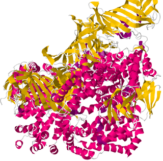
ERAP1 and ERAP2 are Aminopeptidases, able to trim precursors to antigenic peptides in the endoplasmic reticulum. They hydrolyze N-terminal amino acids of proteins or peptide substrates so the Major histocompatibility complex (MHC) class I can present them to CD8+ T cells. [3]
ERAP2 plays a central role in antigen presentation and impacts the response of at least 4 cytokines: It is positively correlated with CCL3, which is involved in the recruitment of neutrophils upon infection, whereas it is inversely correlated with granulocyte colony-stimulating factor , interleukin-1β and IL-10. [4]
Mutations in the ERAP2 gene [5] were found in DNA extracts derived from the skeletons of people who died shortly before, during or soon after the Black Death 1348 in three London cemeteries, including East Smithfield plague cemetery and from Denmark. Per this study, carrying two protective versions of ERAP2 made people 40 percent likelier to survive a Yersinia pestis infection. [4] However this version of the gene also increases the risk of Crohn’s disease. [6]
There is evidence that the Black Death shaped genetic diversity in Europe, in that people who had protective variant of the ERAP2 gene were much more likely to survive Yersinia pestis infection, the causitive agent of Black Death. [7]

Calreticulin also known as calregulin, CRP55, CaBP3, calsequestrin-like protein, and endoplasmic reticulum resident protein 60 (ERp60) is a protein that in humans is encoded by the CALR gene.
An epitope, also known as antigenic determinant, is the part of an antigen that is recognized by the immune system, specifically by antibodies, B cells, or T cells. The part of an antibody that binds to the epitope is called a paratope. Although epitopes are usually non-self proteins, sequences derived from the host that can be recognized are also epitopes.

MHC class I molecules are one of two primary classes of major histocompatibility complex (MHC) molecules and are found on the cell surface of all nucleated cells in the bodies of vertebrates. They also occur on platelets, but not on red blood cells. Their function is to display peptide fragments of proteins from within the cell to cytotoxic T cells; this will trigger an immediate response from the immune system against a particular non-self antigen displayed with the help of an MHC class I protein. Because MHC class I molecules present peptides derived from cytosolic proteins, the pathway of MHC class I presentation is often called cytosolic or endogenous pathway.
Cross-presentation is the ability of certain professional antigen-presenting cells (mostly dendritic cells) to take up, process and present extracellular antigens with MHC class I molecules to CD8 T cells (cytotoxic T cells). Cross-priming, the result of this process, describes the stimulation of naive cytotoxic CD8+ T cells into activated cytotoxic CD8+ T cells. This process is necessary for immunity against most tumors and against viruses that infect dendritic cells and sabotage their presentation of virus antigens. Cross presentation is also required for the induction of cytotoxic immunity by vaccination with protein antigens, for example, tumour vaccination.
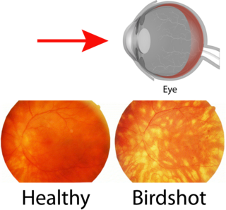
Birdshot chorioretinopathy, now commonly named birdshot uveitis or HLA-A29 uveitis, is a rare form of bilateral posterior uveitis affecting both eyes. It causes severe, progressive inflammation of both the choroid and retina.
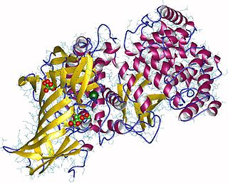
Aminopeptidases are enzymes that catalyze the cleavage of amino acids from the amino terminus (N-terminus) of proteins or peptides (exopeptidases). They are widely distributed throughout the animal and plant kingdoms and are found in many subcellular organelles, in cytosol, and as membrane components. Aminopeptidases are used in essential cellular functions. Many, but not all, of these peptidases are zinc metalloenzymes.
Bare lymphocyte syndrome is a condition caused by mutations in certain genes of the major histocompatibility complex or involved with the processing and presentation of MHC molecules. It is a form of severe combined immunodeficiency.

Antigen presentation is a vital immune process that is essential for T cell immune response triggering. Because T cells recognize only fragmented antigens displayed on cell surfaces, antigen processing must occur before the antigen fragment, now bound to the major histocompatibility complex (MHC), is transported to the surface of the cell, a process known as presentation, where it can be recognized by a T-cell receptor. If there has been an infection with viruses or bacteria, the cell will present an endogenous or exogenous peptide fragment derived from the antigen by MHC molecules. There are two types of MHC molecules which differ in the behaviour of the antigens: MHC class I molecules (MHC-I) bind peptides from the cell cytosol, while peptides generated in the endocytic vesicles after internalisation are bound to MHC class II (MHC-II). Cellular membranes separate these two cellular environments - intracellular and extracellular. Each T cell can only recognize tens to hundreds of copies of a unique sequence of a single peptide among thousands of other peptides presented on the same cell, because an MHC molecule in one cell can bind to quite a large range of peptides. Predicting which antigens will be presented to the immune system by a certain MHC/HLA type is difficult, but the technology involved is improving.

TAP-associated glycoprotein, also known as tapasin or TAPBP, is a protein that in humans is encoded by the TAPBP gene.
CD59 glycoprotein, also known as MAC-inhibitory protein (MAC-IP), membrane inhibitor of reactive lysis (MIRL), or protectin, is a protein that in humans is encoded by the CD59 gene. It is an LU domain and belongs to the LY6/uPAR/alpha-neurotoxin protein family.

Protein disulfide-isomerase A3 (PDIA3), also known as glucose-regulated protein, 58-kD (GRP58), is an isomerase enzyme. This protein localizes to the endoplasmic reticulum (ER) and interacts with lectin chaperones calreticulin and calnexin (CNX) to modulate folding of newly synthesized glycoproteins. It is thought that complexes of lectins and this protein mediate protein folding by promoting formation of disulfide bonds in their glycoprotein substrates.
Human leukocyte histocompatibility complex DO (HLA-DO) is an intracellular, dimeric non-classical Major Histocompatibility Complex (MHC) class II protein composed of α- and β-subunits which interact with HLA-DM in order to fine tune immunodominant epitope selection. As a non-classical MHC class II molecule, HLA-DO is a non-polymorphic accessory protein that aids in antigenic peptide chaperoning and loading, as opposed to its classical counterparts, which are polymorphic and involved in antigen presentation. Though more remains to be elucidated about the function of HLA-DO, its unique distribution in the mammalian body—namely, the exclusive expression of HLA-DO in B cells, thymic medullary epithelial cells, and dendritic cells—indicate that it may be of physiological importance and has inspired further research. Although HLA-DM can be found without HLA-DO, HLA-DO is only found in complex with HLA-DM and exhibits instability in the absence of HLA-DM. The evolutionary conservation of both DM and DO, further denote its biological significance and potential to confer evolutionary benefits to its host.
HLA class II histocompatibility antigen gamma chain also known as HLA-DR antigens-associated invariant chain or CD74, is a protein that in humans is encoded by the CD74 gene. The invariant chain is a polypeptide which plays a critical role in antigen presentation. It is involved in the formation and transport of MHC class II peptide complexes for the generation of CD4+ T cell responses. The cell surface form of the invariant chain is known as CD74. CD74 is a cell surface receptor for the cytokine macrophage migration inhibitory factor (MIF).
HLA class I histocompatibility antigen, alpha chain F is a protein that in humans is encoded by the HLA-F gene. It is an empty intracellular molecule that encodes a non-classical heavy chain anchored to the membrane and forming a heterodimer with a β-2 microglobulin light chain. It belongs to the HLA class I heavy chain paralogues that separate from most of the HLA heavy chains. HLA-F is localized in the endoplasmic reticulum and Golgi apparatus, and is also unique in the sense that it exhibits few polymorphisms in the human population relative to the other HLA genes; however, there have been found different isoforms from numerous transcript variants found for the HLA-F gene. Its pathways include INF-gamma signaling and CDK-mediated phosphorylation and removal of the Saccharomycescerevisiae Cdc6 protein, which is crucial for functional DNA replication.

B-cell receptor-associated protein 31 is a protein that in humans is encoded by the BCAP31 gene.
Type 1 tumor necrosis factor receptor shedding aminopeptidase regulator, also known as endoplasmic reticulum aminopeptidase 1 (ARTS-1), is a protein which in humans is encoded by the ARTS-1 gene.

Sarcoplasmic/endoplasmic reticulum calcium ATPase 3 is an enzyme that in humans is encoded by the ATP2A3 gene.

Minor histocompatibility antigen H13 is a protein that in humans is encoded by the HM13 gene.

Programmed cell death 1 ligand 2 is a protein that in humans is encoded by the PDCD1LG2 gene. PDCD1LG2 has also been designated as CD273. PDCD1LG2 is an immune checkpoint receptor ligand which plays a role in negative regulation of the adaptive immune response. PD-L2 is one of two known ligands for Programmed cell death protein 1 (PD-1).

PNGase also known as N-glycanase 1 or peptide-N(4)-(N-acetyl-beta-glucosaminyl)asparagine amidase is an enzyme that in humans is encoded by the NGLY1 gene. PNGase is a de-N-glycosylating enzyme that removes N-linked or asparagine-linked glycans (N-glycans) from glycoproteins. More specifically, NGLY1 catalyzes the hydrolysis of the amide bond between the innermost N-acetylglucosamine (GlcNAc) and an Asn residue on an N-glycoprotein, generating a de-N-glycosylated protein, in which the N-glycoylated Asn residue is converted to asp, and a 1-amino-GlcNAc-containing free oligosaccharide. Ammonia is then spontaneously released from the 1-amino GlcNAc at physiological pH (<8), giving rise to a free oligosaccharide with an N,N’-diacetylchitobiose structure at the reducing end.
This article incorporates text from the United States National Library of Medicine, which is in the public domain.
| | This article on a gene on human chromosome 5 is a stub. You can help Wikipedia by expanding it. |