Entamoeba coli

Last updated

Entamoeba coli
Scientific classification OOjs UI icon edit-ltr.svg
Domain: Eukaryota
Phylum: Amoebozoa
Family: Entamoebidae
Genus: Entamoeba
Species:
E. coli
Binomial name
Entamoeba coli
(Grassi, 1879)

Entamoeba coli is a non-pathogenic species of Entamoeba that frequently exists as a commensal parasite in the human gastrointestinal tract. E. coli (not to be confused with the bacterium Escherichia coli ) is important in medicine because it can be confused during microscopic examination of stained stool specimens with the pathogenic Entamoeba histolytica . [1] This amoeba does not move much by the use of its pseudopod, and creates a "sur place (non-progressive) movement" inside the large intestine. Usually, the amoeba is immobile, and keeps its round shape. This amoeba, in its trophozoite stage, is only visible in fresh, unfixed stool specimens. Sometimes the Entamoeba coli have parasites as well. One is the fungus Sphaerita spp. This fungus lives in the cytoplasm of the E. coli. [2] While this differentiation is typically done by visual examination of the parasitic cysts via light microscopy, new methods using molecular biology techniques have been developed. [3] The scientific name of the amoeba, E. coli, is often mistaken for the bacterium, Escherichia coli. Unlike the bacterium, the amoeba is mostly harmless, and does not cause as many intestinal problems as some strains of the E. coli bacterium. To make the naming of these organisms less confusing, "alternate contractions" are used to name the species for the purpose making the naming easier; for example, using Esch. coli and Ent. coli for the bacterium and amoeba, instead of using E. coli for both. [4] [5]

Contents

Common lifecycle of various non-pathogenic parasites, including E. coli Common Non-pathogen Parasites Lifecycle.png
Common lifecycle of various non-pathogenic parasites, including E. coli

Clinical significance

Entamoeba coli cyst with more than four nuclei Entaemoeba coli cyst 6 nuclei.png
Entamoeba coli cyst with more than four nuclei

The presence of E.coli is not cause in and of itself to seek treatment as it is considered harmless. [6] However, when a person becomes infected with this benign entamoeba, other pathogenic organisms may have been introduced as well, and these other pathogens might cause infection or illness. [7]

Morphology

Stages of the Entamoeba coli parasite Entamoeba coli Stages.png
Stages of the Entamoeba coli parasite

Entamoeba species all come in monogenetic forms, or having one generation lifecycles. E. coli has "three distinct morphological forms exist airing the life cycle-Trophozoite, Pre-cystic stage and Cystic stage". This lifecycle gives rise to the general way of how Entamoeba species form. This parasite has one large nucleus with a thick membrane surrounding the nucleus. There are many chromatin inside the nucleus, and one large, irregular-shaped karyosome. [8] The chromatin is clumped, and uneven in disperse inside the nucleus. The parasite forms by binary fission like most Entamoeba spp. [9] The mature cyst is the infective stage, and is known to survive longer than those of E. histolytica. The cysts can survive three to four months outside the host's body after desiccation. [8] The cysts cause infection by consuming contaminated food and drinks like waste water. Sometimes insects and rodents carry the parasite to cause infection in the food and drinks. Excystation happens once the cysts are ingested, and travel to the large intestine. [2]

Diagnosis

E. coli trophozoites can be distinguished by their wide and tapered pseudopodia. They are often mistaken for E. histolytica due to their overlap in size. The cysts are distinguished by noticing the eight nuclei found in the mature form. [10] To diagnose for E. coli, a stool sample is usually tested. This is the best method to check to see if the parasite is E. coli and not E. histolytica. This usually involves checking the cysts for the size, shape, and the number of nuclei. E. coli has cysts in size up to 10 to 35 micrometers, the shape is irregular, oval with a shell-like appearance that is more uniformed compared to E. histolytica, and has up to eight nuclei in the cyst compared to the four nuclei of E. histolytica. To the untrained eye of an inexperienced microbiologists, "tetranucleate cysts of Entamoeba coli can be mistaken for mature cysts of Entamoeba histolytica" Often " a tetranucleate Entamoeba coli cyst is larger than a mature cyst of Entamoeba histolytica, can be variable in shape, and has nuclear peripheral chromatin and karyosome composed of irregular granules" in this matter of comparison. To make a diagnosis for any Entamoeba species, usually a wet mount is created "by finding the characteristic cysts in an iodine stained, formol-ether concentration method or by detecting the characteristic trophozoites in a wet preparation or a permanent stained preparation" to see what they may look like. [11] Also, these stains of trichrome can be used to mount the cysts of any Entamoeba spp. [12] Other tests can be used to diagnosis for Entamoeba spp. These tests involve the use of laboratory methods. Some of these laboratory tests include: the use of light microscopy, culture methods, isoenzyme analysis, antibody detection tests, antigen detection tests, immunochromatographic assays, and DNA-based diagnostic tests. [13] Some uses of microscopy also involve the use of transmission electron microscopy and scanning electron microscopy. Usually, the cysts are freeze fractured to insure that the samples are easier to look at to compare Entamoeba spp. [14] The DNA-based diagnostic tests include the use of DNA extraction, PCR, microarrays, and typing methods.For example, one DNA-based diagnostic test that is changing how Entamoeba spp. is being diagnosis faster and more accurate is by using the "Reverse Line Hybridization Assay" test. This test main purpose is to detect and different Entamoeba spp. in stool samples in order to find the causative agent of amoebic dysentery, E. histolytica. This test involves the use of gene sequencing, and seeing what different genomes each Entamoeba spp. has to help detect the deadly E. histolytica. [15]

Pathology

E. coli are mostly harmless parasites, and do not cause harm to the host. However, there have been cases of internal bleeding. Usually, the cytoplasm of the E. coli "does not contain red blood cells, except in the rare case of patients with intestinal hemorrhage" that leads to blood in the stools of these patients. This may lead to intestinal lesions. Other problems that E. coli causes are usually result from having too many in the large intestine. For example, large populations of E. coli may lead to "dyspepsia, hyperacidity, gastritis, and indigestion"; these are common problems of most intestinal parasites. [8]

Treatment

There is generally no need to treat for E. coli, due to the rarity of this parasite becoming infectious. In one exceptional situation, E. coli was found to be infectious: in northern Europe, stool samples and electron microscopy revealed large populations of the amoeba within a group of patients with persistent diarrhea. [16] Some types of treatments may need to be used due to large populations. Some arsenical compounds are shown to treat the trophozoite stage, like carbarsone. [8] Other compounds used to treat large populations of E. coli include diloxanide furoate, and this usually is used in antiamebic therapy. [16]

See also

Related Research Articles

<i>Entamoeba</i> Genus of internal parasites

Entamoeba is a genus of Amoebozoa found as internal parasites or commensals of animals. In 1875, Fedor Lösch described the first proven case of amoebic dysentery in St. Petersburg, Russia. He referred to the amoeba he observed microscopically as Amoeba coli; however, it is not clear whether he was using this as a descriptive term or intended it as a formal taxonomic name. The genus Entamoeba was defined by Casagrandi and Barbagallo for the species Entamoeba coli, which is known to be a commensal organism. Lösch's organism was renamed Entamoeba histolytica by Fritz Schaudinn in 1903; he later died, in 1906, from a self-inflicted infection when studying this amoeba. For a time during the first half of the 20th century the entire genus Entamoeba was transferred to Endamoeba, a genus of amoebas infecting invertebrates about which little is known. This move was reversed by the International Commission on Zoological Nomenclature in the late 1950s, and Entamoeba has stayed 'stable' ever since.

<span class="mw-page-title-main">Dysentery</span> Inflammation of the intestine causing diarrhea with blood

Dysentery, historically known as the bloody flux, is a type of gastroenteritis that results in bloody diarrhea. Other symptoms may include fever, abdominal pain, and a feeling of incomplete defecation. Complications may include dehydration.

<i>Entamoeba histolytica</i> Anaerobic parasitic protist

Entamoeba histolytica is an anaerobic parasitic amoebozoan, part of the genus Entamoeba. Predominantly infecting humans and other primates causing amoebiasis, E. histolytica is estimated to infect about 35-50 million people worldwide. E. histolytica infection is estimated to kill more than 55,000 people each year. Previously, it was thought that 10% of the world population was infected, but these figures predate the recognition that at least 90% of these infections were due to a second species, E. dispar. Mammals such as dogs and cats can become infected transiently, but are not thought to contribute significantly to transmission.

<span class="mw-page-title-main">Isosporiasis</span> Human intestinal disease

Isosporiasis, also known as cystoisosporiasis, is a human intestinal disease caused by the parasite Cystoisospora belli. It is found worldwide, especially in tropical and subtropical areas. Infection often occurs in immuno-compromised individuals, notably AIDS patients, and outbreaks have been reported in institutionalized groups in the United States. The first documented case was in 1915. It is usually spread indirectly, normally through contaminated food or water (CDC.gov).

<span class="mw-page-title-main">Giardiasis</span> Parasitic disease that results in diarrhea

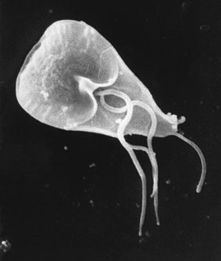
Giardiasis is a parasitic disease caused by Giardia duodenalis. Infected individuals who experience symptoms may have diarrhoea, abdominal pain, and weight loss. Less common symptoms include vomiting and blood in the stool. Symptoms usually begin one to three weeks after exposure and, without treatment, may last two to six weeks or longer.

<span class="mw-page-title-main">Amoebozoa</span> Phylum of protozoans

Amoebozoa is a major taxonomic group containing about 2,400 described species of amoeboid protists, often possessing blunt, fingerlike, lobose pseudopods and tubular mitochondrial cristae. In traditional classification schemes, Amoebozoa is usually ranked as a phylum within either the kingdom Protista or the kingdom Protozoa. In the classification favored by the International Society of Protistologists, it is retained as an unranked "supergroup" within Eukaryota. Molecular genetic analysis supports Amoebozoa as a monophyletic clade. Modern studies of eukaryotic phylogenetic trees identify it as the sister group to Opisthokonta, another major clade which contains both fungi and animals as well as several other clades comprising some 300 species of unicellular eukaryotes. Amoebozoa and Opisthokonta are sometimes grouped together in a high-level taxon, variously named Unikonta, Amorphea or Opimoda.

A trophozoite is the activated, feeding stage in the life cycle of certain protozoa such as malaria-causing Plasmodium falciparum and those of the Giardia group. The complementary form of the trophozoite state is the thick-walled cyst form. They are often different from the cyst stage, which is a protective, dormant form of the protozoa. Trophozoites are often found in the host's body fluids and tissues and in many cases, they are the form of the protozoan that causes disease in the host. In the protozoan, Entamoeba histolytica it invades the intestinal mucosa of its host, causing dysentery, which aid in the trophozoites traveling to the liver and leading to the production of hepatic abscesses.

<span class="mw-page-title-main">Balantidiasis</span> Medical condition

Balantidiasis is a protozoan infection caused by infection with Balantidium coli.

Dientamoebiasis is a medical condition caused by infection with Dientamoeba fragilis, a single-cell parasite that infects the lower gastrointestinal tract of humans. It is an important cause of traveler's diarrhea, chronic abdominal pain, chronic fatigue, and failure to thrive in children.

<span class="mw-page-title-main">Blastocystosis</span> Medical condition

Blastocystosis refers to a medical condition caused by infection with Blastocystis. Blastocystis is a protozoal, single-celled parasite that inhabits the gastrointestinal tracts of humans and other animals. Many different types of Blastocystis exist, and they can infect humans, farm animals, birds, rodents, amphibians, reptiles, fish, and even cockroaches. Blastocystosis has been found to be a possible risk factor for development of irritable bowel syndrome.

<span class="mw-page-title-main">Protozoan infection</span> Parasitic disease caused by a protozoan

Protozoan infections are parasitic diseases caused by organisms formerly classified in the kingdom Protozoa. These organisms are now classified in the supergroups Excavata, Amoebozoa, Harosa, and Archaeplastida. They are usually contracted by either an insect vector or by contact with an infected substance or surface.

<span class="mw-page-title-main">Amoebiasis</span> Human disease caused by amoeba protists

Amoebiasis, or amoebic dysentery, is an infection of the intestines caused by a parasitic amoeba Entamoeba histolytica. Amoebiasis can be present with no, mild, or severe symptoms. Symptoms may include lethargy, loss of weight, colonic ulcerations, abdominal pain, diarrhea, or bloody diarrhea. Complications can include inflammation and ulceration of the colon with tissue death or perforation, which may result in peritonitis. Anemia may develop due to prolonged gastric bleeding.

<span class="mw-page-title-main">Amoebic liver abscess</span> Medical condition

A amoebic liver abscess is a type of liver abscess caused by amebiasis. It is the involvement of liver tissue by trophozoites of the organism Entamoeba histolytica and of its abscess due to necrosis.

<span class="mw-page-title-main">Amoebic brain abscess</span> Medical condition

Amoebic brain abscess is an affliction caused by the anaerobic parasitic protist Entamoeba histolytica. It is extremely rare; the first case being reported in 1849. Brain abscesses resulting from Entamoeba histolytica are difficult to diagnose and very few case reports suggest complete recovery even after the administration of appropriate treatment regimen.

Sappinia is a genus of heterotrophic, lobose amoebae within the family Thecamoebidae. A defining feature of Sappinia, which separates it from its sister genus Thecamoeba, is the presence of two closely apposed nuclei with a central, flattened connection. Sappinia species have two life cycle stages: a trophozoite and a cyst. Up until 2015, only two species had been discovered, Sappinia pedata and Sappinia diploidea. Sequencing of the small subunit rRNA of a particular isolate from a sycamore tree revealed a new species, Sappinia platani.Sappinia species were once thought to be coprozoic, as the first strains were isolated from animal dung. More research has shown that they are typical free-living amoebae, and can be found worldwide in soil, plant litter, and standing decaying plants, as well as freshwater ponds. In 2001, the first and only case of human pathogenesis in Sappinia was confirmed. The patient was a non-immunocompromised 38-year-old male who presented signs of amoebic encephalitis and who patient made a full recovery after treatment with several antimicrobials. The CDC initially classified the causative agent as S. diploidea based on morphological characteristics, but in 2009, Qvarnstrom et al. used molecular data to confirm that the true causative agent was S. pedata.

Entamoeba polecki is an intestinal parasite of the genus Entamoeba. E. polecki is found primarily in pigs and monkeys and is largely considered non-pathogenic in humans, although there have been some reports regarding symptomatic infections of humans. Prevalence is concentrated in New Guinea, with distribution also recorded in areas of southeast Asia, France, and the United States.

<i>Dientamoeba fragilis</i> Parasite of humans, pigs and gorillas

Dientamoeba fragilis is a species of single-celled excavates found in the gastrointestinal tract of some humans, pigs and gorillas. It causes gastrointestinal upset in some people, but not in others. It is an important cause of traveller's diarrhoea, chronic diarrhoea, fatigue and, in children, failure to thrive. Despite this, its role as a "commensal, pathobiont, or pathogen" is still debated. D. fragilis is one of the smaller parasites that are able to live in the human intestine. Dientamoeba fragilis cells are able to survive and move in fresh feces but are sensitive to aerobic environments. They dissociate when in contact or placed in saline, tap water or distilled water.

Entamoeba moshkovskii is part of the genus Entamoeba. It is found in areas with polluted water sources, and is prevalent in places such as Malaysia, India, and Bangladesh, but more recently has made its way to Turkey, Australia, and North America. This amoeba is said to rarely infect humans, but recently this has changed. It is in question as to whether it is pathogenic or not. Despite some sources stating this is a free living amoeba, various studies worldwide have shown it contains the ability to infect humans, with some cases of pathogenic potential being reported. Some of the symptoms that often occur are diarrhea, weight loss, bloody stool, and abdominal pain. The first known human infection also known as the "Laredo strain" of Entamoebic mushkovskii was in Laredo, Texas in 1991, although it was first described by a man named Tshalaia in 1941 in Moscow, Russia. It is known to affect people of all ages and genders.

Entamoeba invadens is an amoebozoa parasite of reptiles, within the genus Entamoeba. It is closely related to the human parasite Entamoeba histolytica, causing similar invasive disease in reptiles, in addition to a similar morphology and lifecycle.

<i>Naegleria fowleri</i> Species of free-living excavate form of protist

Naegleria fowleri, also known as the brain-eating amoeba, is a species of the genus Naegleria. It belongs to the phylum Percolozoa and is technically classified as an amoeboflagellate excavate, rather than a true amoeba. This free-living microorganism primarily feeds on bacteria but can become pathogenic in humans, causing an extremely rare, sudden, severe, and usually fatal brain infection known as naegleriasis or primary amoebic meningoencephalitis (PAM).

References

  1. Sodeman WA (1996). "Intestinal Protozoa: Amebas". In Baron S; et al. (eds.). Baron's Medical Microbiology (4th ed.). Univ of Texas Medical Branch. ISBN   978-0-9631172-1-2.
  2. 1 2 "Atlante dei protozoi intestinali umani - sezione AMEBE identificazione microscopica di Giovanni Swierczynski e Bruno Milanesi". Atlas-protozoa.com. Retrieved 2015-12-03.
  3. Verweij JJ, Laeijendecker D, Brienen EA, van Lieshout L, Polderman AM (2003). "Detection and identification of entamoeba species in stool samples by a reverse line hybridization assay". J. Clin. Microbiol. 41 (11): 5041–5. doi:10.1128/JCM.41.11.5041-5045.2003. PMC   262518 . PMID   14605136.
  4. Pometto, Anthony; Shetty, Kalidas; Paliyath, Gopinadhan; Levin, Robert E. (2005-10-11). Food Biotechnology (Second ed.). p. 9. ISBN   9781420027976 . Retrieved 2015-12-03.
  5. "Entamoeba". Microscope-microscope.org. Retrieved 2015-12-03.
  6. "CDC - Nonpathogenic (Harmless) Intestinal Protozoa - Biology". Cdc.gov. 2015-03-17. Retrieved 2015-12-03.
  7. "CDC - DPDx - Intestinal Amebae". Cdc.gov. 2013-11-29. Retrieved 2015-12-03.
  8. 1 2 3 4 Saritha Pujari (2015-11-14). "Parasite Entamoeba Coli : Life Cycle, Mode of Infection and Treatment". Yourarticlelibrary.com. Retrieved 2015-12-03.
  9. Don Lehman. "Diagnostic Parasitology; E. coli". Udel.edu. Retrieved 2015-12-03.
  10. "Intestinal Protozoa". Tulane.edu. 2015-06-03. Retrieved 2015-12-03.
  11. Jim McMorran; Damian Crowther; Stew McMorran; Steve Youngmin; Ian Wacogne; Jon Pleat; Clive Prince. "Entamoeba coli - General Practice Notebook". Gpnotebook.co.uk. Retrieved 2015-12-03.
  12. "CDC - DPDx - Intestinal Amebae - Images". Cdc.gov. 2013-11-29. Retrieved 2015-12-03.
  13. Fotedar R, Stark D, Beebe N, Marriott D, Ellis J, Harkness J (2015-09-28). "Laboratory Diagnostic Techniques for Entamoeba Species". Clin. Microbiol. Rev. 20 (3): 511–32, table of contents. doi:10.1128/CMR.00004-07. PMC   1932757 . PMID   17630338.
  14. Chávez-Munguía, Bibiana; Martínez-Palomo, Adolfo (2011-08-25). "High-Resolution Electron Microscopical Study of Cyst Walls of Entamoeba spp". Journal of Eukaryotic Microbiology. 58 (6): 480–486. doi:10.1111/j.1550-7408.2011.00576.x. PMID   21883633. S2CID   19124321.
  15. Verweij JJ, Laeijendecker D, Brienen EA, van Lieshout L, Polderman AM (2015-09-28). "Detection and Identification of Entamoeba Species in Stool Samples by a Reverse Line Hybridization Assay". J. Clin. Microbiol. 41 (11): 5041–5. doi:10.1128/JCM.41.11.5041-5045.2003. PMC   262518 . PMID   14605136.
  16. 1 2 Hotez, Peter (July 2000). "The other intestinal protozoa: Enteric infections caused by Blastocystis hominis, Entamoeba coli, and Dientamoeba fragilis". Seminars in Pediatric Infectious Diseases. 11 (3): 178–181. doi:10.1053/pi.2000.6228.

Further reading