Related Research Articles

The spermatic cord is the cord-like structure in males formed by the vas deferens and surrounding tissue that runs from the deep inguinal ring down to each testicle. Its serosal covering, the tunica vaginalis, is an extension of the peritoneum that passes through the transversalis fascia. Each testicle develops in the lower thoracic and upper lumbar region and migrates into the scrotum. During its descent it carries along with it the vas deferens, its vessels, nerves etc. There is one on each side.

The retroperitoneal space (retroperitoneum) is the anatomical space behind (retro) the peritoneum. It has no specific delineating anatomical structures. Organs are retroperitoneal if they have peritoneum on their anterior side only. Structures that are not suspended by mesentery in the abdominal cavity and that lie between the parietal peritoneum and abdominal wall are classified as retroperitoneal.

The rectouterine pouch is the extension of the peritoneum into the space between the posterior wall of the uterus and the rectum in the human female.

The inguinal ligament, also known as Poupart's ligament or groin ligament, is a band running from the pubic tubercle to the anterior superior iliac spine. It forms the base of the inguinal canal through which an indirect inguinal hernia may develop.

The external iliac arteries are two major arteries which bifurcate off the common iliac arteries anterior to the sacroiliac joint of the pelvis.

The abdomen is the part of the body between the thorax (chest) and pelvis, in humans and in other vertebrates. The abdomen is the front part of the abdominal segment of the torso. The area occupied by the abdomen is called the abdominal cavity. In arthropods, it is the posterior tagma of the body; it follows the thorax or cephalothorax.

In human anatomy, the inferior epigastric artery is an artery that arises from the external iliac artery. It is accompanied by the inferior epigastric vein; inferiorly, these two inferior epigastric vessels together travel within the lateral umbilical fold The inferior epigastric artery then traverses the arcuate line of rectus sheath to enter the rectus sheath, then anastomoses with the superior epigastric artery within the rectus sheath.

In anatomy, the abdominal wall represents the boundaries of the abdominal cavity. The abdominal wall is split into the anterolateral and posterior walls.
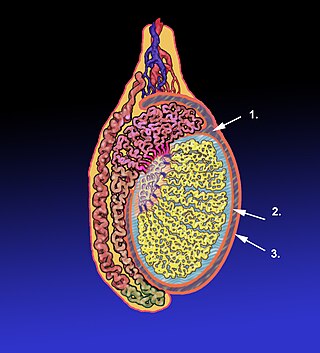
The tunica vaginalis is a pouch of serous membrane within the scrotum that lines the testis and epididymis, and the inner surface of the scrotum. It is the outermost of the three layers that constitute the capsule of the testis, with the tunica albuginea of penis situated beneath it.

The lateral umbilical fold is an elevation of the peritoneum lining the inner/posterior surface of the lower anterior abdominal wall formed by the underlying inferior epigastric artery and inferior epigastric vein which the peritoneum covers. Superiorly, the lateral umbilical fold ends where the vessels reach and enter the rectus sheath at the arcuate line of rectus sheath; in spite of the name, the lateral umbilical folds do not extend as far superiorly as the umbilicus. Inferiorly, it extends to just medial to the deep inguinal ring.

The rectus sheath is a tough fibrous compartment formed by the aponeuroses of the transverse abdominal muscle, and the internal and external oblique muscles. It contains the rectus abdominis and pyramidalis muscles, as well as vessels and nerves.

The perimetrium is the outer serosal layer of the uterus, derived from the peritoneum overlying the uterine fundus, and can be considered a visceral peritoneum. It consists of a superficial layer of mesothelium, and a thin layer of loose connective tissue beneath it.

The pararectal fossa is an inferior-ward extension of the peritoneum on either side of the rectum. It is formed by a (sacrogenital) fold of peritoneum extending inferior-ward from the posterolateral pelvic wall. It represents a lateral extension of the rectouterine pouch in the female, and of rectovesical pouch in the male. It varies in size with the distension of the rectum.
In human female anatomy, the vesicouterine pouch, also uterovesicle pouch, is a fold of peritoneum over the uterus and the bladder. Like the rectouterine pouch, it is a female pelvic recess, but shallower and closer to the anterior fornix of the vagina.

Retropubic space is a potential avascular space located between the pubic symphysis and the urinary bladder. The retropubic space is a preperitoneal space, located behind the transversalis fascia and in front of peritoneum.

The rectovesical pouch is the pocket that lies between the rectum and the bladder in males in humans and other mammals. It is lined by peritoneum.
The development of the gonads is part of the prenatal development of the reproductive system and ultimately forms the testicles in males and the ovaries in females. The gonads initially develop from the mesothelial layer of the peritoneum.
In anatomy, a spatium or anatomic space is a space. Anatomic spaces are often landmarks to find other important structures. When they fill with gases or liquids in pathological ways, they can suffer conditions such as pneumothorax, edema, or pericardial effusion. Many anatomic spaces are potential spaces, which means that they are potential rather than realized. In other words, they are like an empty plastic bag that has not been opened or a balloon that has not been inflated.
Extraperitoneal fascia is a fascial plane - consisting mostly of loose areolar connective tissue - situated between the fascial linings of the walls of the abdominal and pelvic cavities externally, and the parietal peritoneum internally. Its quality and quantity is varies considerably. It occupies the extraperitoneal space.
The retroinguinal space is the extraperitoneal space situated deep to the inguinal ligament. It's limited by the fascia transversalis anteriorly, the peritoneum posteriorly and the iliac fascia laterally. This preperitoneal space communicates with prevesical space of Retzius. It is divided into two compartments. The medial compartment contains vasculature including the femoral artery and vein. The lateral compartment allows for passage of the iliopsoas, allowing attachment to the femur, along with the femoral nerve.
References
- ↑ O'Connell AM, Duddy L, Lee C, Lee MJ (2007). "CT of pelvic extraperitoneal spaces: an anatomical study in cadavers". Clinical Radiology. 62 (5): 432–8. doi:10.1016/j.crad.2006.11.012. PMID 17398268.
| | This anatomy article is a stub. You can help Wikipedia by expanding it. |