
The visual cortex of the brain is the area of the cerebral cortex that processes visual information. It is located in the occipital lobe. Sensory input originating from the eyes travels through the lateral geniculate nucleus in the thalamus and then reaches the visual cortex. The area of the visual cortex that receives the sensory input from the lateral geniculate nucleus is the primary visual cortex, also known as visual area 1 (V1), Brodmann area 17, or the striate cortex. The extrastriate areas consist of visual areas 2, 3, 4, and 5.

The occipital lobe is one of the four major lobes of the cerebral cortex in the brain of mammals. The name derives from its position at the back of the head, from the Latin ob, behind, and caput, the head.

Facial perception is an individual's understanding and interpretation of the face. Here, perception implies the presence of consciousness and hence excludes automated facial recognition systems. Although facial recognition is found in other species, this article focuses on facial perception in humans.
Visual processing is a term that is used to refer to the brain's ability to use and interpret visual information from the world around us. The process of converting light energy into a meaningful image is a complex process that is facilitated by numerous brain structures and higher level cognitive processes. On an anatomical level, light energy first enters the eye through the cornea, where the light is bent. After passing through the cornea, light passes through the pupil and then lens of the eye, where it is bent to a greater degree and focused upon the retina. The retina is where a group of light-sensing cells, called photoreceptors are located. There are two types of photoreceptors: rods and cones. Rods are sensitive to dim light and cones are better able to transduce bright light. Photoreceptors connect to bipolar cells, which induce action potentials in retinal ganglion cells. These retinal ganglion cells form a bundle at the optic disc, which is a part of the optic nerve. The two optic nerves from each eye meet at the optic chiasm, where nerve fibers from each nasal retina cross which results in the right half of each eye's visual field being represented in the left hemisphere and the left half of each eye's visual fields being represented in the right hemisphere. The optic tract then diverges into two visual pathways, the geniculostriate pathway and the tectopulvinar pathway, which send visual information to the visual cortex of the occipital lobe for higher level processing.

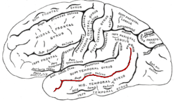
The inferior temporal gyrus is one of three gyri of the temporal lobe and is located below the middle temporal gyrus, connected behind with the inferior occipital gyrus; it also extends around the infero-lateral border on to the inferior surface of the temporal lobe, where it is limited by the inferior sulcus. This region is one of the higher levels of the ventral stream of visual processing, associated with the representation of objects, places, faces, and colors. It may also be involved in face perception, and in the recognition of numbers.
Visual search is a type of perceptual task requiring attention that typically involves an active scan of the visual environment for a particular object or feature among other objects or features. Visual search can take place with or without eye movements. The ability to consciously locate an object or target amongst a complex array of stimuli has been extensively studied over the past 40 years. Practical examples of using visual search can be seen in everyday life, such as when one is picking out a product on a supermarket shelf, when animals are searching for food among piles of leaves, when trying to find a friend in a large crowd of people, or simply when playing visual search games such as Where's Wally?
Nancy Gail Kanwisher FBA is the Ellen Swallow Richards Professor in the Department of Brain and Cognitive Sciences at the Massachusetts Institute of Technology and an investigator at the McGovern Institute for Brain Research. She studies the neural and cognitive mechanisms underlying human visual perception and cognition.

The colour centre is a region in the brain primarily responsible for visual perception and cortical processing of colour signals received by the eye, which ultimately results in colour vision. The colour centre in humans is thought to be located in the ventral occipital lobe as part of the visual system, in addition to other areas responsible for recognizing and processing specific visual stimuli, such as faces, words, and objects. Many functional magnetic resonance imaging (fMRI) studies in both humans and macaque monkeys have shown colour stimuli to activate multiple areas in the brain, including the fusiform gyrus and the lingual gyrus. These areas, as well as others identified as having a role in colour vision processing, are collectively labelled visual area 4 (V4). The exact mechanisms, location, and function of V4 are still being investigated.

The fusiform face area is a part of the human visual system that is specialized for facial recognition. It is located in the inferior temporal cortex (IT), in the fusiform gyrus.
In cognitive neuroscience, visual modularity is an organizational concept concerning how vision works. The way in which the primate visual system operates is currently under intense scientific scrutiny. One dominant thesis is that different properties of the visual world require different computational solutions which are implemented in anatomically/functionally distinct regions that operate independently – that is, in a modular fashion.
Discrete categories of objects such as faces, body parts, tools, animals and buildings have been associated with preferential activation in specialised areas of the cerebral cortex, leading to the suggestion that they may be produced separately in discrete neural regions.
The superior temporal sulcus (STS) is the sulcus separating the superior temporal gyrus from the middle temporal gyrus in the temporal lobe of the brain. A sulcus is a deep groove that curves into the largest part of the brain, the cerebrum, and a gyrus is the a ridge that curves outward of the cerebrum.
Visual object recognition refers to the ability to identify the objects in view based on visual input. One important signature of visual object recognition is "object invariance", or the ability to identify objects across changes in the detailed context in which objects are viewed, including changes in illumination, object pose, and background context.

Functional specialization suggests that different areas in the brain are specialized for different functions.
The N170 is a component of the event-related potential (ERP) that reflects the neural processing of faces, familiar objects or words. Furthermore, the N170 is modulated by prediction error processes.
The neural basis of self is the idea of using modern concepts of neuroscience to describe and understand the biological processes that underlie humans' perception of self-understanding. The neural basis of self is closely related to the psychology of self with a deeper foundation in neurobiology.
Biased competition theory advocates the idea that each object in the visual field competes for cortical representation and cognitive processing. This theory suggests that the process of visual processing can be biased by other mental processes such as bottom-up and top-down systems which prioritize certain features of an object or whole items for attention and further processing. Biased competition theory is, simply stated, the competition of objects for processing. This competition can be biased, often toward the object that is currently attended in the visual field, or alternatively toward the object most relevant to behavior.
In psychology, the face superiority effect refers to the phenomena of how all individuals perceive and encode other human faces in memory. Rather than perceiving and encoding single features of a face, we perceive and encode a human face as one holistic unified element. This phenomenon aids our visual system in the recognition of thousands of faces, a task that would be difficult if it were necessary to recognize sets of individual features and characteristics. However, this effect is limited to perceiving upright faces and does not occur when a face is at an unusual angle, such as when faces are upside-down or contorted in phenomena like the Thatcher effect and Pareidolia.
The Fusiform body area (FBA) is a part of the extrastriate visual cortex, an object representation system involved in the visual processing of human bodies in contrast to body parts. Its function is similar to but distinct from the extrastriate body area (EBA), which perceives bodies in relation body parts, and the fusiform face area (FFA), which is involved in the perception of faces. Marius Peelen and Paul Downing identified this brain region in 2004 through an fMRI study.; in 2005 Rebecca Schwarzlose and a team of cognitive researchers named this brain region the fusiform body area.
The occipital face area (OFA) is a region of the human cerebral cortex which is specialised for face perception. The OFA is located on the lateral surface of the occipital lobe adjacent to the inferior occipital gyrus. The OFA comprises a network of brain regions including the fusiform face area (FFA) and posterior superior temporal sulcus (STS) which support facial processing.

