Related Research Articles

The cingulate cortex is a part of the brain situated in the medial aspect of the cerebral cortex. The cingulate cortex includes the entire cingulate gyrus, which lies immediately above the corpus callosum, and the continuation of this in the cingulate sulcus. The cingulate cortex is usually considered part of the limbic lobe.

In the human brain, the anterior cingulate cortex (ACC) is the frontal part of the cingulate cortex that resembles a "collar" surrounding the frontal part of the corpus callosum. It consists of Brodmann areas 24, 32, and 33.

In neuroanatomy, the precuneus is the portion of the superior parietal lobule on the medial surface of each brain hemisphere. It is located in front of the cuneus. The precuneus is bounded in front by the marginal branch of the cingulate sulcus, at the rear by the parieto-occipital sulcus, and underneath by the subparietal sulcus. It is involved with episodic memory, visuospatial processing, reflections upon self, and aspects of consciousness.

The frontal lobe is the largest of the four major lobes of the brain in mammals, and is located at the front of each cerebral hemisphere. It is parted from the parietal lobe by a groove between tissues called the central sulcus and from the temporal lobe by a deeper groove called the lateral sulcus. The most anterior rounded part of the frontal lobe is known as the frontal pole, one of the three poles of the cerebrum.

Brodmann area 9, or BA9, refers to a cytoarchitecturally defined portion of the frontal cortex in the brain of humans and other primates. It contributes to the dorsolateral and medial prefrontal cortex.
A mental image is an experience that, on most occasions, significantly resembles the experience of 'perceiving' some object, event, or scene, but occurs when the relevant object, event, or scene is not actually present to the senses. There are sometimes episodes, particularly on falling asleep and waking up, when the mental imagery may be dynamic, phantasmagoric and involuntary in character, repeatedly presenting identifiable objects or actions, spilling over from waking events, or defying perception, presenting a kaleidoscopic field, in which no distinct object can be discerned. Mental imagery can sometimes produce the same effects as would be produced by the behavior or experience imagined.

The posterior cingulate cortex (PCC) is the caudal part of the cingulate cortex, located posterior to the anterior cingulate cortex. This is the upper part of the "limbic lobe". The cingulate cortex is made up of an area around the midline of the brain. Surrounding areas include the retrosplenial cortex and the precuneus.
Lie detection is an assessment of a verbal statement with the goal to reveal a possible intentional deceit. Lie detection may refer to a cognitive process of detecting deception by evaluating message content as well as non-verbal cues. It also may refer to questioning techniques used along with technology that record physiological functions to ascertain truth and falsehood in response. The latter is commonly used by law enforcement in the United States, but rarely in other countries because it is based on pseudoscience.
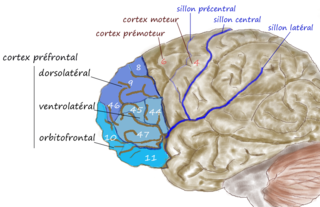
The dorsolateral prefrontal cortex is an area in the prefrontal cortex of the primate brain. It is one of the most recently derived parts of the human brain. It undergoes a prolonged period of maturation which lasts into adulthood. The DLPFC is not an anatomical structure, but rather a functional one. It lies in the middle frontal gyrus of humans. In macaque monkeys, it is around the principal sulcus. Other sources consider that DLPFC is attributed anatomically to BA 9 and 46 and BA 8, 9 and 10.

In neuroscience, the default mode network (DMN), also known as the default network, default state network, or anatomically the medial frontoparietal network (M-FPN), is a large-scale brain network primarily composed of the dorsal medial prefrontal cortex, posterior cingulate cortex/precuneus and angular gyrus. It is best known for being active when a person is not focused on the outside world and the brain is at wakeful rest, such as during daydreaming and mind-wandering. It can also be active during detailed thoughts related to external task performance. Other times that the DMN is active include when the individual is thinking about others, thinking about themselves, remembering the past, and planning for the future.
The biology of obsessive–compulsive disorder (OCD) refers biologically based theories about the mechanism of OCD. Cognitive models generally fall into the category of executive dysfunction or modulatory control. Neuroanatomically, functional and structural neuroimaging studies implicate the prefrontal cortex (PFC), basal ganglia (BG), insula, and posterior cingulate cortex (PCC). Genetic and neurochemical studies implicate glutamate and monoamine neurotransmitters, especially serotonin and dopamine.
The neural basis of self is the idea of using modern concepts of neuroscience to describe and understand the biological processes that underlie humans' perception of self-understanding. The neural basis of self is closely related to the psychology of self with a deeper foundation in neurobiology.

The neuroscience of sex differences is the study of characteristics that separate the male and female brain. Psychological sex differences are thought by some to reflect the interaction of genes, hormones, and social learning on brain development throughout the lifespan.
The dorsal nexus is an area within the dorsal medial prefrontal cortex that serves as an intersection point for multiple brain networks. Research suggests it plays a role in the maintenance and manipulation of information, as well as supporting the control of cognitive functions such as behavior, memory, and conflict resolution. Abnormally increased connectivity between these networks through the Dorsal Nexus has been associated with certain types of depression. The activity generated by this abnormally high level of connectivity during a depressive state can be identified through Magnetic resonance imaging (MRI) and Positron emission tomography (PET).
Cognitive humor processing refers to the neural circuitry and pathways that are involved in detecting incongruities of various situations presented in a humorous manner. Over the past decade, many studies have emerged utilizing fMRI studies to describe the neural correlates associated with how a human processes something that is considered "funny". Conceptually, humor is subdivided into two elements: cognitive and affective. The cognitive element, known as humor detection, refers to understanding the joke. Usually, this is characterized by the perceiver attempting to comprehend the disparities between the punch line and prior experience. The affective element, otherwise known as humor appreciation, is involved with enjoying the joke and producing visceral, emotional responses depending on the hilarity of the joke. This ability to comprehend and appreciate humor is a vital aspect of social functioning and is a significant part of the human condition that is relevant from a very early age. Humor comprehension develops in parallel with growing cognitive and language skills during childhood, while its content is mostly influenced by social and cultural factors. A further approach is described which refers to humor as an attitude related to strains. Humorous responses when confronted with troubles are discussed as a skill often associated with high social competence. The concept of humor has also been shown to have therapeutic effects, improving physiological systems such as the immune and central nervous system. It also has been shown to help cope with stress and pain. In sum, humor proves to be a personal resource throughout the life span, and helps support the coping of everyday tasks.
Daniel Langleben is a psychiatrist, professor, and scientific researcher. He pioneered a technique for using functional magnetic resonance imaging (fMRI) as a means of lie detection. He has also studied the brain effects of packaging and advertising and how infants' cuteness motivates caretaking in adults.
An identity disturbance is a deficiency or inability to maintain one or more major components of identity. These components include a sense of continuity over time; emotional commitment to representations of self, role relationships, core values and self-standards; development of a meaningful world view; and recognition of one's place in the world.
Neuromorality is an emerging field of neuroscience that studies the connection between morality and neuronal function. Scientists use fMRI and psychological assessment together to investigate the neural basis of moral cognition and behavior. Evidence shows that the central hub of morality is the prefrontal cortex guiding activity to other nodes of the neuromoral network. A spectrum of functional characteristics within this network to give rise to both altruistic and psychopathological behavior. Evidence from the investigation of neuromorality has applications in both clinical neuropsychiatry and forensic neuropsychiatry.
Meditation and pain is the study of the physiological mechanisms underlying meditation-specifically its neural components- that implicate it in the reduction of pain perception.
Social cognitive neuroscience is the scientific study of the biological processes underpinning social cognition. Specifically, it uses the tools of neuroscience to study "the mental mechanisms that create, frame, regulate, and respond to our experience of the social world". Social cognitive neuroscience uses the epistemological foundations of cognitive neuroscience, and is closely related to social neuroscience. Social cognitive neuroscience employs human neuroimaging, typically using functional magnetic resonance imaging (fMRI). Human brain stimulation techniques such as transcranial magnetic stimulation and transcranial direct-current stimulation are also used. In nonhuman animals, direct electrophysiological recordings and electrical stimulation of single cells and neuronal populations are utilized for investigating lower-level social cognitive processes.
References
- ↑ Silberman, Steve (2006). "Don't Even Think About Lying". Wired. pp. Issue 14.01. Retrieved 9 July 2014.
- ↑ Prospect: Politics, Essay, Review. C. Seaford. October 2009.
- ↑ Zack Lynch; Byron Laursen (21 July 2009). The Neuro Revolution: How Brain Science Is Changing Our World . St. Martin's Press. pp. 29. ISBN 978-1-4299-8823-0.
- ↑ Bonnier Corporation (August 2002). "Popular Science". The Popular Science Monthly. Bonnier Corporation: 58. ISSN 0161-7370.
- ↑ Boy Scouts of America, Inc. (January 2005). "Boys' Life". Boys' Life. Inkprint Edition. Boy Scouts of America, Inc.: 11. ISSN 0006-8608.
- ↑ Committee on Science, Technology, Law (26 September 2011). Reference Manual on Scientific Evidence:: Third Edition. National Academies Press. p. 803. ISBN 978-0-309-21421-6.
{{cite book}}: CS1 maint: multiple names: authors list (link) - ↑ Allan Pease; Barbara Pease (1 January 2004). Why Men Don't Have a Clue and Women Always Need More Shoes: The Ultimate Guide to the Opposite Sex . Broadway Books. pp. 272. ISBN 978-0-7679-1610-3.
- ↑ Langleben, Daniel (2002). "Brain activity during simulated deception: an event-related functional magnetic resonance study". NeuroImage. 15 (3): 727–32. doi:10.1006/nimg.2001.1003. PMID 11848716. S2CID 14676750.
- 1 2 3 4 5 6 7 8 9 Rusconi, Elena; Mitchener-Nissen, Timothy (2013). "Prospects of functional magnetic resonance imaging as lie detector". Frontiers in Human Neuroscience. 7: 594. doi: 10.3389/fnhum.2013.00594 . PMC 3781577 . PMID 24065912.
- ↑ Simpson JR (2008). "Functional MRI lie detection: too good to be true?". J. Am. Acad. Psychiatry Law. 36 (4): 491–498. PMID 19092066.
- ↑ Douglas PK (2011). "Performance comparison of machine learning algorithms and number of independent components used in fMRI decoding of belief vs. disbelief". NeuroImage. 56 (2): 544–553. doi:10.1016/j.neuroimage.2010.11.002. PMC 3099263 . PMID 21073969.
- 1 2 3 4 Ganis, G.; Kosslyn, S. M.; Stose, S.; Thompson, W. L.; Yurgelun-Todd, D. A. (2003). "Neural Correlates of Different Types of Deception: An fMRI Investigation". Cerebral Cortex. 13 (8): 830–836. doi: 10.1093/cercor/13.8.830 . PMID 12853369.
- 1 2 Langleben, Daniel D.; Loughead, James W.; Bilker, Warren B.; Ruparel, Kosha; Childress, Anna Rose; Busch, Samantha I.; Gur, Ruben C. (2005). "Telling truth from lie in individual subjects with fast event-related fMRI". Human Brain Mapping. 26 (4): 262–272. doi:10.1002/hbm.20191. PMC 6871667 . PMID 16161128.
- 1 2 3 Langleben, D. D.; Dattilio, F. M. (2008). "Commentary: The future of forensic functional brain imaging". The Journal of the American Academy of Psychiatry and the Law. 36 (4): 502–4. PMID 19092068.
- ↑ Langleben, Daniel D.; Moriarty, Jane Campbell (2013-05-01). "Using Brain Imaging for Lie Detection: Where Science, Law and Research Policy Collide". Psychology, Public Policy, and Law. 19 (2): 222–234. doi:10.1037/a0028841. ISSN 1076-8971. PMC 3680134 . PMID 23772173.
- ↑ Abe, Nobuhito (December 2008). "Neural Correlates of True Memory, False Memory, and Deception" (PDF). Cerebral Cortex. 18 (12): 2811–2819. doi:10.1093/cercor/bhn037. PMC 2583150 . PMID 18372290. Archived from the original (PDF) on 2011-01-14.
- ↑ Farah, Martha; Hutchinson, J. Benjamin; Phelps, Elizabeth; Wagner, Anthony (2014-01-01). "Functional MRI-Based Lie Detection: Scientific and Societal Challenges". Nature Reviews Neuroscience. 15 (2): 123–131. doi:10.1038/nrn3665. PMID 24588019. S2CID 8480199.