Femoroacetabular impingement

Last updated

Femoroacetabular impingement
Other namesHip impingement syndrome,
The three recognized types of femoroacetabular impingement. Click image to see video illustration of the three types.
Specialty Orthopedic

Femoroacetabular impingement (FAI) is a condition involving one or more anatomical abnormalities of the hip joint, which is a ball and socket joint. [1] It is a common cause of hip pain and discomfort in young and middle-aged adults. [2] It occurs when the ball shaped femoral head contacts the acetabulum abnormally or does not permit a normal range of motion in the acetabular socket. [3] Damage can occur to the articular cartilage, or labral cartilage (soft tissue, ring-shaped bumper of the socket), or both. [4] The condition may be symptomatic or asymptomatic. It may cause osteoarthritis of the hip. [1] Treatment options range from conservative management to surgery. [5]

Contents

Signs and symptoms

Pain is the most common complaint in those with FAI. [6] It is experienced in a number of areas, making the diagnosis challenging, but commonly occurs in the groin, upper buttock/lower back, the buttock or beneath the buttock, side of the affected hip and posterior upper leg. [6] [3] [7] Onset of symptoms has been reported to present in both an acute and more gradual manner. [6] The pain is often significant enough to cause a decrease in activity level and movement. [6] Some will also describe decreased range of motion of the affected hip. [8] Another symptom is groin pain associated with activity and no prior history of trauma. [9] Inability to perform activities such as high hip flexion or prolong sitting can also be seen in individuals with FAI. [9]

Cause

FAI is characterized by abnormal contact between the proximal femur and rim of the acetabulum (hip socket). In most cases, patients present with a deformity in the femoral head, or acetabulum, a poorly positioned femoral-acetabular junction, or any or all of the foregoing. [3] The cause of FAI is currently unknown, but both congenital and acquired etiologies have been put forth. Studies have shown an increased incidence in siblings, suggesting a genetic component. [10] At least one study has also shown a predilection in the white population. [10] It has also been reported to be more common in males. [10] However, there is no concrete evidence to suggest a genetic trait and instead, the most favored theory currently supports that FAI (the cam type in particular) is due to repetitive movements involving the hip (e.g. squatting) in young athletes. [10] Aggravating activities that are commonly reported include repetitive or prolonged squatting, twisting movements of the hip, like pivoting during athletics, getting in and out of cars, and even sitting for prolonged periods. [6] [11] A combination of these factors may also predispose to a form of FAI; predominantly, a marginal developmental hip abnormality together with environmental factors such as recurrent motion of the legs within a supraphysiologic range. [12]

Three types of FAI are recognized (see title image). The first involves an excess of bone along the upper surface of the femoral head, known as a cam deformity (abbreviation for camshaft, which the shape of the femoral head and neck resembles). The second is due to an excess of growth of the upper lip of the acetabular cup and is known as a 'pincer' deformity. The third is a combination of the two, generally referred to as 'mixed.' The most common type seen, approximately 70% of the time, is the mixed type. [13] A complicating issue is that some of the radiographic findings of FAI have also been described in asymptomatic subjects. [14]

Current literature suggests that the cam type of impingement is associated with the development of hip osteoarthritis. [15] Thus far, no correlation has been seen between the pincer type and development of hip osteoarthritis. [15]

Anatomy

The hip joint is classified as a ball and socket joint. This type of synovial joint allows for multidirectional movement and rotation. There are two bones that make up the hip joint and create an articulation between the femur and pelvis. This articulation connects the axial skeleton with the lower extremity. The pelvic bone, also known as the innominate bone, is formed by three bones fused together: the ilium, ischium, and pubis. The musculature of the hip is divided into anterior hip muscles and posterior hip muscles. The major nerve supply that runs through the hip joint is the femoral nerve and the sciatic nerve. [16]

Diagnosis

Clinical evaluation is the first step in diagnosis, but will rarely lead to the diagnosis on its own, due to inconsistent and vague nature of the pain. [10] Childhood and current activity should be inquired about. Physical exam should also involve assessing passive internal rotation of the hip during flexion, as range of motion is reduced in proportion to the size of a cam lesion. [10] Flexing the hip to 90 degrees, adducting, and internally rotating the hip, known as the FADDIR test, should also be performed. [10] It is positive when it causes pain. The FABER test should also be performed, this test involves flexing, abducting, and externally rotating the hip. The FABER test is useful when diagnosing concurrently with a labral pathology and is considered positive if the position elicits pain. [17] Additional non-invasive ways to observe possible FAI is changes in gait that include a lower peak hip extension and internal rotation to compensate for bony growth. [18]

X-ray

Radiograph of a cam type impingement. X-ray of cam.jpg
Radiograph of a cam type impingement.
Radiograph of a pincer type impingement. X-ray of pincer impingement.jpg
Radiograph of a pincer type impingement.

Projectional radiography ("X-ray")is often considered first line for FAI. [10] Anterior-posterior pelvis and a lateral image of the hip in question should be attained. [10] A 45-degree Dunn view is also recommended. [10] [19]

Measurements of impingement on X-ray. [notes 1] [20]
MeasurementImageTargetNormal value
Center-edge angle of Wiberg
Center-edge angle of Wiberg.jpg
The superior-lateral coverage of the femoral head.
  • >20° (<55 years old) [notes 2]
  • <24° (>55 years old) [notes 2]
  • >40° indicates overcoverage
Crossing ratio Crossing ratio of the hip.jpg Percentage of acetabular walls crossing. Normal acetabulum is oriented in anteversion. Its value ranges from 15 to 20° in the equatorial plane of the acetabulum and decreases gradually towards the acetabular roof, where normal values range from 0 to 5°. Retroversion of the upper part of the acetabulum has been related with pincer type impingement. In radiography the presence of a "crossover sign" is produced when the posterior wall of the acetabulum crosses the anterior wall before reaching the acetabular roof. It is a sign of acetabular retroversion and it has been linked with overcoverage and pincer impingement. Nevertheless, this sign has been described in 6% of the normal population. Therefore, more important than its presence is the percentage of crossing.<20%
  • Higher is significant crossing
Alpha angle
Alpha angle measured in 45 degrees Dunn view.jpg
Measured in 45° Dunn view.
Degree of bulging of the femoral head-neck junction: In normal conditions there is a symmetric concave contour at the junction of the femoral head and neck. Loss of this concavity or bone bulging may lead to cam type impingement. The degree of this deformity can be measured by the alpha angle. Although it can be measured in the cross-lateral view, the 45° Dunn view is considered more sensitive and the frog leg view more specific in determining pathologic values.
  • Normal: ≤68° in men, ≤50° in women
  • Borderline: 69° to 82° in men, 51° to 56° in women
  • Pathological: ≥83° in men and ≥57° in women
Femoral head-neck offset
Hip offset percentage measured in cross-lateral view.jpg
Measured in cross-lateral view.
Offset of the femoral head with regard to most prominent aspect of the femora neck>10 mm
Offset percentageFemoral head-neck offset related to femoral head diameter>0.18
  • less indicates high risk of cam type impingement
Tönnis angle Tonnis angle of the hip.jpg Slope of the sourcil (the sclerotic weight-bearing portion of the acetabulum)0 to 10°
  • >10° is a risk factor for instability
  • <0° is a risk factor for pincer impingement
Caput-sourcil angle [21] Caput-sourcil angle.jpg Superior to the Tönnis angle in cases without joint space narrowing or subluxation. [21] The medial point of the sourcil is at the same height as the most superior point of caput femoris.−6 to 12° [21]
  • >12° is a risk factor for instability
  • <-6° is a risk factor for pincer impingement

Other modalities

MRI imaging may follow, particularly if there is no specific evidence on radiographs, producing a three-dimensional reconstruction of the joint for better definition, to evaluate the hip cartilage, or measure hip socket angles (e.g. the alpha-angle as described by Nötzli [22] in 2-D and by Siebenrock in 3-D [23] ). MR arthrogram had been used in the past, as it was more sensitive for picking up soft tissue lesions; however, due to improvement in technology, MRI is now considered comparable for picking up such lesions. [6] CT is not usually used due to radiation exposure and no benefit above MRI. [6] It is possible to perform dynamic simulation of hip motion with CT or MRI [24] [25] assisting to establish whether, where, and to what extent, impingement is occurring.

The diagnosis is often made in conjunction with a labral tear. [26]

Differential Diagnosis

Source: [8]

Prevention

Prevention is currently being investigated. [27] The goal of prevention would be to avoid joint damage and premature hip osteoarthritis. [27] Studies are examining the effectiveness of screening adolescents in school and targeting at-risk individuals for education, physical therapy and decreasing participation in possibly harmful activities/sports as referenced in the epidemiology section. [27]

Treatment

Treatment of FAI can be divided into those that are non-operative (conservative) and operative. Conservative treatment is often prescribed for those who have not yet received any therapy. [28] Conservative treatment includes physical therapy, avoidance of those activities that produce pain, and nonsteroidal anti-inflammatory drugs. [6] It may also include joint injections with cortisone or hyaluronic acid, particularly for those who wish to avoid surgery. [6]

Physical therapy is implemented for the purpose of improving joint mobility, strengthening muscles surrounding the joint, correcting posture, and treating any other muscle or joint deficits that may be exacerbating the condition. [11] A movement analysis may also be performed to identify specific movement patterns that may be causing injury. [29] Studies to demonstrate the effectiveness of physical therapy are currently underway, with no conclusive results to date. [28]

Operative treatment is generally recommended to those who continue to have symptoms. It involves the surgical correction of any bony abnormalities causing the impingement and correction of any soft tissue lesions, such as labral tears. [6] The primary aim of surgery is to correct the fit of the femoral head and acetabulum to create a hip socket that reduces contact between the two, allowing a greater range of movement. [30] This includes femoral head sculpting and/or trimming of the acetabular rim. [30] [31]

Surgery may be arthroscopic or open. [7] A 2011 study analyzing current surgical methods for management of symptomatic femoral acetabular impingement, suggested that the arthroscopic method had surgical outcomes equal to or better than other methods with a lower rate of major complications when performed by experienced surgeons; [32] consequently, the surgery is now rarely done open. [33]

Outcomes of arthroscopic surgery are currently being studied, but have generally been positive. According to a 2019 meta-analysis, the risk of having surgery fail or need to be re-operated on is about 5.5% whereas the complication rate is 1.7%. [34] Additionally, patient reported outcomes show that approximately three to six months post-operative hip arthroscopy is when pain reduction and activities of daily life are improved. For sport function this timeline is about six months to a year. [35] Failure of hip arthroscopy is more likely to fail in older patients, females, or those who have experienced the symptoms of FAI for a long period of time. [34]

When performed on elite athletes, most are able to return their previous level of competition. [10] These athletes also have a higher rate of return to sport than recreational and collegiate athletes. [36]

Long term, randomized controlled trials evaluating the efficacy of conservative and operative treatments are underway. [37]

Epidemiology

There has been limited research on the prevalence of FAI among a general population according to a literature review by Algarni. [38] On the other hand, there are many sources that discuss the prevalence of athletes with the condition especially those that are younger and white. [39] Hockey, tennis, soccer, and equestrian are all sports where the prevalence of a femoral neck abnormalities are higher due to the nature of the sport to force athletes into forced, loaded flexion and internal rotation. [38]

Cam lesions are more common in males, where pincer lesions are more common in females due to differences in anatomical development of the pelvis. [17]

History

FAI is a relatively recent discovery. Its original description is credited to orthopedic surgeon Dr. Reinhold Ganz, who first proposed the condition as a cause for hip osteoarthritis in a publication in 2003. [40] [41]

While the true diagnosis of FAI can be considered a relatively recent discovery, reports of damage to the femoroaceatabular region date back over a century ago in the orthopedic realm of medicine. It was not until the development of an open surgical dislocation procedure was developed that FAI was discovered as an anatomical difference and cause of osteoarthritis. Orthopedic surgeon Dr. Reinhold Ganz can be credited with this discovery in his 2003 publication that discussed the findings and relation to hip osteoarthritis. [10]

Society

Notable persons who have had hip impingement:

Notes

  1. Unless otherwise specified in boxes, reference is the one marked in header.
  2. 1 2 This can also be used in children. At between 5 and 10 years, the minimum normal value is 15°.

Related Research Articles

<span class="mw-page-title-main">Acetabulum</span> Cavity where the thigh bone (femur) articulates with the pelvis

The acetabulum also called the cotyloid cavity, is a concave surface of the pelvis. The head of the femur meets with the pelvis at the acetabulum, forming the hip joint.

<span class="mw-page-title-main">Arthroscopy</span> Medical procedure

Arthroscopy is a minimally invasive surgical procedure on a joint in which an examination and sometimes treatment of damage is performed using an arthroscope, an endoscope that is inserted into the joint through a small incision. Arthroscopic procedures can be performed during ACL reconstruction.

<span class="mw-page-title-main">Orthopedic surgery</span> Branch of surgery concerned with the musculoskeletal and bones system

Orthopedic surgery or orthopedics is the branch of surgery concerned with conditions involving the musculoskeletal system. Orthopedic surgeons use both surgical and nonsurgical means to treat musculoskeletal trauma, spine diseases, sports injuries, degenerative diseases, infections, tumors, and congenital disorders.

<span class="mw-page-title-main">Osteoarthritis</span> Form of arthritis caused by degeneration of joints

Osteoarthritis (OA) is a type of degenerative joint disease that results from breakdown of joint cartilage and underlying bone. It is believed to be the fourth leading cause of disability in the world, affecting 1 in 7 adults in the United States alone. The most common symptoms are joint pain and stiffness. Usually the symptoms progress slowly over years. Other symptoms may include joint swelling, decreased range of motion, and, when the back is affected, weakness or numbness of the arms and legs. The most commonly involved joints are the two near the ends of the fingers and the joint at the base of the thumbs, the knee and hip joints, and the joints of the neck and lower back. The symptoms can interfere with work and normal daily activities. Unlike some other types of arthritis, only the joints, not internal organs, are affected.

An osteotomy is a surgical operation whereby a bone is cut to shorten or lengthen it or to change its alignment. It is sometimes performed to correct a hallux valgus, or to straighten a bone that has healed crookedly following a fracture. It is also used to correct a coxa vara, genu valgum, and genu varum. The operation is done under a general anaesthetic.

<span class="mw-page-title-main">Hip replacement</span> Surgery replacing hip joint with prosthetic implant

Hip replacement is a surgical procedure in which the hip joint is replaced by a prosthetic implant, that is, a hip prosthesis. Hip replacement surgery can be performed as a total replacement or a hemi/semi(half) replacement. Such joint replacement orthopaedic surgery is generally conducted to relieve arthritis pain or in some hip fractures. A total hip replacement consists of replacing both the acetabulum and the femoral head while hemiarthroplasty generally only replaces the femoral head. Hip replacement is one of the most common orthopaedic operations, though patient satisfaction varies widely. Approximately 58% of total hip replacements are estimated to last 25 years. The average cost of a total hip replacement in 2012 was $40,364 in the United States, and about $7,700 to $12,000 in most European countries.

<span class="mw-page-title-main">Hip</span> Anatomical region between the torso and the legs, holding the buttocks and genital region

In vertebrate anatomy, the hip, or coxa(pl.: coxae) in medical terminology, refers to either an anatomical region or a joint on the outer (lateral) side of the pelvis.

<span class="mw-page-title-main">SLAP tear</span> Medical condition

A SLAP tear or SLAP lesion is an injury to the superior glenoid labrum that initiates in the back of the labrum and stretches toward the front into the attachment point of the long head of the biceps tendon. SLAP is an acronym for "Superior Labrum Anterior and Posterior". SLAP lesions are commonly seen in overhead throwing athletes but middle-aged labor workers can also be affected, and they can be caused by chronic overuse or an acute stretch injury of the shoulder.

Articular cartilage, most notably that which is found in the knee joint, is generally characterized by very low friction, high wear resistance, and poor regenerative qualities. It is responsible for much of the compressive resistance and load bearing qualities of the knee joint and, without it, walking is painful to impossible. Osteoarthritis is a common condition of cartilage failure that can lead to limited range of motion, bone damage and invariably, pain. Due to a combination of acute stress and chronic fatigue, osteoarthritis directly manifests itself in a wearing away of the articular surface and, in extreme cases, bone can be exposed in the joint. Some additional examples of cartilage failure mechanisms include cellular matrix linkage rupture, chondrocyte protein synthesis inhibition, and chondrocyte apoptosis. There are several different repair options available for cartilage damage or failure.

<span class="mw-page-title-main">Zona orbicularis</span>

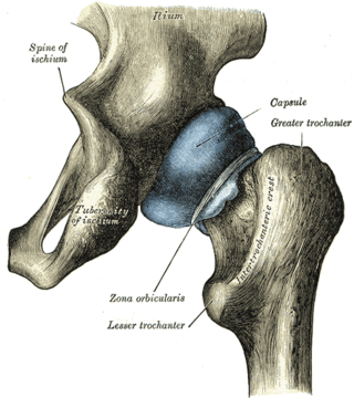
The zona orbicularis or annular ligament is a ligament on the neck of the femur formed by the circular fibers of the articular capsule of the hip joint. It is also known as the orbicular zone, ring ligament, and zonular band.

<span class="mw-page-title-main">Acetabular labrum</span> Ring of cartilage that surrounds the acetabulum of the hip

The acetabular labrum is a fibrocartilaginous ring which surrounds the circumference of the acetabulum of the hip, deepening the acetabulum. The labrum is attached onto the bony rim and transverse acetabular ligament. It is triangular in cross-section.

<span class="mw-page-title-main">Extracorporeal shockwave therapy</span> Ultrasonic, non-invasive, outpatient treatment

Extracorporeal shockwave therapy (ESWT) is a non-invasive, out-patient alternative to surgery for those with many joint and tendon disorders. ESWT sends acoustic shock waves into bone or soft tissue, in effect reinjuring the area on a cellular level and breaking up the scarring that has penetrated tendons and ligaments. The controlled reinjuring of tissue allows the body to regenerate blood vessels and bone cells. The resulting revascularization leads to faster healing and often a return to pre-injury activity levels. ESWT is mostly used for kidney stones removal, in physical therapy and orthopedics.

<span class="mw-page-title-main">Hip dysplasia</span> Joint abnormality

Hip dysplasia is an abnormality of the hip joint where the socket portion does not fully cover the ball portion, resulting in an increased risk for joint dislocation. Hip dysplasia may occur at birth or develop in early life. Regardless, it does not typically produce symptoms in babies less than a year old. Occasionally one leg may be shorter than the other. The left hip is more often affected than the right. Complications without treatment can include arthritis, limping, and low back pain. Females are affected more often than males. Risk factors for hip dysplasia include female sex, family history, certain swaddling practices, and breech presentation whether an infant is delivered vaginally or by cesarean section. If one identical twin is affected, there is a 40% risk the other will also be affected. Screening all babies for the condition by physical examination is recommended. Ultrasonography may also be useful.

<span class="mw-page-title-main">Knee pain</span> Medical condition

Knee pain is pain in or around the knee.

<span class="mw-page-title-main">Hip arthroscopy</span>

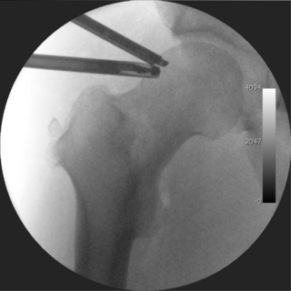
Hip arthroscopy refers to the viewing of the interior of the acetabulofemoral (hip) joint through an arthroscope and the treatment of hip pathology through a minimally invasive approach. This technique is sometimes used to help in the treatment of various joint disorders and has gained popularity because of the small incisions used and shorter recovery times when compared with conventional surgical techniques. Hip arthroscopy was not feasible until recently, new technology in both the tools used and the ability to distract the hip joint has led to a recent surge in the ability to do hip arthroscopy and the popularity of it.

Osteoplasty is the branch of surgery concerned with bone repair or bone grafting. It is the surgical alteration or reshaping of bone. It may be used to relieve pain associated with metastatic bone disease. Percutaneous osteoplasty involves the use of bone cement to reduce pain and improve mobility. Resection osteoplasty is used in joint preserving surgery on the hip and thigh bones.

Radiographic systems to classify osteoarthritis vary by which joint is being investigated. In osteoarthritis, the choice of treatment is based on pain and decreased function, but radiography can be useful before surgery in order to prepare for the procedure.

<span class="mw-page-title-main">Hip pain</span>

Pain in the hip is the experience of pain in the muscles or joints in the hip/ pelvic region, a condition commonly arising from any of a number of factors. Sometimes it is closely associated with lower back pain.

<span class="mw-page-title-main">Labral reconstruction</span> Medical procedure

Labral reconstruction is a type of hip arthroscopy in which the patient's native labrum is partially or completely removed and reconstructed using either autograft or allograft tissue. Originally described in 2009 using the ligamentum teres capitis, arthroscopic labral reconstruction using a variety of graft tissue has demonstrated promising short and mid-term clinical outcomes. Most importantly, labral reconstruction has demonstrated utility when the patient's native labral tissue is far too damaged for debridement or repair.

<span class="mw-page-title-main">Ultrasound-guided hip joint injection</span>

Ultrasound-guided hip joint injection is a joint injection in the hip, assisted by medical ultrasound. Hip and groin pain often presents a diagnostic and therapeutic challenge. The differential diagnosis is extensive, comprising intra-articular and extra-articular pathology and referred pain from lumbar spine, knee and elsewhere in the pelvis. Various ultrasound-guided techniques have been described in the hip and groin region for diagnostic and therapeutic purposes. Ultrasound has many advantages over other imaging modalities, including portability, lack of ionizing radiation and real-time visualization of soft tissues and neurovascular structures. Many studies have demonstrated the safety, accuracy and efficacy of ultrasound-guided techniques, although there is lack of standardization regarding the injectates used and long-term benefit remains uncertain.

References

  1. 1 2 Pun S, Kumar D, Lane NE (January 2015). "Femoroacetabular impingement". Arthritis & Rheumatology. 67 (1): 17–27. doi:10.1002/art.38887. PMC   4280287 . PMID   25308887.
  2. Dooley PJ (January 2008). "Femoroacetabular impingement syndrome: Nonarthritic hip pain in young adults". Canadian Family Physician. 54 (1): 42–7. PMC   2293316 . PMID   18208954.
  3. 1 2 3 Banerjee P, McLean CR (March 2011). "Femoroacetabular impingement: a review of diagnosis and management". Current Reviews in Musculoskeletal Medicine. 4 (1): 23–32. doi:10.1007/s12178-011-9073-z. PMC   3070009 . PMID   21475562.
  4. Agricola R, Weinans H (February 2016). "What is femoroacetabular impingement?". British Journal of Sports Medicine. 50 (4): 196–7. doi:10.1136/bjsports-2015-094766. PMID   26130699. S2CID   12149232.
  5. Egger, Anthony C.; Frangiamore, Salvatore; Rosneck, James (2016). "Wolters Kluwer Health - Article Landing Page". Sports Medicine and Arthroscopy Review. 24 (4): e53–e58. doi:10.1097/jsa.0000000000000126. PMID   27811519.
  6. 1 2 3 4 5 6 7 8 9 10 Menge TJ, Truex NW (May 2018). "Femoroacetabular impingement: a common cause of hip pain". The Physician and Sportsmedicine. 46 (2): 139–144. doi:10.1080/00913847.2018.1436844. PMID   29406812. S2CID   46857494.
  7. 1 2 Chakraverty, J.K.; Snelling, N.J. (2012). "Anterior hip pain – Have you considered femoroacetabular impingement?". International Journal of Osteopathic Medicine. 15: 22–27. doi:10.1016/j.ijosm.2011.09.003.
  8. 1 2 Ghaffari A, Davis I, Storey T, Moser M (November 2018). "Current Concepts of Femoroacetabular Impingement". Radiologic Clinics of North America. 56 (6): 965–982. doi:10.1016/j.rcl.2018.06.009. PMID   30322493. S2CID   53191134.
  9. 1 2 Banerjee, Purnajyoti; Mclean, Christopher R. (March 2011). "Femoroacetabular impingement: a review of diagnosis and management". Current Reviews in Musculoskeletal Medicine. 4 (1): 23–32. doi:10.1007/s12178-011-9073-z. ISSN   1935-973X. PMC   3070009 . PMID   21475562.
  10. 1 2 3 4 5 6 7 8 9 10 11 12 Egger, Anthony C.; Frangiamore, Salvatore; Rosneck, James (December 2016). "Femoroacetabular Impingement: A Review". Sports Medicine and Arthroscopy Review. 24 (4): e53–e58. doi:10.1097/JSA.0000000000000126. ISSN   1538-1951. PMID   27811519.
  11. 1 2 Kuhlman GS, Domb BG (December 2009). "Hip impingement: identifying and treating a common cause of hip pain". American Family Physician. 80 (12): 1429–34. PMID   20000305.
  12. Leunig M, Beaulé PE, Ganz R (March 2009). "The concept of femoroacetabular impingement: current status and future perspectives". Clinical Orthopaedics and Related Research. 467 (3): 616–22. doi:10.1007/s11999-008-0646-0. PMC   2635437 . PMID   19082681.
  13. Beck M, Kalhor M, Leunig M, Ganz R (July 2005). "Hip morphology influences the pattern of damage to the acetabular cartilage: femoroacetabular impingement as a cause of early osteoarthritis of the hip". The Journal of Bone and Joint Surgery. British Volume. 87 (7): 1012–8. doi: 10.1302/0301-620X.87B7.15203 . PMID   15972923.
  14. Chakraverty JK, Sullivan C, Gan C, Narayanaswamy S, Kamath S (February 2013). "Cam and pincer femoroacetabular impingement: CT findings of features resembling femoroacetabular impingement in a young population without symptoms". AJR. American Journal of Roentgenology. 200 (2): 389–95. doi:10.2214/AJR.12.8546. PMID   23345362. S2CID   35426349.
  15. 1 2 van Klij, Pim; Heerey, Joshua; Waarsing, Jan H.; Agricola, Rintje (April 2018). "The Prevalence of Cam and Pincer Morphology and Its Association With Development of Hip Osteoarthritis". The Journal of Orthopaedic and Sports Physical Therapy. 48 (4): 230–238. doi: 10.2519/jospt.2018.7816 . hdl:1765/105591. ISSN   1938-1344. PMID   29548271. S2CID   4528621.
  16. Prentice, William E. (November 21, 2016). Principles of athletic training : a guide to evidence-based clinical practice (Sixteenth ed.). New York, NY. ISBN   978-1-259-82400-5. OCLC   953984958.{{cite book}}: CS1 maint: location missing publisher (link)
  17. 1 2 Starkey, Chad, 1959- (2015). Examination of orthopedic & athletic injuries. Brown, Sara D. (Fourth ed.). Philadelphia. ISBN   978-0-8036-3918-8. OCLC   893974504.{{cite book}}: CS1 maint: location missing publisher (link) CS1 maint: multiple names: authors list (link) CS1 maint: numeric names: authors list (link)
  18. King, Matthew G; Lawrenson, Peter R; Semciw, Adam I; Middleton, Kane J; Crossley, Kay M (May 2018). "Lower limb biomechanics in femoroacetabular impingement syndrome: a systematic review and meta-analysis". British Journal of Sports Medicine. 52 (9): 566–580. doi: 10.1136/bjsports-2017-097839 . ISSN   0306-3674. PMID   29439949. S2CID   4859307.
  19. Clohisy JC, Carlisle JC, Beaulé PE, Kim YJ, Trousdale RT, Sierra RJ, Leunig M, Schoenecker PL, Millis MB (November 2008). "A systematic approach to the plain radiographic evaluation of the young adult hip". The Journal of Bone and Joint Surgery. American Volume. 90 (Suppl 4): 47–66. doi:10.2106/JBJS.H.00756. PMC   2682767 . PMID   18984718.
  20. Ruiz Santiago, Fernando; Santiago Chinchilla, Alicia; Ansari, Afshin; Guzmán Álvarez, Luis; Castellano García, Maria del Mar; Martínez Martínez, Alberto; Tercedor Sánchez, Juan (2016). "Imaging of Hip Pain: From Radiography to Cross-Sectional Imaging Techniques". Radiology Research and Practice. 2016: 1–15. doi: 10.1155/2016/6369237 . ISSN   2090-1941. PMC   4738697 . PMID   26885391.
  21. 1 2 3 Fa, Liangguo; Wang, Qing; Ma, Xiangxing (2014). "Superiority of the modified Tönnis angle over the Tönnis angle in the radiographic diagnosis of acetabular dysplasia". Experimental and Therapeutic Medicine. 8 (6): 1934–1938. doi:10.3892/etm.2014.2009. ISSN   1792-0981. PMC   4218684 . PMID   25371759.
  22. Nötzli HP, Wyss TF, Stoecklin CH, Schmid MR, Treiber K, Hodler J (May 2002). "The contour of the femoral head-neck junction as a predictor for the risk of anterior impingement". The Journal of Bone and Joint Surgery. British Volume. 84 (4): 556–60. doi: 10.1302/0301-620X.84B4.12014 . PMID   12043778.
  23. Siebenrock KA, Ferner F, Noble PC, Santore RF, Werlen S, Mamisch TC (November 2011). "The cam-type deformity of the proximal femur arises in childhood in response to vigorous sporting activity". Clinical Orthopaedics and Related Research. 469 (11): 3229–40. doi:10.1007/s11999-011-1945-4. PMC   3183218 . PMID   21761254.
  24. Tannast M, Kubiak-Langer M, Langlotz F, Puls M, Murphy SB, Siebenrock KA (January 2007). "Noninvasive three-dimensional assessment of femoroacetabular impingement". Journal of Orthopaedic Research. 25 (1): 122–31. doi:10.1002/jor.20309. PMID   17054112. S2CID   26697099.
  25. http://www.clinicalgraphics.com Dynamic motion simulation for hip impingement.
  26. Bedi, Asheesh; Chen, Neal; Robertson, William; Kelly, Bryan T. (2008). "The Management of Labral Tears and Femoroacetabular Impingement of the Hip in the Young, Active Patient". Arthroscopy: The Journal of Arthroscopic & Related Surgery. 24 (10): 1135–1145. doi:10.1016/j.arthro.2008.06.001. PMID   19028166.
  27. 1 2 3 Cakic, J.; Patricios, J. (July 2014). "Femoroacetabular impingement: prevention or intervention? The sports physician's quandary". British Journal of Sports Medicine. 48 (14): 1073–1074. doi:10.1136/bjsports-2014-093792. hdl: 2263/42041 . ISSN   1473-0480. PMID   24858362. S2CID   7104674.
  28. 1 2 Casartelli, Nicola C.; Bizzini, Mario; Kemp, Joanne; Naal, Florian D.; Leunig, Michael; Maffiuletti, Nicola A. (May 2018). "What treatment options exist for patients with femoroacetabular impingement syndrome but without surgical indication?". British Journal of Sports Medicine. 52 (9): 552–553. doi:10.1136/bjsports-2017-098059. ISSN   1473-0480. PMID   28935688. S2CID   4817032.
  29. Loudon, Janice K.; Reiman, Michael P. (May 2014). "Conservative management of femoroacetabular impingement (FAI) in the long distance runner". Physical Therapy in Sport. 15 (2): 82–90. doi:10.1016/j.ptsp.2014.02.004. ISSN   1873-1600. PMID   24629576.
  30. 1 2 Nasser, Rima; Domb, Benjamin (2018). "Bone & Joint". EFORT Open Reviews. 3 (4): 121–129. doi:10.1302/2058-5241.3.170041. PMC   5941972 . PMID   29780619.
  31. Bhatia, Sanjeev; Lee, Simon; Shewman, Elizabeth; Mather, Richard C.; Salata, Michael J.; Bush-Joseph, Charles A.; Nho, Shane J. (September 2015). "Effects of acetabular rim trimming on hip joint contact pressures: how much is too much?". The American Journal of Sports Medicine. 43 (9): 2138–2145. doi:10.1177/0363546515590400. ISSN   1552-3365. PMID   26180260. S2CID   1172435.
  32. Matsuda DK, Carlisle JC, Arthurs SC, Wierks CH, Philippon MJ (February 2011). "Comparative systematic review of the open dislocation, mini-open, and arthroscopic surgeries for femoroacetabular impingement". Arthroscopy. 27 (2): 252–69. doi:10.1016/j.arthro.2010.09.011. PMID   21266276.
  33. Kierkegaard, Signe; Langeskov-Christensen, Martin; Lund, Bent; Naal, Florian D.; Mechlenburg, Inger; Dalgas, Ulrik; Casartelli, Nicola C. (April 2017). "Pain, activities of daily living and sport function at different time points after hip arthroscopy in patients with femoroacetabular impingement: a systematic review with meta-analysis". British Journal of Sports Medicine. 51 (7): 572–579. doi: 10.1136/bjsports-2016-096618 . ISSN   1473-0480. PMID   27845683. S2CID   11254041.
  34. 1 2 Minkara, Anas A.; Westermann, Robert W.; Rosneck, James; Lynch, T. Sean (February 2019). "Systematic Review and Meta-analysis of Outcomes After Hip Arthroscopy in Femoroacetabular Impingement". The American Journal of Sports Medicine. 47 (2): 488–500. doi:10.1177/0363546517749475. ISSN   0363-5465. PMID   29373805. S2CID   25695274.
  35. Kierkegaard, Signe; Langeskov-Christensen, Martin; Lund, Bent; Naal, Florian D; Mechlenburg, Inger; Dalgas, Ulrik; Casartelli, Nicola C (April 2017). "Pain, activities of daily living and sport function at different time points after hip arthroscopy in patients with femoroacetabular impingement: a systematic review with meta-analysis". British Journal of Sports Medicine. 51 (7): 572–579. doi: 10.1136/bjsports-2016-096618 . ISSN   0306-3674. PMID   27845683. S2CID   11254041.
  36. Casartelli, Nicola C; Leunig, Michael; Maffiuletti, Nicola A; Bizzini, Mario (June 2015). "Return to sport after hip surgery for femoroacetabular impingement: a systematic review". British Journal of Sports Medicine. 49 (12): 819–824. doi:10.1136/bjsports-2014-094414. ISSN   0306-3674. PMID   25841163. S2CID   5811818.
  37. Wall PD, Brown JS, Parsons N, Buchbinder R, Costa ML, Griffin D (September 2014). "Surgery for treating hip impingement (femoroacetabular impingement)". The Cochrane Database of Systematic Reviews. 9 (9): CD010796. doi:10.1002/14651858.CD010796.pub2. PMID   25198064.
  38. 1 2 Algarni, Abdulrahman D. (July 1, 2013). "Femoroacetabular impingement in athletes: A review of the current literature". Saudi Journal of Sports Medicine. 13 (2): 63. doi: 10.4103/1319-6308.123370 . ISSN   1319-6308. S2CID   72759055.
  39. Mallets, Emma; Turner, Ann; Durbin, Jeremy; Bader, Alexander; Murray, Leigh (July 2019). "Short-Term Outcomes of Conservative Treatment for Femoroacetabular Impingement: A Systematic Review and Meta-Analysis". International Journal of Sports Physical Therapy. 14 (4): 514–524. doi:10.26603/ijspt20190514. ISSN   2159-2896. PMC   6670054 . PMID   31440404.
  40. 1 2 3 4 Hessel, Joy A. (May 2014). "Femoroacetabular impingement in athletes". Orthopedic Nursing. 33 (3): 137–139, quiz 140–141. doi:10.1097/NOR.0000000000000045. ISSN   1542-538X. PMID   24845836. S2CID   22837401.
  41. Ganz, Reinhold; Parvizi, Javad; Beck, Martin; Leunig, Michael; Nötzli, Hubert; Siebenrock, Klaus A. (December 2003). "Femoroacetabular impingement: a cause for osteoarthritis of the hip". Clinical Orthopaedics and Related Research (417): 112–120. doi: 10.1097/01.blo.0000096804.78689.c2 . ISSN   0009-921X. PMID   14646708. S2CID   37330685.
  42. "Surgery now, flexibility later for Banner". USCFootball.com.
  43. "Hiljemark måste opereras - blir borta i tre månader". fotbollskanalen (in Swedish). Retrieved October 28, 2019.
  44. "Hip issue lands struggling Cardinals reliever Holland on DL". AP NEWS. May 26, 2018. Retrieved January 19, 2019.
  45. "Reds catcher Devin Mesoraco holding off on hip surgery for now". May 5, 2015.
  46. "/ccpa/". TribLIVE.com.
  47. Marston, Robert (January 2, 2018). "Medical opinion: Andy Murray may have degenerative arthritis, leaving hip replacement as the likely optionÂ". The Telegraph. ISSN   0307-1235 . Retrieved January 19, 2019.
  48. "Isaiah Thomas Ruled Out for Remainder of Playoffs". NBA.com .

Further reading