Fish coloration

Last updated

Fish coloration, a subset of animal coloration, is extremely diverse. Fish across all taxa vary greatly in their coloration through special mechanisms, mainly pigment cells called chromatophores. [1] Fish can have any colors of the visual spectrum on their skin, evolutionarily derived for many reasons. There are three factors to coloration, brightness (intensity of light), hue (mixtures of wavelengths), and saturation (the purity of wavelengths). [2] Fish coloration has three proposed functions: thermoregulation, intraspecific communication, and interspecific communication. [3] Fishes' diverse coloration is possibly derivative of the fact that "fish most likely see colors very differently than humans". [4]

Contents

Mechanisms

Close-up of fish melanophores.

Fish coloration is produced through specialized cells called chromatophores. The dermal chromatophore is a basic color unit in amphibians, reptiles, and fish which has three cell layers: "the xanthophore (contains carotenoid and pteridine pigments), the iridophore (reflects color structurally), and the melanophore (contains melanin)". [5] The pigments in the chromatophores are generally classified into two groups: melanin (makes browns, grays, and blacks), and carotenoids (makes reds, oranges, and yellows). [5] Xanthophores, iridophores, and melanophores "originate from neural crest‐derived stem cells associated with the dorsal root ganglia of the peripheral nervous system". [6]

Specific mechanisms by color

Thalassoma purpureum, of the family Labridae showing bright and varied coloration. Thalassoma purpureum Reunion 2.jpg
Thalassoma purpureum, of the family Labridae showing bright and varied coloration.

An example of a family of fish that is widely known for their highly varied and bright coloration are the Labridae (wrasses) and Scaridae (parrotfish). [4] These fishes are known to possess all of the above pigments in different ratios depending on where they live in relation to the coral reef environment. Different wavelengths, and thus different colors, travel differently and therefore appear differently depending on the depth of the water and the things on which they are reflecting. [4]

Evolutionary function

Ex. of dynamic display in a blue Betta splendens flaring its gills in an aggressive display to show the bright red coloration inside. Siamese fighting fish flaring its gills..jpg
Ex. of dynamic display in a blue Betta splendens flaring its gills in an aggressive display to show the bright red coloration inside.

Signalling

One way that fish coloration can be categorized is into "static" or "dynamic" coloration/displays. [2] Static coloration often serves as an "identification badge" for information such as species, reproductive condition, sex, or age. [2] An example of a type of static coloration that conveys clear information to predators of different species is aposematic coloration. An example of aposematic coloration is in the lionfish (Pterois sp.). [7] Dynamic displays consist of either changes of color or "rapid exposure of colored, previously hidden structures" such as colored fins that can be erected at will, colored mouth opening and closing, or flaring gills with bright coloration on the gill margins. [2] For example, grunts have a bright red lining on their mouth that they can show by opening it in a head-to-head encounter. [2] Another common example is the betta fish, or Siamese fighting fish, that will flare its gills as an aggressive behavior. [2] These gills have brightly colored margins that contrast the rest of the body.

Camouflage

Some fish are famous for their camouflage, and it comes in many forms. Camouflage is when a fish is trying to blend in with its background, or not look obvious. Some major forms of camouflage in fish include protective resemblance, disruptive coloration, countershading, mirror-siding, and transparency. [8]

Protective resemblance

Protective resemblance is blending in, or resembling an object that is not of interest to a predator and is thus inconspicuous. [8] One example is the juvenile Platax orbicularis that resembles a leaf floating in the water. Another example is the Hippocampus bargibanti that resembles the coral it hooks to.

Ex. of disruptive coloration in the Bamboo Shark (Chiloscyllium punctatum). Bamboo Shark.jpg
Ex. of disruptive coloration in the Bamboo Shark (Chiloscyllium punctatum).

Disruptive coloration

Disruptive coloration in fish functions to break up the fishlike outline. This can be done with stripes, bars, or spot patterns on the fish. [9] [10] Bars are lines that go dorsal to ventral, for example in the blackbanded sunfish. Stripes are lines that go from the snout to the tail, such as in Aeoliscus strigatus . Stripes and bars often continue through the eye to break up the easily recognized vertebrate eye. [10]

Countershading

Countershading (dark on top and light on the bottom) in fish works well in conjunction with how light comes into the water from above. Looking from below up at a countershaded fish, the light belly will blend in with the light surface of the water. Looking from above down at a countershaded fish, the dark back will blend with the dark water below. An example of countershading in fish is the Atlantic bluefin tuna. Some fish are even known to have reverse countershading, being light on the dorsal side and dark on the ventral side. An example of this is Tyrannochromis macrostoma , which turns upside-down right before it strikes, essentially disappearing. [8]

Mimicry

Ex. of Batesian mimicry in false scorpionfish (Centrogenys vaigiensis). CentrogenyVaigiensisRLS.jpg
Ex. of Batesian mimicry in false scorpionfish (Centrogenys vaigiensis).

Mimicry is defined as an animal resembling a different animal that is avoided or not commonly preyed upon and is thus conspicuous. [8] There are two types of mimicry: Müllerian mimicry and Batesian mimicry. An example of Batesian mimicry in fishes are the Centrogeniidae (false scorpionfishes), that resemble the Scorpaenidae (scorpionfishes). [8] Another example of Batesian mimicry is the ringed snake eel ( Myrichthys colubrinus ) that mimics the venomous sea snake Laticauda colubrina . [8] An example of Müllerian mimicry is in saber-toothed blennies. [8] The Meiacanthus atrodorsalis and the Plagiotremus laudandus , both venomous, resemble each other and the Meiacanthus oualanensis and the Plagiotremus laudandus flavus, also both venomous, resemble each other.

Color change

Midas cichlid with the gold polymorphism. Amphilophus citrinellus 2015 G5.jpg
Midas cichlid with the gold polymorphism.

Color change in fishes can be roughly divided into two categories: physiological color change and morphological color change. [1] Physiological color change is considered to be more rapid and consist of motile chromatophore responses, while morphological color change consists of the density and morphology of chromatophores changing. [1] Overall, morphological color changes are considered to be a "physiological phenomena involved in the balance between differentiation [of melanophores] and apoptosis of chromatophores" but are still being studied; that is to say it has to do with the synthesis of pigment. [1] [11] The genetic factors behind natural morph variants of color in fish are still mostly undiscovered. [12] Some hormonal factors of morphological color change in fish include α-MSH, prolactin, estrogen, noradrenaline, MCH, and possibly melatonin. [1] Some of these are also involved in physiological color change. In physiological color change, there is also neurohumoral regulation of chromatophores in fish. Additionally, there have been found to be "differences at the intracellular level where fish chromatophores show smaller, better coordinated, and higher speed of the pigment organelles" in comparison to color-changing frogs. [13]

An example of physiological color change is found in the black-spotted rockskipper (Entomacrodus striatus). They are known to change color rapidly using their chromatophores, which is thought to enhance their crypsis in the "high-contrast environment of the rock wall". [11] Another example of physiological color change is in the body and the eyes of guppy juveniles and Nile tilapia. [13] An example of morphological color change is in the Midas cichlid (Amphilophus citrinellus), that has "normal" and "gold" polymorphisms. Most of these cichlids maintain a "normal" grayish color pattern from juvenile to adult. However some of these species undergo morphological color change over their lifetimes, growing to be a gold or white color pattern as an adult. [14] Another example of a fish that undergo morphological color change is the Hyphessobrycon myrmex sp. nov.. Juveniles are pale yellow and females maintain that color as adults. Males undergo morphological color change and become red or orange [15]

Related Research Articles

<span class="mw-page-title-main">Camouflage</span> Concealment in plain sight by any means, e.g. colour, pattern and shape

Camouflage is the use of any combination of materials, coloration, or illumination for concealment, either by making animals or objects hard to see, or by disguising them as something else. Examples include the leopard's spotted coat, the battledress of a modern soldier, and the leaf-mimic katydid's wings. A third approach, motion dazzle, confuses the observer with a conspicuous pattern, making the object visible but momentarily harder to locate, as well as making general aiming easier. The majority of camouflage methods aim for crypsis, often through a general resemblance to the background, high contrast disruptive coloration, eliminating shadow, and countershading. In the open ocean, where there is no background, the principal methods of camouflage are transparency, silvering, and countershading, while the ability to produce light is among other things used for counter-illumination on the undersides of cephalopods such as squid. Some animals, such as chameleons and octopuses, are capable of actively changing their skin pattern and colours, whether for camouflage or for signalling. It is possible that some plants use camouflage to evade being eaten by herbivores.

<span class="mw-page-title-main">Cephalopod</span> Class of mollusks

A cephalopod is any member of the molluscan class Cephalopoda such as a squid, octopus, cuttlefish, or nautilus. These exclusively marine animals are characterized by bilateral body symmetry, a prominent head, and a set of arms or tentacles modified from the primitive molluscan foot. Fishers sometimes call cephalopods "inkfish", referring to their common ability to squirt ink. The study of cephalopods is a branch of malacology known as teuthology.

<span class="mw-page-title-main">Melanin</span> Group of natural pigments found in most organisms

Melanin is a broad term for a group of natural pigments found in most organisms. The melanin pigments are produced in a specialized group of cells known as melanocytes.

<span class="mw-page-title-main">Chromatophore</span> Cells with a primary function of coloration found in a wide range of animals

Chromatophores are cells that produce color, of which many types are pigment-containing cells, or groups of cells, found in a wide range of animals including amphibians, fish, reptiles, crustaceans and cephalopods. Mammals and birds, in contrast, have a class of cells called melanocytes for coloration.

<span class="mw-page-title-main">Mimicry</span> Imitation of another species for selective advantage

In evolutionary biology, mimicry is an evolved resemblance between an organism and another object, often an organism of another species. Mimicry may evolve between different species, or between individuals of the same species. Often, mimicry functions to protect a species from predators, making it an anti-predator adaptation. Mimicry evolves if a receiver perceives the similarity between a mimic and a model and as a result changes its behaviour in a way that provides a selective advantage to the mimic. The resemblances that evolve in mimicry can be visual, acoustic, chemical, tactile, or electric, or combinations of these sensory modalities. Mimicry may be to the advantage of both organisms that share a resemblance, in which case it is a form of mutualism; or mimicry can be to the detriment of one, making it parasitic or competitive. The evolutionary convergence between groups is driven by the selective action of a signal-receiver or dupe. Birds, for example, use sight to identify palatable insects and butterflies, whilst avoiding the noxious ones. Over time, palatable insects may evolve to resemble noxious ones, making them mimics and the noxious ones models. In the case of mutualism, sometimes both groups are referred to as "co-mimics". It is often thought that models must be more abundant than mimics, but this is not so. Mimicry may involve numerous species; many harmless species such as hoverflies are Batesian mimics of strongly defended species such as wasps, while many such well-defended species form Müllerian mimicry rings, all resembling each other. Mimicry between prey species and their predators often involves three or more species.

<span class="mw-page-title-main">Cat coat genetics</span> Genetics responsible for the appearance of a cats fur

Cat coat genetics determine the coloration, pattern, length, and texture of feline fur. The variations among cat coats are physical properties and should not be confused with cat breeds. A cat may display the coat of a certain breed without actually being that breed. For example, a Neva Masquerade could wear point coloration, the stereotypical coat of a Siamese.

<span class="mw-page-title-main">Melanosome</span> Organelle found in animal cells used for the synthesis, storage and transport of melanin

A melanosome is an organelle found in animal cells and is the site for synthesis, storage and transport of melanin, the most common light-absorbing pigment found in the animal kingdom. Melanosomes are responsible for color and photoprotection in animal cells and tissues.

<span class="mw-page-title-main">Müllerian mimicry</span> Mutually beneficial mimicry of strongly defended species

Müllerian mimicry is a natural phenomenon in which two or more well-defended species, often foul-tasting and sharing common predators, have come to mimic each other's honest warning signals, to their mutual benefit. The benefit to Müllerian mimics is that predators only need one unpleasant encounter with one member of a set of Müllerian mimics, and thereafter avoid all similar coloration, whether or not it belongs to the same species as the initial encounter. It is named after the German naturalist Fritz Müller, who first proposed the concept in 1878, supporting his theory with the first mathematical model of frequency-dependent selection, one of the first such models anywhere in biology.

<span class="mw-page-title-main">Crypsis</span> Aspect of animal behaviour and morphology

In ecology, crypsis is the ability of an animal or a plant to avoid observation or detection by other animals. It may be a predation strategy or an antipredator adaptation. Methods include camouflage, nocturnality, subterranean lifestyle and mimicry. Crypsis can involve visual, olfactory or auditory concealment. When it is visual, the term cryptic coloration, effectively a synonym for animal camouflage, is sometimes used, but many different methods of camouflage are employed by animals or plants.

<span class="mw-page-title-main">Biological pigment</span> Substances produced by living organisms

Biological pigments, also known simply as pigments or biochromes, are substances produced by living organisms that have a color resulting from selective color absorption. Biological pigments include plant pigments and flower pigments. Many biological structures, such as skin, eyes, feathers, fur and hair contain pigments such as melanin in specialized cells called chromatophores. In some species, pigments accrue over very long periods during an individual's lifespan.

<span class="mw-page-title-main">Animal coloration</span> General appearance of an animal

Animal colouration is the general appearance of an animal resulting from the reflection or emission of light from its surfaces. Some animals are brightly coloured, while others are hard to see. In some species, such as the peafowl, the male has strong patterns, conspicuous colours and is iridescent, while the female is far less visible.

<span class="mw-page-title-main">Axanthism</span> Mutation that interferes with yellow pigment

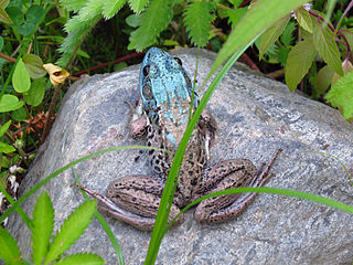
Axanthism is a mutation that interferes with an animal's ability to produce yellow pigment. The mutation affects the amount of xanthophores and carotenoid vesicles, sometimes causing them to be completely absent. Erythrophores and iridophores, which are responsible for red coloration and light reflecting pigments respectively, may also be affected. Axanthism is most obvious in green animals, specifically amphibians, making them appear blue. Green coloration in animals is caused by iridiphores reflecting blue wavelengths of light back through the carotenoids in the xanthophores. In the absence of xanthophores and carotenoids, the blue light is unaltered and reflected back normally. Animals that are normally yellow will appear white if affected with axanthism.

<span class="mw-page-title-main">Cuttlefish</span> Order of molluscs

Cuttlefish, or cuttles, are marine molluscs of the order Sepiida. They belong to the class Cephalopoda which also includes squid, octopuses, and nautiluses. Cuttlefish have a unique internal shell, the cuttlebone, which is used for control of buoyancy.

<span class="mw-page-title-main">Amelanism</span> Pigmentation abnormality

Amelanism is a pigmentation abnormality characterized by the lack of pigments called melanins, commonly associated with a genetic loss of tyrosinase function. Amelanism can affect fish, amphibians, reptiles, birds, and mammals including humans. The appearance of an amelanistic animal depends on the remaining non-melanin pigments. The opposite of amelanism is melanism, a higher percentage of melanin.

<span class="mw-page-title-main">Scale (insect anatomy)</span>

Scales are present on the bodies of various insects. A notable example are the Lepidoptera, the insect order comprising moths and butterflies, which have scales on their wings and on the head, parts of the thorax and abdomen, and parts of the genitalia. The name is derived from Ancient Greek λεπίδος (scale) and πτερόν (wing).

<span class="mw-page-title-main">Underwater camouflage</span> Camouflage in water, mainly by transparency, reflection, counter-illumination

Underwater camouflage is the set of methods of achieving crypsis—avoidance of observation—that allows otherwise visible aquatic organisms to remain unnoticed by other organisms such as predators or prey.

<i>Concealing-Coloration in the Animal Kingdom</i> Book by Abbott Handerson Thayer

Concealing-Coloration in the Animal Kingdom: An Exposition of the Laws of Disguise Through Color and Pattern; Being a Summary of Abbott H. Thayer’s Discoveries is a book published ostensibly by Gerald H. Thayer in 1909, and revised in 1918, but in fact a collaboration with and completion of his father Abbott Handerson Thayer's major work.

Deception in animals is the transmission of misinformation by one animal to another, of the same or different species, in a way that propagates beliefs that are not true.

<span class="mw-page-title-main">Albinism</span> Disorder causing lack of pigmentation

Albinism is the congenital absence of melanin in an animal or plant resulting in white hair, feathers, scales and skin and reddish pink or blue eyes. Individuals with the condition are referred to as albinos.

In evolutionary biology, mimicry in vertebrates is mimicry by a vertebrate of some model, deceiving some other animal, the dupe. Mimicry differs from camouflage as it is meant to be seen, while animals use camouflage to remain hidden. Visual, olfactory, auditory, biochemical, and behavioral modalities of mimicry have been documented in vertebrates.

References

  1. 1 2 3 4 5 Sugimoto, Masazumi (2002). "Morphological color changes in fish: Regulation of pigment cell density and morphology". Microscopy Research and Technique. 58 (6): 496–503. doi: 10.1002/jemt.10168 . ISSN   1097-0029. PMID   12242707. S2CID   26654746.
  2. 1 2 3 4 5 6 7 8 9 10 11 The diversity of fishes : biology, evolution, and ecology. Helfman, Gene S., Helfman, Gene S. (2nd ed.). Chichester, UK: Blackwell. 2009. ISBN   978-1-4051-2494-2. OCLC   233283748.{{cite book}}: CS1 maint: others (link)
  3. Endler, John A. (1978), "A Predator's View of Animal Color Patterns", in Hecht, Max K.; Steere, William C.; Wallace, Bruce (eds.), Evolutionary Biology, Springer US, pp. 319–364, doi:10.1007/978-1-4615-6956-5_5, ISBN   978-1-4615-6956-5
  4. 1 2 3 "Significance of colors and patterns of coral reef fishes: an overview". ReefCI. Retrieved 2020-04-28.
  5. 1 2 3 Price, Anna; Weadick, Cameron; Shim, Janet; Rodd, Frieda (2009-01-01). "Pigments, Patterns, and Fish Behavior". Zebrafish. 5 (4): 297–307. doi:10.1089/zeb.2008.0551. PMID   19133828.
  6. Nüsslein‐Volhard, Christiane; Singh, Ajeet Pratap (2017). "How fish color their skin: A paradigm for development and evolution of adult patterns". BioEssays. 39 (3): 1600231. doi: 10.1002/bies.201600231 . ISSN   1521-1878. PMID   28176337.
  7. Ulman, Aylin; Tunçer, Sezginer; Kizilkaya, Inci Tuney; Zilifli, Aytuğ; Alford, Polly; Giovos, Ioannis (2020-04-01). "The lionfish expansion in the Aegean Sea in Turkey: A looming potential ecological disaster". Regional Studies in Marine Science. 36: 101271. Bibcode:2020RSMS...3601271U. doi:10.1016/j.rsma.2020.101271. ISSN   2352-4855. S2CID   216281896.
  8. 1 2 3 4 5 6 7 Randall, John E. (April 3, 2005). "A Review of Mimicry in Marine Fishes" (PDF). Zoological Studies. 44 (3): 299–328.
  9. Parichy, David M (December 2003). "Pigment patterns: fish in stripes and spots". Current Biology. 13 (24): R947–R950. doi: 10.1016/j.cub.2003.11.038 . PMID   14680649.
  10. 1 2 Barlow, George W. (1972-03-08). "The Attitude of Fish Eye-Lines in Relation to Body Shape and to Stripes and Bars". Copeia. 1972 (1): 4–12. doi:10.2307/1442777. JSTOR   1442777.
  11. 1 2 Heflin, B.; Young, L.; Londraville, R. L. (2009). "Short-term cycling of skin colouration in the blackspotted rockskipper Entomacrodus striatus". Journal of Fish Biology. 74 (7): 1635–1641. doi:10.1111/j.1095-8649.2009.02200.x. ISSN   1095-8649. PMID   20735660.
  12. Henning, Frederico; Renz, Adina Josepha; Fukamachi, Shoji; Meyer, Axel (May 2010). "Genetic, Comparative Genomic, and Expression Analyses of the Mc1r Locus in the Polychromatic Midas Cichlid Fish (Teleostei, Cichlidae Amphilophus sp.) Species Group". Journal of Molecular Evolution. 70 (5): 405–412. Bibcode:2010JMolE..70..405H. doi:10.1007/s00239-010-9340-4. ISSN   0022-2844. PMID   20449580. S2CID   6080241.
  13. 1 2 Sköld, Helen Nilsson; Aspengren, Sara; Wallin, Margareta (2013). "Rapid color change in fish and amphibians – function, regulation, and emerging applications". Pigment Cell & Melanoma Research. 26 (1): 29–38. doi: 10.1111/pcmr.12040 . ISSN   1755-148X. PMID   23082932. S2CID   39162509.
  14. Henning, Frederico; Jones, Julia C.; Franchini, Paolo; Meyer, Axel (2013-03-13). "Transcriptomics of morphological color change in polychromatic Midas cichlids". BMC Genomics. 14 (1): 171. doi: 10.1186/1471-2164-14-171 . ISSN   1471-2164. PMC   3623868 . PMID   23497064.
  15. Pastana, M. N. L.; Dagosta, F. C. P.; Esguícero, A. L. H. (2017). "A new sexually dichromatic miniature Hyphessobrycon (Teleostei: Characiformes: Characidae) from the Rio Formiga, upper Rio Juruena basin, Mato Grosso, Brazil, with a review of sexual dichromatism in Characiformes". Journal of Fish Biology. 91 (5): 1301–1318. doi:10.1111/jfb.13449. ISSN   1095-8649. PMID   28905382.