Functional ultrasound imaging

Last updated
Main applications and features of functional ultrasound (fUS) imaging Main applications and features of functional ultrasound (fUS) imaging.svg
Main applications and features of functional ultrasound (fUS) imaging

Functional ultrasound imaging (fUS) is a medical ultrasound imaging technique of detecting or measuring changes in neural activities or metabolism, for example, the loci of brain activity, typically through measuring blood flow or hemodynamic changes. The method can be seen as an extension of Doppler imaging.

Contents

Background

Main brain functional imaging technique resolutions Main brain functional imaging technique resolutions.svg
Main brain functional imaging technique resolutions

Brain activation can either be directly measured by imaging electrical activity of neurons using voltage sensitive dyes, calcium imaging, electroencephalography, or magnetoencephalography, or indirectly by detecting hemodynamic changes in blood flow in the neurovascular systems through functional magnetic resonance imaging (fMRI), positron emission tomography (PET), Functional near-infrared spectroscopy (fNIRS), or Doppler ultrasonography )... [1]

Optical based methods generally provide the highest spatial and temporal resolutions; however, due to scattering, they are intrinsically limited to the investigation of the cortex. Thus, they are often used on animal models after partially removing or thinning the skull to allow for the light to penetrate into tissue. fMRI and PET, which measure the blood-oxygen level dependent (BOLD) signal, were the only techniques capable of imaging brain activation in depth. BOLD signal increases when neuronal activation exceeds oxygen consumption, where blood flow increases significantly. In fact, in-depth imaging of cerebral hemodynamic responses by fMRI, being noninvasive, paved the way for major discoveries in neurosciences in the early stage, and is applicable on humans. However, fMRI also suffers limitations. First, the cost and size of MR machines can be prohibitive. Also, spatially resolved fMRI is achieved at the expense of a substantial drop in temporal resolution and/or SNR. As a result, the imaging of transient events such as epilepsy is particularly challenging. Finally, fMRI is not appropriate for all clinical applications. For example, fMRI is rarely performed on infants because of specific issues concerning infant sedation. [2]

Like fMRI, Doppler based functional ultrasound approach are based on the neurovascular coupling and are thus limited by the spatiotemporal features of neurovascular coupling as they measure cerebral blood volume (CBV) changes. CBV is a pertinent parameter for functional imaging that is already used by other modalities such as intrinsic optical imaging or CBV-weighted fMRI. The spatiotemporal extent of CBV response was extensively studied. The spatial resolution of sensory-evoked CBV response can go down to cortical column (~100 μm). Temporally, the CBV impulse response function was measured to typically start at ~0.3 s and peak at ~1 s in response to ultrashort stimuli (300μs), which is much slower than the underlying electrical activity. [3]

Conventional doppler based approaches

Hemodynamic changes in the brain are often used as a surrogate indicator of neuronal activity to map the loci of brain activity. Major part of the hemodynamic response occurs in small vessels; however, conventional Doppler ultrasound is not sensitive enough to detect the blood flow in such small vessels. [2]

Functional transcranial doppler (fTCD)

Ultrasound Doppler imaging can be used to obtain basic functional measurements of brain activity using blood flow. In functional transcranial Doppler sonography, a low frequency (1-3 MHz) transducer is used through the temporal bone window with a conventional pulse Doppler mode to estimate blood flow at a single focal location. The temporal profile of blood velocity is usually acquired in main large arteries such as the middle cerebral artery (MCA). The peak velocity is compared between rest and task conditions or between right and left sides when studying lateralization. [4]

Power doppler

Power Doppler is a Doppler sequence that measures the ultrasonic energy backscattered from red blood cells in each pixel of the image. It provides no information on blood velocity but is proportional to blood volume within the pixel. However, conventional power Doppler imaging lacks sensitivity to detect small arterioles/venules and thus is unable to provide local neurofunctional information through neurovascular coupling. [2]

Ultrasensitive doppler

Functional ultrasound imaging was pioneered at ESPCI by Mickael Tanter  [ fr ]'s team [5] following work on ultrafast imaging [6] and ultrafast Doppler. [7]

Ultrasensitive doppler principle

Ultrasensitive Doppler relies on ultrafast imaging scanners [6] able to acquire images at thousands of frames per second, thus boosting the power Doppler SNR without any contrast agents. Instead of the line by line acquisition of conventional ultrasound devices, ultra-fast ultrasound takes advantage of successive tilted plane wave transmissions that afterward coherently compounded to form images at high frame rates. Coherent Compound Beamforming consists of the recombination of backscattered echoes from different illuminations achieved on the acoustic pressure field with various angles (as opposed to the acoustic intensity for the incoherent case). All images are added coherently to obtain a final compounded image. This very addition is produced without taking the envelope of the beamformed signals or any other nonlinear procedure to ensure a coherent addition. As a result, coherent adding of several echo waves leads to cancellation of out-of-phase waveforms, narrowing the point spread function (PSF), and thus increasing spatial resolution. A theoretical model demonstrates that the gain in sensitivity of the ultrasensitive Doppler method is due to the combination of the high signal-to-noise ratio (SNR) of the gray scale images, due to the synthetic compounding of backscattered echoes and the extensive signal samples averaging due to the high temporal resolution of ultrafast frame rates. [2] The sensitivity was recently further improved using multiple plane wave transmissions [8] and advanced spatiotemporal clutter filters for better discrimination between low blood flow and tissue motion. Ultrasound researchers have been using ultrafast imaging research platforms with parallel acquisition of channels and custom sequences programming to investigate ultrasensitive Doppler/fUS modalities. A custom real-time high-performance GPU beamforming code with a high data transfer rate (several GBytes per second) must then be implemented to perform imaging at high frame rate. Acquisitions can also typically easily provide gigabytes of data depending on acquisition duration.

Ultrasensitive Doppler has a typical 50-200 μm spatial resolution depending on the ultrasound frequency used. [2] It features a temporal resolution in the tens of milliseconds, can image the full depth of the brain and can provide 3D angiography. [9]

functional Ultrasound imaging

This signal boost enables the sensitivity required to map subtle blood variations in small arterioles (down to 1mm/s) related to neuronal activity. By applying an external stimulus such as a sensory, auditory or visual stimulation, it is then possible to construct a map of brain activation from the ultrasensitive Doppler movie.

fUS measures indirectly cerebral blood volume which provides an effect size close to 20% and as such is quite more sensitive than fMRI whose BOLD response is typically only a couple of percents. Correlation maps or statistical parametric maps can be constructed to highlight the activated areas. fUS has been shown to have a spatial resolution on the order 100 micrometers at 15 MHz in ferrets [10] and is sensitive enough to perform single trial detection in awake primates. [11] Other fMRI-like modalities such as functional connectivity can also be implemented.

Commercial scanners with specialized hardware and software [12] are enabling fUS to rapidly expand behind ultrasound research labs to the neuroscience community.

4D functional ultrasound imaging

Some researchers conducted 4D functional ultrasound imaging of whole-brain activity in rodents. Currently, two different technological solutions are proposed for the acquisition of 3D and 4D fUS data, each with its own advantages and drawbacks. [13] The first is a tomographic approach based on motorized translation of linear probes. This approach proved to be a successful method for several applications such as 3D retinotopic mapping in the rodent brain [14] [15] and 3D tonotopic mapping of the auditory system in ferrets. [10] The second approach relies on high frequency 2D matrix array transducer technology coupled with a high channel count electronic system for fast 3D imaging. To counterbalance the intrinsically poor sensitivity of matrix elements, they devised a 3D multiplane-wave scheme with 3D spatiotemporal encoding of transmit signals using Hadamard coefficients. For each transmission, the backscattered signals containing mixed echoes from the different plane waves are decoded using the summation of echoes from successive receptions with appropriate Hadamard coefficients. This summation enables the synthetic building of echoes from a virtual individual plane wave transmission with a higher amplitude. Finally, they perform coherent compounding beamforming of decoded echoes to produce 3D ultrasonic images and apply a spatiotemporal clutter filter separating blood flow from tissue motion to compute a power Doppler volume, which is proportional to the cerebral blood volume. [16]

Applications

Preclinical

Preclinical applications of fUS imaging Preclinical applications of fUS imaging.svg
Preclinical applications of fUS imaging

fUS can benefit in monitoring cerebral function in the whole brain which is important to understanding how the brain works on a large scale under normal or pathological conditions. The ability to image cerebral blood volume at high spatiotemporal resolution and with high sensitivity using fUS could be of great interest for applications in which fMRI reaches its limits, such as imaging of epileptic-induced changes in blood volume. [5] fUS can be applied for chronic studies in animal models through a thinned-skull [17] or smaller cranial window or directly through the skull in mice.

Brain activity mapping

Tonotopics or retinotopics maps [18] can be constructed by mapping the response of frequency-varying sounds [10] or moving visual targets. [14] [18] [15]

Functional connectivity / resting state

When no stimulus is applied, fUS can be used to study functional connectivity during resting state. The method has been demonstrated in rats [19] and awake mice [20] and can be used for pharmacological studies when testing drugs. [21] Seed-based maps, independent component analysis of resting states modes or functional connectivity matrix between atlas-based regions of interests can be constructed with high resolution.

Awake fUS imaging

Using dedicated ultralight probes, it is possible to perform freely-moving experiments in rats or mice. [22] [23] The size of the probes and electromagnetic-compatibility of fUS means it can also be used easily on head-fixed setups for mice [15] or in electrophysiology chambers in primate. [11]

Clinical

Clinical neuroimaging using ultrasound Clinical neuroimaging using ultrasound.svg
Clinical neuroimaging using ultrasound

Neonates

Thanks to its portability, fUS has also been used in clinics in awake neonates. [24] Functional ultrasound imaging can be applied to neonatal brain imaging in a non-invasive manner through the fontanel window. Ultrasound is usually performed in this case, which means that the current procedures does not have to be changed. High quality angiographic images could help diagnose vascular diseases such as perinatal ischemia or ventricular hemorrhage.

Adults / intraoperative

For adults, this method can be used during neurosurgery to guide the surgeon through the vasculature and to monitor the patient's brain function prior to tumor resection [25] [26]

See also

Related Research Articles

<span class="mw-page-title-main">Medical ultrasound</span> Diagnostic and therapeutic technique

Medical ultrasound includes diagnostic techniques using ultrasound, as well as therapeutic applications of ultrasound. In diagnosis, it is used to create an image of internal body structures such as tendons, muscles, joints, blood vessels, and internal organs, to measure some characteristics or to generate an informative audible sound. The usage of ultrasound to produce visual images for medicine is called medical ultrasonography or simply sonography, or echography. The practice of examining pregnant women using ultrasound is called obstetric ultrasonography, and was an early development of clinical ultrasonography. The machine used is called an ultrasound machine, a sonograph or an echograph. The visual image formed using this technique is called an ultrasonogram, a sonogram or an echogram.

<span class="mw-page-title-main">Functional magnetic resonance imaging</span> MRI procedure that measures brain activity by detecting associated changes in blood flow

Functional magnetic resonance imaging or functional MRI (fMRI) measures brain activity by detecting changes associated with blood flow. This technique relies on the fact that cerebral blood flow and neuronal activation are coupled. When an area of the brain is in use, blood flow to that region also increases.

<span class="mw-page-title-main">Functional neuroimaging</span>

Functional neuroimaging is the use of neuroimaging technology to measure an aspect of brain function, often with a view to understanding the relationship between activity in certain brain areas and specific mental functions. It is primarily used as a research tool in cognitive neuroscience, cognitive psychology, neuropsychology, and social neuroscience.

Functional integration is the study of how brain regions work together to process information and effect responses. Though functional integration frequently relies on anatomic knowledge of the connections between brain areas, the emphasis is on how large clusters of neurons – numbering in the thousands or millions – fire together under various stimuli. The large datasets required for such a whole-scale picture of brain function have motivated the development of several novel and general methods for the statistical analysis of interdependence, such as dynamic causal modelling and statistical linear parametric mapping. These datasets are typically gathered in human subjects by non-invasive methods such as EEG/MEG, fMRI, or PET. The results can be of clinical value by helping to identify the regions responsible for psychiatric disorders, as well as to assess how different activities or lifestyles affect the functioning of the brain.

<span class="mw-page-title-main">Functional near-infrared spectroscopy</span> Optical technique for monitoring brain activity

Functional near-infrared spectroscopy (fNIRS) is an optical brain monitoring technique which uses near-infrared spectroscopy for the purpose of functional neuroimaging. Using fNIRS, brain activity is measured by using near-infrared light to estimate cortical hemodynamic activity which occur in response to neural activity. Alongside EEG, fNIRS is one of the most common non-invasive neuroimaging techniques which can be used in portable contexts. The signal is often compared with the BOLD signal measured by fMRI and is capable of measuring changes both in oxy- and deoxyhemoglobin concentration, but can only measure from regions near the cortical surface. fNIRS may also be referred to as Optical Topography (OT) and is sometimes referred to simply as NIRS.

<span class="mw-page-title-main">Transcranial Doppler</span>

Transcranial Doppler (TCD) and transcranial color Doppler (TCCD) are types of Doppler ultrasonography that measure the velocity of blood flow through the brain's blood vessels by measuring the echoes of ultrasound waves moving transcranially. These modes of medical imaging conduct a spectral analysis of the acoustic signals they receive and can therefore be classified as methods of active acoustocerebrography. They are used as tests to help diagnose emboli, stenosis, vasospasm from a subarachnoid hemorrhage, and other problems. These relatively quick and inexpensive tests are growing in popularity. The tests are effective for detecting sickle cell disease, ischemic cerebrovascular disease, subarachnoid hemorrhage, arteriovenous malformations, and cerebral circulatory arrest. The tests are possibly useful for perioperative monitoring and meningeal infection. The equipment used for these tests is becoming increasingly portable, making it possible for a clinician to travel to a hospital, to a doctor's office, or to a nursing home for both inpatient and outpatient studies. The tests are often used in conjunction with other tests such as MRI, MRA, carotid duplex ultrasound and CT scans. The tests are also used for research in cognitive neuroscience.

<span class="mw-page-title-main">Neuroimaging</span> Set of techniques to measure and visualize aspects of the nervous system

Neuroimaging is the use of quantitative (computational) techniques to study the structure and function of the central nervous system, developed as an objective way of scientifically studying the healthy human brain in a non-invasive manner. Increasingly it is also being used for quantitative research studies of brain disease and psychiatric illness. Neuroimaging is highly multidisciplinary involving neuroscience, computer science, psychology and statistics, and is not a medical specialty. Neuroimaging is sometimes confused with neuroradiology.

<span class="mw-page-title-main">Susceptibility weighted imaging</span>

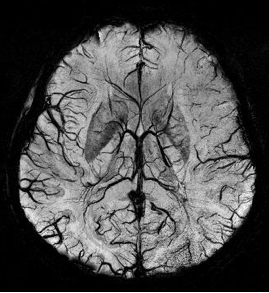
Susceptibility weighted imaging (SWI), originally called BOLD venographic imaging, is an MRI sequence that is exquisitely sensitive to venous blood, hemorrhage and iron storage. SWI uses a fully flow compensated, long echo, gradient recalled echo (GRE) pulse sequence to acquire images. This method exploits the susceptibility differences between tissues and uses the phase image to detect these differences. The magnitude and phase data are combined to produce an enhanced contrast magnitude image. The imaging of venous blood with SWI is a blood-oxygen-level dependent (BOLD) technique which is why it was referred to as BOLD venography. Due to its sensitivity to venous blood SWI is commonly used in traumatic brain injuries (TBI) and for high resolution brain venographies but has many other clinical applications. SWI is offered as a clinical package by Philips and Siemens but can be run on any manufacturer's machine at field strengths of 1.0 T, 1.5 T, 3.0 T and higher.

Connectomics is the production and study of connectomes: comprehensive maps of connections within an organism's nervous system. More generally, it can be thought of as the study of neuronal wiring diagrams with a focus on how structural connectivity, individual synapses, cellular morphology, and cellular ultrastructure contribute to the make up of a network. The nervous system is a network made of billions of connections and these connections are responsible for our thoughts, emotions, actions, memories, function and dysfunction. Therefore, the study of connectomics aims to advance our understanding of mental health and cognition by understanding how cells in the nervous system are connected and communicate. Because these structures are extremely complex, methods within this field use a high-throughput application of functional and structural neural imaging, most commonly magnetic resonance imaging (MRI), electron microscopy, and histological techniques in order to increase the speed, efficiency, and resolution of these nervous system maps. To date, tens of large scale datasets have been collected spanning the nervous system including the various areas of cortex, cerebellum, the retina, the peripheral nervous system and neuromuscular junctions.

Psychophysiological interaction (PPI) is a brain connectivity analysis method for functional brain imaging data, mainly functional magnetic resonance imaging (fMRI). It estimates context-dependent changes in effective connectivity (coupling) between brain regions. Thus, PPI analysis identifies brain regions whose activity depends on an interaction between psychological context and physiological state of the seed region.

Preclinical imaging is the visualization of living animals for research purposes, such as drug development. Imaging modalities have long been crucial to the researcher in observing changes, either at the organ, tissue, cell, or molecular level, in animals responding to physiological or environmental changes. Imaging modalities that are non-invasive and in vivo have become especially important to study animal models longitudinally. Broadly speaking, these imaging systems can be categorized into primarily morphological/anatomical and primarily molecular imaging techniques. Techniques such as high-frequency micro-ultrasound, magnetic resonance imaging (MRI) and computed tomography (CT) are usually used for anatomical imaging, while optical imaging, positron emission tomography (PET), and single photon emission computed tomography (SPECT) are usually used for molecular visualizations.

Anders Martin Dale is a prominent neuroscientist and professor of radiology, neurosciences, psychiatry, and cognitive science at the University of California, San Diego (UCSD), and is one of the world's leading developers of sophisticated computational neuroimaging techniques. He is the founding Director of the Center for Multimodal Imaging Genetics (CMIG) at UCSD.

A silent stroke is a stroke that does not have any outward symptoms associated with stroke, and the patient is typically unaware they have suffered a stroke. Despite not causing identifiable symptoms, a silent stroke still causes damage to the brain and places the patient at increased risk for both transient ischemic attack and major stroke in the future. In a broad study in 1998, more than 11 million people were estimated to have experienced a stroke in the United States. Approximately 770,000 of these strokes were symptomatic and 11 million were first-ever silent MRI infarcts or hemorrhages. Silent strokes typically cause lesions which are detected via the use of neuroimaging such as MRI. The risk of silent stroke increases with age but may also affect younger adults. Women appear to be at increased risk for silent stroke, with hypertension and current cigarette smoking being amongst the predisposing factors.

<span class="mw-page-title-main">Resting state fMRI</span> Type of functional magnetic resonance imaging

Resting state fMRI is a method of functional magnetic resonance imaging (fMRI) that is used in brain mapping to evaluate regional interactions that occur in a resting or task-negative state, when an explicit task is not being performed. A number of resting-state brain networks have been identified, one of which is the default mode network. These brain networks are observed through changes in blood flow in the brain which creates what is referred to as a blood-oxygen-level dependent (BOLD) signal that can be measured using fMRI.

Functional magnetic resonance spectroscopy of the brain (fMRS) uses magnetic resonance imaging (MRI) to study brain metabolism during brain activation. The data generated by fMRS usually shows spectra of resonances, instead of a brain image, as with MRI. The area under peaks in the spectrum represents relative concentrations of metabolites.

<span class="mw-page-title-main">MRI pulse sequence</span>

An MRI pulse sequence in magnetic resonance imaging (MRI) is a particular setting of pulse sequences and pulsed field gradients, resulting in a particular image appearance.

Arterial spin labeling (ASL), also known as arterial spin tagging, is a magnetic resonance imaging technique used to quantify cerebral blood perfusion by labelling blood water as it flows throughout the brain. ASL specifically refers to magnetic labeling of arterial blood below or in the imaging slab, without the need of gadolinium contrast. A number of ASL schemes are possible, the simplest being flow alternating inversion recovery (FAIR) which requires two acquisitions of identical parameters with the exception of the out-of-slice saturation; the difference in the two images is theoretically only from inflowing spins, and may be considered a 'perfusion map'. The ASL technique was developed by Alan P. Koretsky, Donald S. Williams, John A. Detre and John S. Leigh Jr in 1992.

<span class="mw-page-title-main">Neurovascular unit</span>

The neurovascular unit (NVU) comprises the components of the brain that collectively regulate cerebral blood flow in order to deliver the requisite nutrients to activated neurons. The NVU addresses the brain's unique dilemma of having high energy demands yet low energy storage capacity. In order to function properly, the brain must receive substrates for energy metabolism–mainly glucose–in specific areas, quantities, and times. Neurons do not have the same ability as, for example, muscle cells, which can use up their energy reserves and refill them later; therefore, cerebral metabolism must be driven in the moment. The neurovascular unit facilitates this ad hoc delivery and, thus, ensures that neuronal activity can continue seamlessly.

Ultrasound Localization Microscopy (ULM) is an advanced ultrasound imaging technique. By localizing microbubbles, ULM overcomes the physical limit of diffraction, achieving sub-wavelength level resolution and qualifying as a super-resolution technique.

References

  1. Petersen CC (October 2007). "The functional organization of the barrel cortex". Neuron. 56 (2): 339–55. doi:10.1016/j.neuron.2007.09.017. PMID   17964250.
  2. 1 2 3 4 5 Mace E, Montaldo G, Osmanski BF, Cohen I, Fink M, Tanter M (March 2013). "Functional ultrasound imaging of the brain: theory and basic principles". IEEE Transactions on Ultrasonics, Ferroelectrics, and Frequency Control. 60 (3): 492–506. doi:10.1109/tuffc.2013.2592. PMID   23475916. S2CID   27482186.
  3. Deffieux T, Demene C, Pernot M, Tanter M (June 2018). "Functional ultrasound neuroimaging: a review of the preclinical and clinical state of the art". Current Opinion in Neurobiology. 50: 128–135. doi:10.1016/j.conb.2018.02.001. PMID   29477979.
  4. Knecht S, Deppe M, Ebner A, Henningsen H, Huber T, Jokeit H, et al. (January 1998). "Noninvasive determination of language lateralization by functional transcranial Doppler sonography: a comparison with the Wada test". Stroke. 29 (1): 82–6. doi:10.1161/01.str.29.1.82. PMID   9445333.
  5. 1 2 Macé E, Montaldo G, Cohen I, Baulac M, Fink M, Tanter M (July 2011). "Functional ultrasound imaging of the brain". Nature Methods. 8 (8): 662–664. doi:10.1038/nmeth.1641. PMID   21725300.
  6. 1 2 Tanter M, Fink M (January 2014). "Ultrafast imaging in biomedical ultrasound". IEEE Transactions on Ultrasonics, Ferroelectrics, and Frequency Control. 61 (1): 102–19. doi:10.1109/TUFFC.2014.6689779. PMID   24402899.
  7. Bercoff J, Montaldo G, Loupas T, Savery D, Mézière F, Fink M, et al. (January 2011). "Ultrafast compound Doppler imaging: providing full blood flow characterization". IEEE Transactions on Ultrasonics, Ferroelectrics, and Frequency Control. 58 (1): 134–47. doi:10.1109/TUFFC.2011.1780. PMID   21244981.
  8. Tiran E, Deffieux T, Correia M, Maresca D, Osmanski BF, Sieu LA, et al. (November 2015). "Multiplane wave imaging increases signal-to-noise ratio in ultrafast ultrasound imaging". Physics in Medicine and Biology. 60 (21): 8549–66. Bibcode:2015PMB....60.8549T. doi:10.1088/0031-9155/60/21/8549. PMID   26487501.
  9. Demené C, Tiran E, Sieu LA, Bergel A, Gennisson JL, Pernot M, et al. (February 2016). "4D microvascular imaging based on ultrafast Doppler tomography". NeuroImage. 127: 472–483. doi:10.1016/j.neuroimage.2015.11.014. PMID   26555279.
  10. 1 2 3 Bimbard C, Demene C, Girard C, Radtke-Schuller S, Shamma S, Tanter M, et al. (June 2018). "Multi-scale mapping along the auditory hierarchy using high-resolution functional UltraSound in the awake ferret". eLife. 7. doi: 10.7554/eLife.35028 . PMC   6039176 . PMID   29952750.
  11. 1 2 Dizeux A, Gesnik M, Ahnine H, Blaize K, Arcizet F, Picaud S, et al. (March 2019). "Functional ultrasound imaging of the brain reveals propagation of task-related brain activity in behaving primates". Nature Communications. 10 (1): 1400. Bibcode:2019NatCo..10.1400D. doi:10.1038/s41467-019-09349-w. PMC   6438968 . PMID   30923310.
  12. "Iconeus Functional Ultrasound (fUS) preclinical imaging systems - Iconeus". iconeus.com. 2021-08-13. Retrieved 2024-06-15.
  13. "The path to 4D fUS" (PDF). Iconeus. Retrieved 25 May 2020.
  14. 1 2 Gesnik M, Blaize K, Deffieux T, Gennisson JL, Sahel JA, Fink M, et al. (April 2017). "3D functional ultrasound imaging of the cerebral visual system in rodents". NeuroImage. 149: 267–274. doi:10.1016/j.neuroimage.2017.01.071. PMC   5387157 . PMID   28167348.
  15. 1 2 3 Macé É, Montaldo G, Trenholm S, Cowan C, Brignall A, Urban A, et al. (December 2018). "Whole-Brain Functional Ultrasound Imaging Reveals Brain Modules for Visuomotor Integration". Neuron. 100 (5): 1241–1251.e7. doi:10.1016/j.neuron.2018.11.031. PMC   6292977 . PMID   30521779.
  16. Rabut C, Correia M, Finel V, Pezet S, Pernot M, Deffieux T, et al. (October 2019). "4D functional ultrasound imaging of whole-brain activity in rodents". Nature Methods. 16 (10): 994–997. doi:10.1038/s41592-019-0572-y. PMC   6774790 . PMID   31548704.
  17. Drew PJ, Shih AY, Driscoll JD, Knutsen PM, Blinder P, Davalos D, et al. (December 2010). "Chronic optical access through a polished and reinforced thinned skull". Nature Methods. 7 (12): 981–4. doi:10.1038/nmeth.1530. PMC   3204312 . PMID   20966916.
  18. 1 2 Blaize K, Arcizet F, Gesnik M, Ahnine H, Ferrari U, Deffieux T, et al. (June 2020). "Functional ultrasound imaging of deep visual cortex in awake nonhuman primates". Proceedings of the National Academy of Sciences of the United States of America. 117 (25): 14453–14463. Bibcode:2020PNAS..11714453B. doi: 10.1073/pnas.1916787117 . PMC   7321983 . PMID   32513717.
  19. Osmanski BF, Pezet S, Ricobaraza A, Lenkei Z, Tanter M (October 2014). "Functional ultrasound imaging of intrinsic connectivity in the living rat brain with high spatiotemporal resolution". Nature Communications. 5: 5023. Bibcode:2014NatCo...5.5023O. doi:10.1038/ncomms6023. PMC   4205893 . PMID   25277668.
  20. Ferrier J, Tiran E, Deffieux T, Tanter M, Lenkei Z (June 2020). "Functional imaging evidence for task-induced deactivation and disconnection of a major default mode network hub in the mouse brain". Proceedings of the National Academy of Sciences of the United States of America. 117 (26): 15270–15280. Bibcode:2020PNAS..11715270F. doi: 10.1073/pnas.1920475117 . PMC   7334502 . PMID   32541017.
  21. Rabut C, Ferrier J, Bertolo A, Osmanski B, Mousset X, Pezet S, et al. (November 2020). "Pharmaco-fUS: Quantification of pharmacologically-induced dynamic changes in brain perfusion and connectivity by functional ultrasound imaging in awake mice". NeuroImage. 222: 117231. doi:10.1016/j.neuroimage.2020.117231. PMID   32795659.
  22. Sieu LA, Bergel A, Tiran E, Deffieux T, Pernot M, Gennisson JL, et al. (September 2015). "EEG and functional ultrasound imaging in mobile rats". Nature Methods. 12 (9): 831–834. doi:10.1038/nmeth.3506. PMC   4671306 . PMID   26237228.
  23. Tiran E, Ferrier J, Deffieux T, Gennisson JL, Pezet S, Lenkei Z, et al. (August 2017). "Transcranial Functional Ultrasound Imaging in Freely Moving Awake Mice and Anesthetized Young Rats without Contrast Agent". Ultrasound in Medicine & Biology. 43 (8): 1679–1689. doi:10.1016/j.ultrasmedbio.2017.03.011. PMC   5754333 . PMID   28476311.
  24. Demené C, Mairesse J, Baranger J, Tanter M, Baud O (January 2019). "Ultrafast Doppler for neonatal brain imaging". NeuroImage. 185: 851–856. doi:10.1016/j.neuroimage.2018.04.016. PMID   29649559.
  25. Imbault M, Chauvet D, Gennisson JL, Capelle L, Tanter M (August 2017). "Intraoperative Functional Ultrasound Imaging of Human Brain Activity". Scientific Reports. 7 (1): 7304. Bibcode:2017NatSR...7.7304I. doi:10.1038/s41598-017-06474-8. PMC   5544759 . PMID   28779069.
  26. Soloukey S, Vincent AJ, Satoer DD, Mastik F, Smits M, Dirven CM, et al. (2019). "Functional Ultrasound (fUS) During Awake Brain Surgery: The Clinical Potential of Intra-Operative Functional and Vascular Brain Mapping". Frontiers in Neuroscience. 13: 1384. doi: 10.3389/fnins.2019.01384 . PMC   6962116 . PMID   31998060.