Gene therapy for color blindness

Last updated

Gene therapy for color blindness is an experimental gene therapy of the human retina aiming to grant typical trichromatic color vision to individuals with congenital color blindness by introducing typical alleles for opsin genes. Animal testing for gene therapy began in 2007 with a 2009 breakthrough in squirrel monkeys suggesting an imminent gene therapy in humans. While the research into gene therapy for red-green colorblindness has lagged since then, successful human trials are ongoing for achromatopsia. Congenital color vision deficiency affects upwards of 200 million people in the world, which represents a large demand for this gene therapy.

Contents

Color vision

The retina of the human eye contains photoreceptive cells called cones that allow color vision. A normal trichromat possesses three different types of cones to distinguish different colors within the visible spectrum. The three types of cones are designated L, M, and S cones, each containing an opsin sensitive to a different portion of the visible spectrum. More specifically, the L cone absorbs around 560 nm, the M cone absorbs near 530 nm, and the S cone absorbs near 420 nm. [1] These cones transduce the absorbed light into electrical information to be relayed through other cells along the phototransduction pathway, before reaching the visual cortex in the brain. [1]

The signals from the 3 cones are compared to each other to generate 3 opponent process channels. The channels are perceived as balances between red-green, blue-yellow and black-white. [1]

Color vision deficiency

Color vision deficiency (CVD) is the deviation of an individual's color vision from typical human trichromatic vision. Relevant to gene therapy, CVD can be classified in 2 groups.

Dichromacy

Dichromats have partial color vision. The most common form of dichromacy is red-green colorblindness. Dichromacy usually arises when one of the three opsin genes is deleted or otherwise fully nonfunctional. The effects and diagnosis depend on the missing opsin. Protanopes (very common) have no L-opsin, Deuteranopes (very common) have no M-opsin, and Tritanopes (rare) have no S-opsin. Accordingly, a missing cone means one of the opponent channels is inactive: red-green for protanopes/deuteranopes and blue-yellow for tritanopes. They therefore perceive a much reduced color space. Although dichromacy poses few critical problems in daily life, a lack of access to many occupations (where color vision may be safety-critical) is a large disadvantage.

Anomalous Trichromats are not missing an opsin gene, but rather have a mutated (or chimeric) gene. They have trichromatic vision, but with a smaller color gamut than typical color vision. Regarding gene therapy, they are equivalent to dichromats.

Blue Cone Monochromats are missing both the L- and M-opsin and therefore have no color vision. They are treated as a subset of dichromacy since a combination of gene therapies for protanopia and deuteranopia would be used.

Achromatopsia

Individuals with congenital achromatopsia tend to have typical opsin genes, but have a mutation in another gene downstream in the phototransduction pathway (e.g. GNAT2 protein) that prevents their cones (and therefore photopic vision) from functioning. Achromats rely solely on their scotopic vision. The severity of achromatopsia is much higher than dichromacy, not only in the lack of color vision, but also in co-occurring symptoms photophobia, nystagmus and poor visual acuity.

Retinal gene therapy

Gene therapies aim to inject functional copies of missing or mutated genes into affected individuals by the use of viral vectors. Using a replication-defective recombinant adeno-associated virus (rAAV) as a vector, the cDNA of the affected gene can be delivered to the cones at the back of the retina typically via subretinal injection. Intravitreal injections are much less invasive, but not yet as effective as subretinal injections. Upon gaining the gene, the cone begins to express the new photopigment. The effect is ideally permanent.

Research

The first retinal gene therapy to be approved by the FDA was Voretigene neparvovec in 2017, which treats Leber's congenital amaurosis, a genetic disorder that can lead to blindness. These treatments also use subretinal injections of AAV vector and are therefore foundational to research in gene therapy for color blindness. [2] [3]

Human L-cone photopigment have been introduced into mice. Since the mice possess only S cones and M cones, they are dichromats. [4] M-opsin was replaced with a cDNA of L-opsin in the X chromosome of some mice. By breeding these "knock-in" transgenic mice, they generated heterozygous females with both an M cone and an L cone. These mice had improved range of color vision and have gained trichromacy, as tested by electroretinogram and behavioral tests. However, this is more difficult to apply in the form of gene therapy.

Recombinant AAV vector was to introduce the green fluorescent protein (GFP) gene in the cones of gerbils. [5] The genetic insert was designed to only be expressed in S or M cones, and the expression of GFP in vivo was observed over time. Gene expression could stabilize if a sufficiently high dose of the viral vector is given.

In 2009, adult dichromatic squirrel monkeys were converted into trichromats using gene therapy. [6] New world monkeys are polymorphic in their M-opsin, such that females can be trichromatic, but all males are dichromatic. [6] Recombinant AAV vector was used to deliver a human L-opsin gene subretinally. A subset of the monkey's M-cones gained the L-opsin genes and began co-expressing the new and old photopigments. [6] Electroretinograms demonstrated that the cones were expressing the new opsin and after 20 weeks a pseudoisochromatic color vision test demonstrated that the treated monkeys had indeed developed functional trichromatic vision. [6]

Gene therapy was to restore some of the sight of mice with achromatopsia. The results were positive for 80% of the mice treated. [7]

In 2010, gene therapy for a form of achromatopsia was performed in dogs. Cone function and day vision have been restored for at least 33 months in two young dogs with achromatopsia. However, this therapy was less efficient for older dogs. [8]

In 2022, 4 young human ACHM2 and ACHM3 achromats were shown to have neurological responses (as measured with fMRI) to photopic vision that matched patterns generated by their scotopic vision after gene therapy. This inferred a photopic cone-driven system that was at least marginally functional. The methodology did not investigate novel color vision, though one respondent claimed to more easily interpret traffic lights. [9] This may be considered the first case of a cure for colorblindness in humans.

In July 2023, a study found positive but limited improvements on congenital CNGA3 achromatopsia. [10] [11]

Challenges

While the benefits of gene therapy to achromats typically outweigh the current risks, there are several challenges before large acceptance of gene therapy in dichromats can occur.

Safety

The procedure – namely the subretinal injection – is quite invasive, requiring several incisions and punctures in the eyeball. This poses a significant risk of infection and other complications. Subretinal injections methods promise to become less invasive with their application in other retinal gene therapies. They could also be replaced by intravitreal injections, which are significantly less invasive and can in theory be performed by a family doctor, but are less effective. [12]

The permanence of these therapies is also in question. Mancuso et al. reported that the treated squirrel monkeys maintained 2 years of color vision after the treatment. [6] However, if repeat injections are needed, there is also the concern of the body developing an immune reaction to the virus. If a body develops sensitivity to the viral vector, the success of the therapy could be jeopardized and/or the body may respond unfavorably. An editorial by J. Bennett points to Mancuso et al.'s use of an "unspecified postinjection corticosteroid therapy". [13] Bennett suggests that the monkeys may have experienced inflammation due to the injection. [13] However, the AAV virus that is commonly used for this study is non-pathogenic, and the body is less likely to develop an immune reaction. [14]

Neuroplasticity

According to research by David H. Hubel and Torsten Wiesel, suturing shut one eye of monkeys at an early age resulted in an irreversible loss of vision in that eye, even after the suture was removed. [1] [15] The study concluded that the neural circuitry for vision is wired during a "critical period" in childhood, after which the visual circuitry can no longer be rewired to process new sensory input. Contrary to this finding, Mancuso et al.’s success in conferring trichromacy to adult squirrel monkeys suggests that it is possible to adapt the preexisting circuit to allow greater acuity in color vision. The researchers concluded that integrating the stimulus from the new photopigment as an adult was not analogous to vision loss following visual deprivation. [6]

It is yet unknown how the animals that gain a new photopigment are perceiving the new color. While the article by Mancuso et al. states that the monkey has indeed gained trichromacy and gained the ability to discriminate between red and green, they claim no knowledge of how the animal internally perceives the sensation. [6]

Ethics

As a way to introduce new genetic information to change a person's phenotype, a gene therapy for color blindness is open to the same ethical questions and criticisms as gene therapy in general. These include issues around the governance of the therapy, whether treatment should be available only to those who can afford it, and whether the availability of treatment creates a stigma for those with color blindness. Given the large number of people with color blindness, there is also the question of whether color blindness is a disorder. [16] Furthermore, even if gene therapy succeeds in converting incomplete colorblind individuals to trichromats, the degree of satisfaction among the subjects is unknown. It is uncertain how the quality of life will improve (or worsen) after the therapy.

The gene therapy for converting dichromats to trichromats can also be used hypothetically to "upgrade" typical trichromats to tetrachromats by introducing a new opsin genes. This begs the ethics of designer babies that contain genes not available naturally in the human gene pool. In 2022, the lab of Jay Neitz engineered a novel opsin sensitive to wavelengths between the typical human S- (420 nm) and M- (530 nm) opsins, i.e. the novel opsin at 493 nm. This allowed the opsin to be clearly visible in ERGs, but could be used to create tetrachromacy. [12]

See also

Related Research Articles

<span class="mw-page-title-main">Color blindness</span> Decreased ability to see color or color differences

Color blindness or color vision deficiency (CVD) is the decreased ability to see color or differences in color. The severity of color blindness ranges from mostly unnoticeable to full absence of color perception. Color blindness is usually an inherited problem or variation in the functionality of one or more of the three classes of cone cells in the retina, which mediate color vision. The most common form is caused by a genetic condition called congenital red–green color blindness, which affects up to 1 in 12 males (8%) and 1 in 200 females (0.5%). The condition is more prevalent in males, because the opsin genes responsible are located on the X chromosome. Rarer genetic conditions causing color blindness include congenital blue–yellow color blindness, blue cone monochromacy, and achromatopsia. Color blindness can also result from physical or chemical damage to the eye, the optic nerve, parts of the brain, or from medication toxicity. Color vision also naturally degrades in old age.

<span class="mw-page-title-main">Retina</span> Part of the eye

The retina is the innermost, light-sensitive layer of tissue of the eye of most vertebrates and some molluscs. The optics of the eye create a focused two-dimensional image of the visual world on the retina, which then processes that image within the retina and sends nerve impulses along the optic nerve to the visual cortex to create visual perception. The retina serves a function which is in many ways analogous to that of the film or image sensor in a camera.

Achromatopsia, also known as Rod monochromacy, is a medical syndrome that exhibits symptoms relating to five conditions, most notably monochromacy. Historically, the name referred to monochromacy in general, but now typically refers only to an autosomal recessive congenital color vision condition. The term is also used to describe cerebral achromatopsia, though monochromacy is usually the only common symptom. The conditions include: monochromatic color blindness, poor visual acuity, and day-blindness. The syndrome is also present in an incomplete form that exhibits milder symptoms, including residual color vision. Achromatopsia is estimated to affect 1 in 30,000 live births worldwide.

<span class="mw-page-title-main">Color vision</span> Ability to perceive differences in light frequency

Color vision, a feature of visual perception, is an ability to perceive differences between light composed of different frequencies independently of light intensity.

<span class="mw-page-title-main">Tetrachromacy</span> Type of color vision with four types of cone cells

Tetrachromacy is the condition of possessing four independent channels for conveying color information, or possessing four types of cone cell in the eye. Organisms with tetrachromacy are called tetrachromats.

<span class="mw-page-title-main">Trichromacy</span> Possessing of three independent channels for conveying color information

Trichromacy or trichromatism is the possession of three independent channels for conveying color information, derived from the three different types of cone cells in the eye. Organisms with trichromacy are called trichromats.

<span class="mw-page-title-main">Cone cell</span> Photoreceptor cells responsible for color vision made to function in bright light

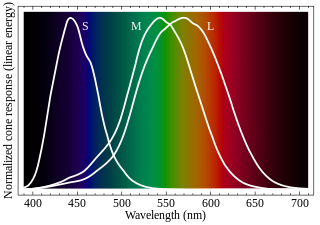
Cone cells or cones are photoreceptor cells in the retinas of vertebrates' eyes. They respond differently to light of different wavelengths, and the combination of their responses is responsible for color vision. Cones function best in relatively bright light, called the photopic region, as opposed to rod cells, which work better in dim light, or the scotopic region. Cone cells are densely packed in the fovea centralis, a 0.3 mm diameter rod-free area with very thin, densely packed cones which quickly reduce in number towards the periphery of the retina. Conversely, they are absent from the optic disc, contributing to the blind spot. There are about six to seven million cones in a human eye, with the highest concentration being towards the macula.

Dichromacy is the state of having two types of functioning photoreceptors, called cone cells, in the eyes. Organisms with dichromacy are called dichromats. Dichromats require only two primary colors to be able to represent their visible gamut. By comparison, trichromats need three primary colors, and tetrachromats need four. Likewise, every color in a dichromat's gamut can be evoked with monochromatic light. By comparison, every color in a trichromat's gamut can be evoked with a combination of monochromatic light and white light.

<span class="mw-page-title-main">Monochromacy</span> Type of color vision

Monochromacy is the ability of organisms to perceive only light intensity without respect to spectral composition. Organisms with monochromacy lack color vision and can only see in shades of grey ranging from black to white. Organisms with monochromacy are called monochromats. Many mammals, such as cetaceans, the owl monkey and the Australian sea lion are monochromats. In humans, monochromacy is one among several other symptoms of severe inherited or acquired diseases, including achromatopsia or blue cone monochromacy, together affecting about 1 in 30,000 people.

<span class="mw-page-title-main">Melanopsin</span> Mammalian protein found in Homo sapiens

Melanopsin is a type of photopigment belonging to a larger family of light-sensitive retinal proteins called opsins and encoded by the gene Opn4. In the mammalian retina, there are two additional categories of opsins, both involved in the formation of visual images: rhodopsin and photopsin in the rod and cone photoreceptor cells, respectively.

<span class="mw-page-title-main">OPN1SW</span> Protein-coding gene in the species Homo sapiens

Blue-sensitive opsin is a protein that in humans is encoded by the OPN1SW gene.

<span class="mw-page-title-main">OPN1MW</span> Protein-coding gene in the species Homo sapiens

Green-sensitive opsin is a protein that in humans is encoded by the OPN1MW gene. OPN1MW2 is a similar opsin.

<span class="mw-page-title-main">OPN1LW</span> Protein-coding gene in humans

OPN1LW is a gene on the X chromosome that encodes for long wave sensitive (LWS) opsin, or red cone photopigment. It is responsible for perception of visible light in the yellow-green range on the visible spectrum. The gene contains 6 exons with variability that induces shifts in the spectral range. OPN1LW is subject to homologous recombination with OPN1MW, as the two have very similar sequences. These recombinations can lead to various vision problems, such as red-green colourblindness and blue monochromacy. The protein encoded is a G-protein coupled receptor with embedded 11-cis-retinal, whose light excitation causes a cis-trans conformational change that begins the process of chemical signalling to the brain.

<span class="mw-page-title-main">Evolution of color vision in primates</span> Loss and regain of colour vision during the evolution of primates

The evolution of color vision in primates is highly unusual compared to most eutherian mammals. A remote vertebrate ancestor of primates possessed tetrachromacy, but nocturnal, warm-blooded, mammalian ancestors lost two of four cones in the retina at the time of dinosaurs. Most teleost fish, reptiles and birds are therefore tetrachromatic while most mammals are strictly dichromats, the exceptions being some primates and marsupials, who are trichromats, and many marine mammals, who are monochromats.

Color vision, a proximate adaptation of the vision sensory modality, allows for the discrimination of light based on its wavelength components.

Gene therapy using lentiviral vectors was being explored in early stage trials as of 2009.

Retinal gene therapy holds a promise in treating different forms of non-inherited and inherited blindness.

Blue cone monochromacy (BCM) is an inherited eye disease that causes severe color blindness, poor visual acuity, nystagmus and photophobia due to the absence of functional red (L) and green (M) cone photoreceptor cells in the retina. BCM is a recessive X-linked disease and almost exclusively affects XY karyotypes.

<span class="mw-page-title-main">Color blind glasses</span> Light filters to alleviate color blindness

Color blind glasses or color correcting lenses are light filters, usually in the form of glasses or contact lenses, that attempt to alleviate color blindness, by bringing deficient color vision closer to normal color vision or to make certain color tasks easier to accomplish. Despite its viral status, the academic literature is generally skeptical of the efficacy of color correcting lenses.

<span class="mw-page-title-main">Congenital red–green color blindness</span> Most common genetic condition leading to color blindness

Congenital red–green color blindness is an inherited condition that is the root cause of the majority of cases of color blindness. It has no significant symptoms aside from its minor to moderate effect on color vision. It is caused by variation in the functionality of the red and/or green opsin proteins, which are the photosensitive pigment in the cone cells of the retina, which mediate color vision. Males are more likely to inherit red–green color blindness than females, because the genes for the relevant opsins are on the X chromosome. Screening for congenital red–green color blindness is typically performed with the Ishihara or similar color vision test. There is no cure for color blindness.

References

  1. 1 2 3 4 Kandel ER, Schwartz JH, Jessel TM, eds. (2000). "Ch. 29: Color Vision". Principles of Neural Science . McGraw-Hill Professional. ISBN   978-0-8385-7701-1.
  2. Bennicelli J, Wright JF, Komaromy A, Jacobs JB, Hauck B, Zelenaia O, et al. (March 2008). "Reversal of blindness in animal models of leber congenital amaurosis using optimized AAV2-mediated gene transfer". Molecular Therapy. 16 (3): 458–65. doi:10.1038/sj.mt.6300389. PMC   2842085 . PMID   18209734.
  3. Jacobson SG, Boye SL, Aleman TS, Conlon TJ, Zeiss CJ, Roman AJ, et al. (August 2006). "Safety in nonhuman primates of ocular AAV2-RPE65, a candidate treatment for blindness in Leber congenital amaurosis". Human Gene Therapy. 17 (8): 845–58. doi:10.1089/hum.2006.17.845. PMID   16942444.
  4. Jacobs GH, Williams GA, Cahill H, Nathans J (March 2007). "Emergence of novel color vision in mice engineered to express a human cone photopigment". Science. 315 (5819): 1723–5. Bibcode:2007Sci...315.1723J. doi:10.1126/science.1138838. PMID   17379811. S2CID   85273369.
  5. Mauck MC, Mancuso K, Kuchenbecker JA, Connor TB, Hauswirth WW, Neitz J, Neitz M (2008). "Longitudinal evaluation of expression of virally delivered transgenes in gerbil cone photoreceptors". Visual Neuroscience. 25 (3): 273–82. doi:10.1017/S0952523808080577. PMC   2643299 . PMID   18598398.
  6. 1 2 3 4 5 6 7 Mancuso K, Hauswirth WW, Li Q, Connor TB, Kuchenbecker JA, Mauck MC, et al. (October 2009). "Gene therapy for red-green colour blindness in adult primates". Nature. 461 (7265): 784–7. Bibcode:2009Natur.461..784M. doi:10.1038/nature08401. PMC   2782927 . PMID   19759534.
  7. Alexander JJ, Umino Y, Everhart D, Chang B, Min SH, Li Q, et al. (June 2007). "Restoration of cone vision in a mouse model of achromatopsia". Nature Medicine. 13 (6): 685–7. doi:10.1038/nm1596. PMC   3985124 . PMID   17515894.
  8. Komáromy AM, Alexander JJ, Rowlan JS, Garcia MM, Chiodo VA, Kaya A, et al. (July 2010). "Gene therapy rescues cone function in congenital achromatopsia". Human Molecular Genetics. 19 (13): 2581–93. doi:10.1093/hmg/ddq136. PMC   2883338 . PMID   20378608.
  9. Farahbakhsh M, Anderson EJ, Maimon-Mor RO, Rider A, Greenwood JA, Hirji N, et al. (August 2022). "A demonstration of cone function plasticity after gene therapy in achromatopsia". Brain. 145 (11): 3803–3815. doi:10.1093/brain/awac226. PMC   9679164 . PMID   35998912.
  10. McKyton, Ayelet; Marks Ohana, Devora; Nahmany, Einav; Banin, Eyal; Levin, Netta (July 2023). "Seeing color following gene augmentation therapy in achromatopsia". Current Biology. 33 (16): 3489–3494.e2. Bibcode:2023CBio...33E3489M. doi:10.1016/j.cub.2023.06.041. PMID   37433300. S2CID   259504295.
  11. Jackson, Justin; Xpress, Medical. "Gene therapy to restore color vision in complete achromatopsia patients shows modest improvement". medicalxpress.com. Retrieved 2023-08-28.
  12. 1 2 Giarmarco MM, Barborek R, Rowlan J, Rezeanu D, Bembry B, Neitz J, Neitz M, Kuchenbecker JA (June 2022). "Efficiency of gene therapy via intravitreal injection in primate cones". Investigative Ophthalmology & Visual Science.
  13. 1 2 Bennett J (December 2009). "Gene therapy for color blindness". The New England Journal of Medicine. 361 (25): 2483–4. doi:10.1056/NEJMcibr0908643. PMID   20018970.
  14. Grieger JC, Samulski RJ (2005). "Adeno-associated Virus as a Gene Therapy Vector: Vector Development, Production and Clinical Applications". Gene Therapy and Gene Delivery Systems. Advances in Biochemical Engineering/Biotechnology. Vol. 99. pp. 119–45. doi:10.1007/10_005. ISBN   3-540-28404-4. PMID   16568890.
  15. Hubel DH, Wiesel TN, LeVay S (April 1977). "Plasticity of ocular dominance columns in monkey striate cortex". Philosophical Transactions of the Royal Society of London. Series B, Biological Sciences. 278 (961): 377–409. Bibcode:1977RSPTB.278..377H. doi: 10.1098/rstb.1977.0050 . PMID   19791.
  16. Morgan MJ, Adam A, Mollon JD (June 1992). "Dichromats detect colour-camouflaged objects that are not detected by trichromats". Proceedings. Biological Sciences. 248 (1323): 291–5. Bibcode:1992RSPSB.248..291M. doi:10.1098/rspb.1992.0074. PMID   1354367. S2CID   35694740.