Grid cell

Last updated
Trajectory of a rat through a square environment is shown in black. Red dots indicate locations at which a particular entorhinal grid cell fired. Grid cell image V2.jpg
Trajectory of a rat through a square environment is shown in black. Red dots indicate locations at which a particular entorhinal grid cell fired.

A grid cell is a type of neuron within the entorhinal cortex that fires at regular intervals as an animal navigates an open area, allowing it to understand its position in space by storing and integrating information about location, distance, and direction. [1] Grid cells have been found in many animals, including rats, [1] mice, [2] bats, [3] monkeys, [4] and humans. [5] [6]

Contents

Grid cells were discovered in 2005 by Edvard Moser, May-Britt Moser, and their students Torkel Hafting, Marianne Fyhn, and Sturla Molden at the Centre for the Biology of Memory (CBM) in Norway. They were awarded the 2014 Nobel Prize in Physiology or Medicine together with John O'Keefe for their discoveries of cells that constitute a positioning system in the brain. The arrangement of spatial firing fields, all at equal distances from their neighbors, led to a hypothesis that these cells encode a neural representation of Euclidean space. [1] The discovery also suggested a mechanism for dynamic computation of self-position based on continuously updated information about position and direction.

Grid cells derive their name from the fact that connecting the centers of their firing fields gives a triangular grid. Uniform tiling 63-t2.png
Grid cells derive their name from the fact that connecting the centers of their firing fields gives a triangular grid.

To detect grid cell activity in a typical rat experiment, an electrode which can record single-neuron activity is implanted in the dorsomedial entorhinal cortex and collects recordings as the rat moves around freely in an open arena. The resulting data can be visualized by marking the rat's position on a map of the arena every time that neuron fires an action potential. These marks accumulate over time to form a set of small clusters, which in turn form the vertices of a grid of equilateral triangles. The regular triangle pattern distinguishes grid cells from other types of cells that show spatial firing. By contrast, if a place cell from the rat hippocampus is examined in the same way, then the marks will frequently only form one cluster (one "place field") in a given environment, and even when multiple clusters are seen, there is no perceptible regularity in their arrangement.

Background of discovery

In 1971 John O'Keefe and Jonathon Dostrovsky reported the discovery of place cells in the rat hippocampus—cells that fire action potentials when an animal passes through a specific small region of space, which is called the place field of the cell. [7] This discovery, although controversial at first, led to a series of investigations that culminated in the 1978 publication of a book by O'Keefe and his colleague Lynn Nadel called The Hippocampus as a Cognitive Map (a phrase that also appeared in the title of the 1971 paper) [8] —the book argued that the hippocampal neural network instantiates cognitive maps as hypothesized by the psychologist Edward C. Tolman. This theory aroused a great deal of interest, and motivated hundreds of experimental studies aimed at clarifying the role of the hippocampus in spatial memory and spatial navigation.

Because the entorhinal cortex provides by far the largest input to the hippocampus, it was clearly important to understand the spatial firing properties of entorhinal neurons. The earliest studies, such as Quirk et al. (1992), described neurons in the entorhinal cortex as having relatively large and fuzzy place fields. [9] But the Mosers thought it was possible that a different result would be obtained if recordings were made from a different part of the entorhinal cortex. The entorhinal cortex is a strip of tissue running along the back edge of the rat brain from the ventral to the dorsal sides. Anatomical studies had shown that different sectors of the entorhinal cortex project to different levels of the hippocampus: the dorsal end of the EC projects to the dorsal hippocampus, the ventral end to the ventral hippocampus. [10] This was relevant because several studies had shown that place cells in the dorsal hippocampus have considerably sharper place fields than cells from more ventral levels. [11] But every study of entorhinal spatial activity before 2004 had made use of electrodes implanted near the ventral end of the EC. Accordingly, together with Marianne Fyhn, Sturla Molden, and Menno Witter, the Mosers set out to examine spatial firing from the different dorsal-to-ventral levels of the entorhinal cortex. They found that in the dorsal part of medial entorhinal cortex (MEC), cells had sharply defined place fields like in the hippocampus but the cells fired at multiple locations. [12] The arrangement of the firing fields showed hints of regularity, but the size of the environment was too small for spatial periodicity to be visible in this study.

The next set of experiments, reported in 2005, made use of a larger environment, which led to the recognition that the cells were actually firing in a hexagonal grid pattern. [1] The study showed that cells at similar dorsal-to-ventral MEC levels had similar grid spacing and grid orientation, but that the phase of the grid (the offset of the grid vertices relative to the x and y axes) appeared to be randomly distributed between cells. The periodic firing pattern was expressed independently of the configuration of landmarks, in darkness as well as in the presence of visible landmarks and independently of changes in the animal’s speed and direction, leading the authors to suggest that grid cells expressed a path-integration-dependent dynamic computation of the animal’s location.

For their discovery of grid cells, May-Britt Moser, and Edvard Moser were awarded the Nobel Prize in Physiology or Medicine in 2014, alongside John O'Keefe.

Properties

Spatial autocorrelogram of the neuronal activity of the grid cell from the first figure. Autocorrelation image.jpg
Spatial autocorrelogram of the neuronal activity of the grid cell from the first figure.

Grid cells are neurons that fire when a freely moving animal traverses a set of small regions (firing fields) which are roughly equal in size and arranged in a periodic triangular array that covers the entire available environment. [1] Cells with this firing pattern have been found in all layers of the dorsocaudal medial entorhinal cortex (dMEC), but cells in different layers tend to differ in other respects. Layer II contains the largest density of pure grid cells, in the sense that they fire equally regardless of the direction in which an animal traverses a grid location. Grid cells from deeper layers are intermingled with conjunctive cells and head direction cells (i.e. in layers III, V and VI there are cells with a grid-like pattern that fire only when the animal is facing a particular direction). [13]

Grid cells that lie next to one another (i.e., cells recorded from the same electrode) usually show the same grid spacing and orientation, but their grid vertices are displaced from one another by apparently random offsets. But cells recorded from separate electrodes at a distance from one another typically show different grid spacings. Cells located more ventrally (farther from the dorsal border of the MEC) generally have larger firing fields at each grid vertex, and correspondingly greater spacing between the grid vertices. [1] The total range of grid spacings is not well established: the initial report described a roughly twofold range of grid spacings (from 39 cm to 73 cm) across the dorsalmost part (upper 25%) of the MEC, [1] but there are indications of considerably larger grid scales in more ventral zones. Brun et al. (2008) recorded grid cells from multiple levels in rats running along an 18-meter track, and found that the grid spacing expanded from about 25 cm in their dorsalmost sites to about 3 m at the ventralmost sites. [14] These recordings only extended 3/4 of the way to the ventral tip, so it is possible that even larger grids exist. Such multi-scale representations have been shown to be information theoretically desirable. [15]

Grid-cell activity does not require visual input, since grid patterns remain unchanged when all the lights in an environment are turned off. [1] But when visual cues are present they exert strong control over the alignment of the grids: rotating a cue card on the wall of a cylinder causes grid patterns to rotate by the same amount. [1] Grid patterns appear on the first entrance of an animal into a novel environment, and then usually remain stable. [1] When an animal is moved into a completely different environment, grid cells maintain their grid spacing, and the grids of neighboring cells maintain their relative offsets. [1]

Interactions with hippocampal place cells

When a rat is moved to a different environment, the spatial activity patterns of hippocampal place cells usually show "complete remapping"—that is, the pattern of place fields reorganizes in a way that bears no detectable resemblance to the pattern in the original environment. [16] If the features of an environment are altered less radically, however, the place field pattern may show a lesser degree of change, referred to as "rate remapping", in which many cells alter their firing rates but the majority of cells retain place fields in the same locations as before. This was examined using simultaneous recordings of hippocampal and entorhinal cells, and found that in situations where the hippocampus shows rate remapping, grid cells show unaltered firing patterns, whereas when the hippocampus shows complete remapping, grid cell firing patterns show unpredictable shifts and rotations. [17]

Theta rhythmicity

Neural activity in nearly every part of the hippocampal system is modulated by the hippocampal theta rhythm, which has a frequency range of about 6–9 Hz in rats. The entorhinal cortex is no exception: like the hippocampus, it receives cholinergic and GABAergic input from the medial septal area, the central controller of theta. Grid cells, like hippocampal place cells, show strong theta modulation. [1] Grid cells from layer II of the MEC also resemble hippocampal place cells in that they show phase precession—that is, their spike activity advances from late to early phases of the theta cycle as an animal passes through a grid vertex. A recent model of grid cell activity explained this phase precession by assuming the presence of 1-dimensional attractor network composed of stellate cells. [18] Most grid cells from layer III do not precess, but their spike activity is largely confined to half of the theta cycle. The grid cell phase precession is not derived from the hippocampus, because it continues to appear in animals whose hippocampus has been inactivated by an agonist of GABA. [19]

Possible functions

Many species of mammals can keep track of spatial location even in the absence of visual, auditory, olfactory, or tactile cues, by integrating their movements—the ability to do this is referred to in the literature as path integration. A number of theoretical models have explored mechanisms by which path integration could be performed by neural networks. In most models, such as those of Samsonovich and McNaughton (1997) [20] or Burak and Fiete (2009), [21] the principal ingredients are (1) an internal representation of position, (2) internal representations of the speed and direction of movement, and (3) a mechanism for shifting the encoded position by the right amount when the animal moves. Because cells in the MEC encode information about position (grid cells [1] ) and movement (head direction cells and conjunctive position-by-direction cells [13] ), this area is currently viewed as the most promising candidate for the place in the brain where path integration occurs. However, the question remains unresolved, as in humans the entorhinal cortex does not appear to be required for path integration. [22] Burak and Fiete (2009) showed that a computational simulation of the grid cell system was capable of performing path integration to a high level of accuracy. [21] However, more recent theoretical work has suggested that grid cells might perform a more general denoising process not necessarily related to spatial processing. [23]

Hafting et al. (2005) [1] suggested that a place code is computed in the entorhinal cortex and fed into the hippocampus, which may make the associations between place and events that are needed for the formation of memories.

A hexagonal lattice. Equilateral Triangle Lattice.svg
A hexagonal lattice.

In contrast to a hippocampal place cell, a grid cell has multiple firing fields, with regular spacing, which tessellate the environment in a hexagonal pattern. The unique properties of grid cells are as follows:

  1. Grid cells have firing fields dispersed over the entire environment (in contrast to place fields which are restricted to certain specific regions of the environment)
  2. The firing fields are organized into a hexagonal lattice
  3. Firing fields are generally equally spaced apart, such that the distance from one firing field to all six adjacent firing fields is approximately the same (though when an environment is resized, the field spacing may shrink or expand differently in different directions; Barry et al. 2007)
  4. Firing fields are equally positioned, such that the six neighboring fields are located at approximately 60 degree increments

The grid cells are anchored to external landmarks, but persist in darkness, suggesting that grid cells may be part of a self-motion–based map of the spatial environment.

See also

Related Research Articles

<span class="mw-page-title-main">Entorhinal cortex</span> Area of the temporal lobe of the brain

The entorhinal cortex (EC) is an area of the brain's allocortex, located in the medial temporal lobe, whose functions include being a widespread network hub for memory, navigation, and the perception of time. The EC is the main interface between the hippocampus and neocortex. The EC-hippocampus system plays an important role in declarative (autobiographical/episodic/semantic) memories and in particular spatial memories including memory formation, memory consolidation, and memory optimization in sleep. The EC is also responsible for the pre-processing (familiarity) of the input signals in the reflex nictitating membrane response of classical trace conditioning; the association of impulses from the eye and the ear occurs in the entorhinal cortex.

<span class="mw-page-title-main">Hippocampus</span> Vertebrate brain region involved in memory consolidation

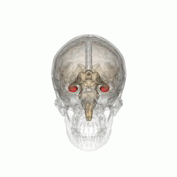
The hippocampus is a major component of the brain of humans and other vertebrates. Humans and other mammals have two hippocampi, one in each side of the brain. The hippocampus is part of the limbic system, and plays important roles in the consolidation of information from short-term memory to long-term memory, and in spatial memory that enables navigation. The hippocampus is located in the allocortex, with neural projections into the neocortex, in humans as well as other primates. The hippocampus, as the medial pallium, is a structure found in all vertebrates. In humans, it contains two main interlocking parts: the hippocampus proper, and the dentate gyrus.

<span class="mw-page-title-main">Dentate gyrus</span> Region of the hippocampus in the brain

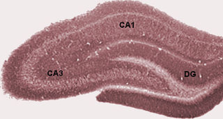
The dentate gyrus (DG) is part of the hippocampal formation in the temporal lobe of the brain, which also includes the hippocampus and the subiculum. The dentate gyrus is part of the hippocampal trisynaptic circuit and is thought to contribute to the formation of new episodic memories, the spontaneous exploration of novel environments and other functions.

<span class="mw-page-title-main">Spatial memory</span> Memory about ones environment and spatial orientation

In cognitive psychology and neuroscience, spatial memory is a form of memory responsible for the recording and recovery of information needed to plan a course to a location and to recall the location of an object or the occurrence of an event. Spatial memory is necessary for orientation in space. Spatial memory can also be divided into egocentric and allocentric spatial memory. A person's spatial memory is required to navigate around a familiar city. A rat's spatial memory is needed to learn the location of food at the end of a maze. In both humans and animals, spatial memories are summarized as a cognitive map.

<span class="mw-page-title-main">Place cell</span> Place-activated hippocampus cells found in some mammals

A place cell is a kind of pyramidal neuron in the hippocampus that becomes active when an animal enters a particular place in its environment, which is known as the place field. Place cells are thought to act collectively as a cognitive representation of a specific location in space, known as a cognitive map. Place cells work with other types of neurons in the hippocampus and surrounding regions to perform this kind of spatial processing. They have been found in a variety of animals, including rodents, bats, monkeys and humans.

<span class="mw-page-title-main">Cognitive map</span> Mental representation of information

A cognitive map is a type of mental representation which serves an individual to acquire, code, store, recall, and decode information about the relative locations and attributes of phenomena in their everyday or metaphorical spatial environment. The concept was introduced by Edward Tolman in 1948. He tried to explain the behavior of rats that appeared to learn the spatial layout of a maze, and subsequently the concept was applied to other animals, including humans. The term was later generalized by some researchers, especially in the field of operations research, to refer to a kind of semantic network representing an individual's personal knowledge or schemas.

<span class="mw-page-title-main">Motion perception</span> Inferring the speed and direction of objects

Motion perception is the process of inferring the speed and direction of elements in a scene based on visual, vestibular and proprioceptive inputs. Although this process appears straightforward to most observers, it has proven to be a difficult problem from a computational perspective, and difficult to explain in terms of neural processing.

<span class="mw-page-title-main">Subiculum</span> Most inferior part of the hippocampal formation

The subiculum is the most inferior component of the hippocampal formation. It lies between the entorhinal cortex and the CA1 subfield of the hippocampus proper.

Head direction (HD) cells are neurons found in a number of brain regions that increase their firing rates above baseline levels only when the animal's head points in a specific direction. They have been reported in rats, monkeys, mice, chinchillas and bats, but are thought to be common to all mammals, perhaps all vertebrates and perhaps even some invertebrates, and to underlie the "sense of direction". When the animal's head is facing in the cell's "preferred firing direction" these neurons fire at a steady rate, but firing decreases back to baseline rates as the animal's head turns away from the preferred direction.

<span class="mw-page-title-main">Path integration</span> Means of dead reckoning used by animals

Path integration is the method thought to be used by animals for dead reckoning.

<span class="mw-page-title-main">Hippocampal formation</span> Region of the temporal lobe in mammalian brains

The hippocampal formation is a compound structure in the medial temporal lobe of the brain. It forms a c-shaped bulge on the floor of the temporal horn of the lateral ventricle. There is no consensus concerning which brain regions are encompassed by the term, with some authors defining it as the dentate gyrus, the hippocampus proper and the subiculum; and others including also the presubiculum, parasubiculum, and entorhinal cortex. The hippocampal formation is thought to play a role in memory, spatial navigation and control of attention. The neural layout and pathways within the hippocampal formation are very similar in all mammals.

<span class="mw-page-title-main">Perforant path</span>

In the brain, the perforant path or perforant pathway provides a connectional route from the entorhinal cortex to all fields of the hippocampal formation, including the dentate gyrus, all CA fields, and the subiculum.

<span class="mw-page-title-main">Michael Hasselmo</span> American neuroscientist

Michael Hasselmo is an American neuroscientist and professor in the Department of Psychological and Brain Sciences at Boston University. He is the director of the Center for Systems Neuroscience and is editor-in-chief of Hippocampus (journal). Hasselmo studies oscillatory dynamics and neuromodulatory regulation in cortical mechanisms for memory guided behavior and spatial navigation using a combination of neurophysiological and behavioral experiments in conjunction with computational modeling. In addition to his peer-reviewed publications, Hasselmo wrote the book How We Remember: Brain Mechanisms of Episodic Memory.

<span class="mw-page-title-main">Hippocampus anatomy</span>

Hippocampus anatomy describes the physical aspects and properties of the hippocampus, a neural structure in the medial temporal lobe of the brain. It has a distinctive, curved shape that has been likened to the sea-horse monster of Greek mythology and the ram's horns of Amun in Egyptian mythology. This general layout holds across the full range of mammalian species, from hedgehog to human, although the details vary. For example, in the rat, the two hippocampi look similar to a pair of bananas, joined at the stems. In primate brains, including humans, the portion of the hippocampus near the base of the temporal lobe is much broader than the part at the top. Due to the three-dimensional curvature of this structure, two-dimensional sections such as shown are commonly seen. Neuroimaging pictures can show a number of different shapes, depending on the angle and location of the cut.

<span class="mw-page-title-main">Boundary cell</span>

Boundary cells are neurons found in the hippocampal formation that respond to the presence of an environmental boundary at a particular distance and direction from an animal. The existence of cells with these firing characteristics were first predicted on the basis of properties of place cells. Boundary cells were subsequently discovered in several regions of the hippocampal formation: the subiculum, presubiculum and entorhinal cortex.

<span class="mw-page-title-main">Edvard Moser</span> Norwegian psychologist and neuroscientist

Edvard Ingjald Moser is a Norwegian psychologist and neuroscientist, who is a professor at the Norwegian University of Science and Technology (NTNU) in Trondheim. In 2005, he and his then-wife May-Britt Moser discovered grid cells in the brain's medial entorhinal cortex. Grid cells are specialized neurons that provide the brain with a coordinate system and a metric for space. In 2018, he discovered a neural network that expresses a person's sense of time in experiences and memories located in the brain's lateral entorhinal cortex.

<span class="mw-page-title-main">May-Britt Moser</span> Norwegian psychologist and neuroscientist

May-Britt Moser is a Norwegian psychologist and neuroscientist, who is a Professor of Psychology and Neuroscience at the Norwegian University of Science and Technology (NTNU). She and her former husband, Edvard Moser, shared half of the 2014 Nobel Prize in Physiology or Medicine, awarded for work concerning the grid cells in the entorhinal cortex, as well as several additional space-representing cell types in the same circuit that make up the positioning system in the brain. Together with Edvard Moser she established the Moser research environment at NTNU, which they lead. Since 2012 she has headed the Centre for Neural Computation.

<span class="mw-page-title-main">John O'Keefe (neuroscientist)</span> American–British neuroscientist

John O'Keefe, is an American-British neuroscientist, psychologist and a professor at the Sainsbury Wellcome Centre for Neural Circuits and Behaviour and the Research Department of Cell and Developmental Biology at University College London. He discovered place cells in the hippocampus, and that they show a specific kind of temporal coding in the form of theta phase precession. He shared the Nobel Prize in Physiology or Medicine in 2014, together with May-Britt Moser and Edvard Moser; he has received several other awards. He has worked at University College London for his entire career, but also held a part-time chair at the Norwegian University of Science and Technology at the behest of his Norwegian collaborators, the Mosers.

<span class="mw-page-title-main">Phase precession</span> Neural mechanism

Phase precession is a neurophysiological process in which the time of firing of action potentials by individual neurons occurs progressively earlier in relation to the phase of the local field potential oscillation with each successive cycle. In place cells, a type of neuron found in the hippocampal region of the brain, phase precession is believed to play a major role in the neural coding of information. John O'Keefe, who later shared the 2014 Nobel Prize in Physiology or Medicine for his discovery that place cells help form a "map" of the body's position in space, co-discovered phase precession with Michael Recce in 1993.

Lisa Giocomo is an American neuroscientist who is a Professor in the Department of Neurobiology at Stanford University School of Medicine. Giocomo probes the molecular and cellular mechanisms underlying cortical neural circuits involved in spatial navigation and memory.

References

  1. 1 2 3 4 5 6 7 8 9 10 11 12 13 14 Hafting, T.; Fyhn, M.; Molden, S.; Moser, M. B.; Moser, E. I. (2005). "Microstructure of a spatial map in the entorhinal cortex". Nature. 436 (7052): 801–806. Bibcode:2005Natur.436..801H. doi:10.1038/nature03721. PMID   15965463. S2CID   4405184.
  2. Fyhn, M.; Hafting, T.; Witter, M. P.; Moser, E. I.; Moser, M. B. (2008). "Grid cells in mice". Hippocampus. 18 (12): 1230–1238. doi: 10.1002/hipo.20472 . PMID   18683845. S2CID   25196865.
  3. Yartsev, M. M.; Witter, M. P.; Ulanovsky, N. (2011). "Grid cells without theta oscillations in the entorhinal cortex of bats". Nature. 479 (7371): 103–107. Bibcode:2011Natur.479..103Y. doi:10.1038/nature10583. PMID   22051680. S2CID   1978069.
  4. Killian, N. J.; Jutras, M. J.; Buffalo, E. A. (2012). "A map of visual space in the primate entorhinal cortex". Nature. 491 (7426): 761–4. Bibcode:2012Natur.491..761K. doi:10.1038/nature11587. PMC   3565234 . PMID   23103863.
  5. Jacobs, J.; Weidemann, C. T.; Miller, J. F.; Solway, A.; Burke, J. F.; Wei, X. X.; Suthana, N.; Sperling, M. R.; Sharan, A. D.; Fried, I.; Kahana, M. J. (2013). "Direct recordings of grid-like neuronal activity in human spatial navigation". Nature Neuroscience. 16 (9): 1188–90. doi:10.1038/nn.3466. PMC   3767317 . PMID   23912946.
  6. Doeller, C. F.; Barry, C.; Burgess, N. (2010). "Evidence for grid cells in a human memory network". Nature. 463 (7281): 657–661. Bibcode:2010Natur.463..657D. doi:10.1038/nature08704. PMC   3173857 . PMID   20090680.
  7. O'Keefe, D. J. (1971). "The hippocampus as a spatial map. Preliminary evidence from unit activity in the freely-moving rat". Brain Research. 34 (1): 171–175. doi:10.1016/0006-8993(71)90358-1. PMID   5124915.
  8. O'Keefe, J.; Nadel, L. (1978). The Hippocampus as a Cognitive Map. Oxford University Press. Retrieved 2009-11-05.
  9. Quirk, M. R. (1992). "The positional firing properties of medial entorhinal neurons: description and comparison with hippocampal place cells". Journal of Neuroscience . 12 (5): 1945–1963. doi:10.1523/JNEUROSCI.12-05-01945.1992. PMC   6575876 . PMID   1578279.
  10. Moser MB, Moser EI (1998). "Functional differentiation in the hippocampus". Hippocampus. 8 (6): 608–19. doi:10.1002/(SICI)1098-1063(1998)8:6<608::AID-HIPO3>3.0.CO;2-7. PMID   9882018. S2CID   32384692.
  11. Maurer, A. P.; Vanrhoads, S. R.; Sutherland, G. R.; Lipa, P.; McNaughton, B. L. (2005). "Self-motion and the origin of differential spatial scaling along the septo-temporal axis of the hippocampus". Hippocampus. 15 (7): 841–852. doi:10.1002/hipo.20114. PMID   16145692. S2CID   7237113.
  12. Fyhn, M.; Molden, S.; Witter, M. P.; Moser, E. I.; Moser, M. -B. (2004). "Spatial Representation in the Entorhinal Cortex". Science. 305 (5688): 1258–1264. Bibcode:2004Sci...305.1258F. doi:10.1126/science.1099901. PMID   15333832.
  13. 1 2 Sargolini, F.; Fyhn, M.; Hafting, T.; McNaughton, B. L.; Witter, M. P.; Moser, M. -B.; Moser, E. I. (2006). "Conjunctive Representation of Position, Direction, and Velocity in Entorhinal Cortex". Science. 312 (5774): 758–762. Bibcode:2006Sci...312..758S. doi:10.1126/science.1125572. PMID   16675704.
  14. Brun, V. H.; Solstad, T.; Kjelstrup, K. B.; Fyhn, M.; Witter, M. P.; Moser, E. I.; Moser, M. B. (2008). "Progressive increase in grid scale from dorsal to ventral medial entorhinal cortex". Hippocampus. 18 (12): 1200–1212. doi: 10.1002/hipo.20504 . PMID   19021257.
  15. Mathis, A.; Herz, A. V. M.; Stemmler, M. B. (2012). "Optimal Population Codes for Space: Grid Cells Outperform Place Cells". Neural Computation. 24 (9): 2280–2317. doi:10.1162/NECO_a_00319. PMID   22594833. S2CID   15755674.
  16. Muller, RU; Kubie, JL (1987). "The effects of changes in the environment on the spatial firing of hippocampal complex-spike cells". J Neurosci. 7 (7): 1951–68. doi:10.1523/JNEUROSCI.07-07-01951.1987. PMC   6568940 . PMID   3612226.
  17. Fyhn, M.; Hafting, T.; Treves, A.; Moser, M. B.; Moser, E. I. (2007). "Hippocampal remapping and grid realignment in entorhinal cortex". Nature. 446 (7132): 190–194. Bibcode:2007Natur.446..190F. doi:10.1038/nature05601. PMID   17322902. S2CID   4431307.
  18. Kovács KA (September 2020). "Episodic Memories: How do the Hippocampus and the Entorhinal Ring Attractors Cooperate to Create Them?". Frontiers in Systems Neuroscience. 14: 68. doi: 10.3389/fnsys.2020.559186 . PMC   7511719 . PMID   33013334.
  19. Hafting, T.; Fyhn, M.; Bonnevie, T.; Moser, M. B.; Moser, E. I. (2008). "Hippocampus-independent phase precession in entorhinal grid cells". Nature. 453 (7199): 1248–1252. Bibcode:2008Natur.453.1248H. doi:10.1038/nature06957. PMID   18480753. S2CID   597353.
  20. Samsonovich a, M. A. B. (1997). "Path integration and cognitive mapping in a continuous attractor neural network model". Journal of Neuroscience. 17 (15): 5900–5920. doi:10.1523/JNEUROSCI.17-15-05900.1997. PMC   6573219 . PMID   9221787.
  21. 1 2 Burak, Y.; Fiete, I. R.; Sporns, O. (2009). Sporns, Olaf (ed.). "Accurate Path Integration in Continuous Attractor Network Models of Grid Cells". PLOS Computational Biology. 5 (2): e1000291. arXiv: 0811.1826 . Bibcode:2009PLSCB...5E0291B. doi:10.1371/journal.pcbi.1000291. PMC   2632741 . PMID   19229307.{{cite journal}}: CS1 maint: unflagged free DOI (link)
  22. Shrager, Y.; Kirwan, C. B.; Squire, L. R. (2008). "Neural basis of the cognitive map: Path integration does not require hippocampus or entorhinal cortex". Proceedings of the National Academy of Sciences. 105 (33): 12034–8. Bibcode:2008PNAS..10512034S. doi: 10.1073/pnas.0805414105 . PMC   2575247 . PMID   18687893.
  23. Sreenivasan, S; Fiete, I (2011). "Grid cells generate an analog error-correcting code for singularly precise neural computation". Nature Neuroscience. 14 (10): 1330–7. doi:10.1038/nn.2901. PMID   21909090. S2CID   17158393.