Hathewaya histolytica

Last updated

Hathewaya histolytica
Scientific classification OOjs UI icon edit-ltr.svg
Domain: Bacteria
Phylum: Bacillota
Class: Clostridia
Order: Eubacteriales
Family: Clostridiaceae
Genus: Hathewaya
Species:
H. histolytica
Binomial name
Hathewaya histolytica
(Weinberg and Séguin 1916) Lawson and Rainey 2016 [1]
Synonyms
  • Bacillus histolyticus
  • Clostridium histolyticum(Weinberg and Séguin 1916)
    Bergey, Harrison, Breed, Hammer, and Huntoon 1923
  • Weinbergillus histolyticus

Hathewaya histolytica (formerly Clostridium histolyticum) is a species of bacteria found in feces and the soil. It is a motile, gram-positive, aerotolerant anaerobe. H. histolytica is pathogenic in many species, including guinea pigs, mice, and rabbits, and humans. H. histolytica has been shown to cause gas gangrene, often in association with other bacteria species.

Contents

History

In 1916, Weinberg and Séguin isolated this bacterium from patients with gas gangrene and called it Bacillus histolyticus. They discovered this bacterium was pathogenic for guinea pigs, mice, and rabbits, but less so for rats. Intramuscular injection of culture caused extensive local tissue destruction, extrusion of a hemorrhagic muscle pulp, splitting of the skin, denudation of the bone, and sometimes autoamputation. In 1922, Heller renamed the bacterium Weinbergillus histolyticus, and a year later Bergey, Harrison, et al. reclassified it as Clostridium histolyticum. [2]

Microbiology

Hathewaya histolytica can be isolated from soil during the early stage of soil cultivation, by heating the sample at 60 °C for 30 minutes. H. histolytica can be plated on Zeissler plate agar, and appear as dewlike colonies of either rough or smooth morphology, surrounded by a zone of weak hemolysis. [3] On blood agar, colonies appear small, rough, irregularly round, and are surrounded by a zone of weak hemolysis. [4] These bacteria tend to clump in pairs or short chains and are rods of 3-5μm x 0.5-0.7μm. Cells are richly flagellate and very motile. Hathewaya histolytica produces large endospores and are asaccharolytic and proteolytic. [2] This bacterium is anaerobic, however minimal growth may be obtained through aerobic culture. [4]

Hathewaya histolytica is difficult to culture because growth is inhibited by sugars, and spores are not very heat resistant. [5] In wound smears, Hathewaya histolytica closely resembles the comparable Clostridium perfringens, but without the capsule of C. perfringens. This may interfere with diagnosis of H. histolytica infection. [4]

Toxigenicity

Studies have shown that the toxigenicity of a strain of Hathewaya histolytica is directly related to its sporulating potency: the higher the sporulating potency, the more toxigenic the strain. Additionally, toxigenic strains possess a stronger potential for growth than less toxigenic or nontoxigenic strains. Smooth substrains of H. histolytica seem to show higher toxigenicity than rough substrains. [3]

Toxins of Hathewaya histolytica

Hathewaya histolytica produces five toxins: alpha, beta, gamma, delta, and epsilon.[ citation needed ]

Alpha-toxin

The alpha-toxin is the major toxigenic factor of H. histolytica. When injected into muscle, it can cause death in laboratory animals within hours. Alpha-toxin is a necrotizing, but not hemolytic, toxin. This toxin is secreted, as it is isolated from filtrates of H. histolytica cultures. It is neutralized by antisera produced against toxic filtrates of C. septicum cultures through cross-neutralization. [5] Additionally, alpha-toxin is readily inactivated by proteolytic enzymes. It has been shown that only about 29% of H. histolytica strains isolated from soil actually produce this alpha-toxin. [3]

Beta-toxin

The beta-toxin of H. histolytica is a group of seven collagenases. Collagenases are zinc metalloproteases that cleave collagen and gelatin into small fragments. The seven collagenases are alpha, beta, gamma, delta, epsilon, zeta, and eta. They are further identified by their molecular masses (68, 115,79,110, 125, and 130 kDa, respectively) to distinguish them from the five toxins. Beta-toxin plays a major role in the pathogenicity of H. histolytica, due to its ability to destroy collagen fibers in the body and cause necrosis. Beta-toxin has been shown to induce hemorrhage when placed on the surface of lungs of animals, hemorrhage and edema when injected into rat paws, and lethal intrapulmonary hemorrhage when injected intravenously into animals. [5]

Gamma-toxin

Gamma-toxin is a cysteine-activated proteinase that digests hide powder, gelatin, and casein. It is not active against collagen. The molecular weight of gamma-toxin is 50,000 Da. [5]

Delta-toxin

Delta- toxin is an elastase activated by Ca2+ ions. This proteolytic enzyme is inhibited by cysteine and reversibly inactivated by reducing agents. The molecular weight is between 10 and 50 kDa. [5]

Epsilon-toxin

Epsilon-toxin is an oxygen-labile hemolysin similar to the θ-toxin of C. perfringens and the δ-toxins of C. septicum, and C. novyi. [5]

Proteinases

Toxigenic strains of Hathewaya histolytica secrete proteinases and collagenases that can degrade and necrotize organs and muscles in the human body. H. histolytica proteinases, including gamma- and delta-toxin, digest native and denatured proteins to amino acids with the production of ammonia. They can also clot milk and later digest the clot, and hemolyze sheep blood. [2] The proteinases are capable of digesting burn eschars, and may be useful for removal of burn tissue from wounds. [6] By themselves, the H. histolytica proteinases gamma-toxin and delta-toxin are not active in the human body. It seems they only attack collagenous material already partially degraded by the beta-toxin, and aid in the nutrition of H. histolytica. [4]

Hathewaya histolytica proteinases are unique in their efficiency of converting tissue proteins to amino acids and peptides. Further, these enzymes are unique in that they do not produce isoacids (isobutyric acid and isocaproic acid) as metabolic end products. [3]

Collagenase

Collagenase clostridium histolyticum is secreted by the bacterium and can destroy connective tissue of muscles. This collagenase has been used to treat Dupuytren's contracture, a disease of pathological collagen production and deposition in the hands. This disease causes flexion contractures of the joints, severely limiting hand function, most often in the ring and little fingers. Studies have shown that injection of collagenase clostridium histolyticum significantly reduces the contractures by lysing the collagen and disrupting the contracted cords. This treatment has restored contractured fingers to full extension 30 days after the last injection in 64% of joints injected with collagenase costridium histolyticum. Side effects are mild, and this treatment is preferred to surgical options because no extensive hand therapy is required post-treatment. Collagenase clostridium histolyticum is manufactured and marketed by Endo Pharmaceuticals in the US, and marketed by SOBI in the EU Auxilium / SOBI agreement, and by Actelion in Canada and Australia Auxilium / Actelion agreement.

Role in disease

Hathewaya histolytica secretes potent exotoxins that have proteolytic and necrotizing properties, causing severe local necrosis. However, there have been few cases of human infection by this species. From 1984 to 2004, only one case of H. histolytica necrotizing infection was reported, in an agricultural worker with a crushed-hand injury. [7]

In 2000, an 18-year-old female drug user in Turkey was diagnosed with infective endocarditis caused by H. histolytica. This was the first documented case where this bacterium has been identified as the cause of infective endocarditis. All diagnostic tests in this case were negative or normal until an anaerobic blood culture identified H. histolytica as the infectious agent isolated from the heart valve tissue. [8]

A 2002 study of the intestinal flora of inflammatory bowel disease (IBD) researchers found that in patients who had ulcerative colitis, a form of IBD, 21% of the total bacteria in the colon were Hathewaya histolytica. Control specimen did not contain this species at all. In healthy adults, the amount of clostridia species in the feces is rarely higher than 106 cells/g feces, less than 1% of the total flora. This suggests that C. histolyticum may play a role in the pathogenesis of ulcerative colitis as putative pathogens. [9]

In 2004, there was an outbreak of H. histolytica infections in necrotic lesions in 11 injecting drug users in England and Scotland. All patients were heroin users, and had injected the drug into the muscle rather than a vein. It was thought that H. histolytica spores had contaminated a batch of heroin early in production, and survived the production process before being distributed across the United Kingdom. [7]

Diagnosis and treatment of infection

Hathewaya histolytica can cause gas gangrene, an acute infection of pain, fever, myonecrosis, and massive edema. If not controlled, the infection can result in systemic toxemia, multiorgan failure, and even death. Further, this infection can progress to life-threatening in a matter of hours. [10]

While gas gangrene is easily diagnosed by the visible necrosis and characteristic smell, identifying Hathewaya histolytica as the causative agent is more difficult. With the exception of protein tests, all commonly used culture tests are negative. [5] There is little or no gas production, and this bacterium is negative for lecithinase and lipase reactions. Anaerobic blood cultures are necessary for identification of H. histolytica infection. [11]

For most infections, clindamycin and penicillin treatment is recommended. Further, a cocktail of antimicrobials targeting aerobic and anaerobic gram-positive and gram-negative bacteria may be used in necrotizing infections caused by multiple species, including H. histolytica. [11] H. histolytica is also susceptible to metronidazole and imipenem. [8] However, advanced gas gangrene infections caused by H. histolytica may only be treatable through amputation of the infected limb.[ citation needed ]

See also

Related Research Articles

<span class="mw-page-title-main">Gangrene</span> Type of tissue death by a lack of blood supply

Gangrene is a type of tissue death caused by a lack of blood supply. Symptoms may include a change in skin color to red or black, numbness, swelling, pain, skin breakdown, and coolness. The feet and hands are most commonly affected. If the gangrene is caused by an infectious agent, it may present with a fever or sepsis.

<span class="mw-page-title-main">Necrotizing fasciitis</span> Infection that results in the death of the bodys soft tissue

Necrotizing fasciitis (NF), also known as flesh-eating disease, is a bacterial infection that results in the death of parts of the body's soft tissue. It is a severe disease of sudden onset that spreads rapidly. Symptoms usually include red or purple skin in the affected area, severe pain, fever, and vomiting. The most commonly affected areas are the limbs and perineum.

<i>Clostridium</i> Genus of Gram-positive bacteria, which includes several significant human pathogens

Clostridium is a genus of anaerobic, Gram-positive bacteria. Species of Clostridium inhabit soils and the intestinal tract of animals, including humans. This genus includes several significant human pathogens, including the causative agents of botulism and tetanus. It also formerly included an important cause of diarrhea, Clostridioides difficile, which was reclassified into the Clostridioides genus in 2016.

<i>Clostridium perfringens</i> Species of bacterium

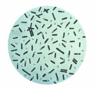
Clostridium perfringens is a Gram-positive, rod-shaped, anaerobic, spore-forming pathogenic bacterium of the genus Clostridium. C. perfringens is ever-present in nature and can be found as a normal component of decaying vegetation, marine sediment, the intestinal tract of humans and other vertebrates, insects, and soil. It has the shortest reported generation time of any organism at 6.3 minutes in thioglycolate medium.

<i>Corynebacterium diphtheriae</i> Species of prokaryote

Corynebacterium diphtheriae is the pathogenic bacterium that causes diphtheria. It is also known as the Klebs–Löffler bacillus, because it was discovered in 1884 by German bacteriologists Edwin Klebs (1834–1912) and Friedrich Löffler (1852–1915). The bacteria are usually harmless unless they are infected by a bacteriophage that carries a gene that gives rise to a toxin. This toxin causes the disease. Diphtheria is caused by the adhesion and infiltration of the bacteria into the mucosal layers of the body, primarily affecting the respiratory tract and the subsequent release of an endotoxin. The toxin has a localized effect on skin lesions, as well as a metastatic, proteolytic effects on other organ systems in severe infections. Originally a major cause of childhood mortality, diphtheria has been almost entirely eradicated due to the vigorous administration of the diphtheria vaccination in the 1910s.

<span class="mw-page-title-main">Gas gangrene</span> Human bacterial infection

Gas gangrene is a bacterial infection that produces tissue gas in gangrene. This deadly form of gangrene usually is caused by Clostridium perfringens bacteria. About 1,000 cases of gas gangrene are reported yearly in the United States.

<span class="mw-page-title-main">Hemolysis (microbiology)</span> Breakdown of red blood cells

Hemolysis is the breakdown of red blood cells. The ability of bacterial colonies to induce hemolysis when grown on blood agar is used to classify certain microorganisms. This is particularly useful in classifying streptococcal species. A substance that causes hemolysis is a hemolysin.

Collagenases are enzymes that break the peptide bonds in collagen. They assist in destroying extracellular structures in the pathogenesis of bacteria such as Clostridium. They are considered a virulence factor, facilitating the spread of gas gangrene. They normally target the connective tissue in muscle cells and other body organs.

Microbial collagenase is an enzyme. This enzyme catalyses the following chemical reaction

<i>Clostridium perfringens</i> alpha toxin Toxin produced by the bacterium Clostridium perfringens

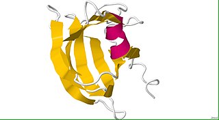
Clostridium perfringens alpha toxin is a toxin produced by the bacterium Clostridium perfringens and is responsible for gas gangrene and myonecrosis in infected tissues. The toxin also possesses hemolytic activity.

<i>Clostridium septicum</i> Species of bacterium

Clostridium septicum is a gram positive, spore forming, obligate anaerobic bacterium.

<span class="mw-page-title-main">Clostridium enterotoxin</span>

Clostridium enterotoxins are toxins produced by Clostridium species. Clostridial species are one of the major causes of food poisoning/gastrointestinal illnesses. They are anaerobic, gram-positive, spore-forming rods that occur naturally in the soil. Among the family are: Clostridium botulinum, which produces one of the most potent toxins in existence; Clostridium tetani, causative agent of tetanus; and Clostridium perfringens, commonly found in wound infections and diarrhea cases.

Microbial toxins are toxins produced by micro-organisms, including bacteria, fungi, protozoa, dinoflagellates, and viruses. Many microbial toxins promote infection and disease by directly damaging host tissues and by disabling the immune system. Endotoxins most commonly refer to the lipopolysaccharide (LPS) or lipooligosaccharide (LOS) that are in the outer plasma membrane of Gram-negative bacteria. The botulinum toxin, which is primarily produced by Clostridium botulinum and less frequently by other Clostridium species, is the most toxic substance known in the world. However, microbial toxins also have important uses in medical science and research. Currently, new methods of detecting bacterial toxins are being developed to better isolate and understand these toxin. Potential applications of toxin research include combating microbial virulence, the development of novel anticancer drugs and other medicines, and the use of toxins as tools in neurobiology and cellular biology.

Clostridium novyi (oedematiens) a Gram-positive, endospore- forming, obligate anaerobic bacteria of the class Clostridia. It is ubiquitous, being found in the soil and faeces. It is pathogenic, causing a wide variety of diseases in man and animals.

Collagenase clostridium histolyticum is an enzyme produced by the bacterium Clostridium histolyticum that dismantles collagen. It is used as a powder-and-solvent injection kit for the treatment of Dupuytren's contracture, a condition where the fingers bend towards the palm and cannot be fully straightened, and Peyronie's disease, a connective tissue disorder involving the growth of fibrous plaques in the soft tissue of the penis. BioSpecifics Technologies developed the preparation, which is manufactured and marketed by Endo Pharmaceuticals as Xiaflex in the US and by Sobi as Xiapex in Europe.

Clostridium perfringens beta toxin is one of the four major lethal protein toxins produced by Clostridium perfringens Type B and Type C strains. It is a necrotizing agent and it induces hypertension by release of catecholamine. It has been shown to cause necrotic enteritis in mammals and induces necrotizing intestinal lesions in the rabbit ileal loop model. C. perfringens beta toxin is susceptible to breakdown by proteolytic enzymes, particularly trypsin. Beta toxin is therefore highly lethal to infant mammals because of trypsin inhibitors present in the colostrum.

Clostridium innocuum is an anaerobic, non-motile, gram-positive bacterium that reproduces by sporulation. While there are over 130 species of Clostridium, C. innocuum is the third most commonly isolated. Although it is not normally considered an aggressive human pathogen, it has been isolated in some disease processes. C. innocuum and other Clostridium line the oropharynx and gastrointestinal tract, and are considered normal gut flora.

Anaerobic infections are caused by anaerobic bacteria. Obligately anaerobic bacteria do not grow on solid media in room air ; facultatively anaerobic bacteria can grow in the presence or absence of air. Microaerophilic bacteria do not grow at all aerobically or grow poorly, but grow better under 10% carbon dioxide or anaerobically. Anaerobic bacteria can be divided into strict anaerobes that can not grow in the presence of more than 0.5% oxygen and moderate anaerobic bacteria that are able of growing between 2 and 8% oxygen. Anaerobic bacteria usually do not possess catalase, but some can generate superoxide dismutase which protects them from oxygen.

<i>Clostridium tertium</i> Species of bacterium

Clostridium tertium is an anaerobic, motile, gram-positive bacterium. Although it can be considered an uncommon pathogen in humans, there has been substantial evidence of septic episodes in human beings. C. tertium is easily decolorized in Gram-stained smears and can be mistaken for a Gram-negative organism. However, C.tertium does not grow on selective media for Gram-negative organisms.

Clostridium novyi-NT is an attenuated form of Clostridium novyi that is under investigation as a cancer treatment. It is one of several pathogenic species of Clostridium bacteria that have been examined for this purpose. The modification eliminated the secretion of α-toxin.

References

  1. Page Species: Hathewaya histolytica on "LPSN - List of Prokaryotic names with Standing in Nomenclature". Deutsche Sammlung von Mikroorganismen und Zellkulturen . Retrieved 2022-09-27.
  2. 1 2 3 Oakley CL, Warrack GH (1950). "The alpha, beta and gamma antigens of Clostridium histolyticum (Weinberg & Séguin, 1916)". J. Gen. Microbiol. 4 (3): 365–373. doi: 10.1099/00221287-4-3-365 . PMID   14778941.
  3. 1 2 3 4 Nishida S, Imaizumi M (1966). "Toxigenicity of Clostridium histolyticum". J Bacteriol. 91 (2): 477–483. doi:10.1128/JB.91.2.477-483.1966. PMC   314883 . PMID   5935337.
  4. 1 2 3 4 MacLennan JD (1962). "The histotoxic clostridial infections of man". Bacteriol Rev. 26 (2_Pt_1–2): 117–274. doi:10.1128/MMBR.26.2_Pt_1-2.177-274.1962. PMC   441149 . PMID   14468017.
  5. 1 2 3 4 5 6 7 Hatheway, CL (1990). "Toxigenic Clostridia". Clin Microbiol Rev. 3 (1): 86–87. doi:10.1128/CMR.3.1.66. PMC   358141 . PMID   2404569.
  6. Jozwiak, J; Komar A; Jankowska E; Martirosian G (2005). "Determination of the cytotoxic effect of Clostridium histolyticum culture supernatant on HeLa cells in the presence of protease inhibitors". FEMS Immunol Med Microbiol. 45 (2): 137–142. doi: 10.1016/j.femsim.2005.03.005 . PMID   16051065.
  7. 1 2 Brazier, JS; Gal M; Hall V; Morris TE (2004). "Outbreak of Clostridium histolyticum infections in injecting drug users in England and Scotland". Euro Surveill. 9 (9): 15–16. doi: 10.2807/esm.09.09.00475-en . PMID   15381836.
  8. 1 2 Durmaz, B; Agel HE; Sonmez E; Turkoz R; Aydin E (2000). "Infective endocarditis due to clostridium histolyticum". Clin Microbiol Infect. 6 (10): 561–563. doi: 10.1046/j.1469-0691.2000.00110.x . PMID   11168053.
  9. Kleessen, B; Kroesen AJ; Buhr HJ; Blunt M (2002). "Mucosal and invading bacteria in patients with inflammatory bowel disease compared with controls". Scand J Gastroenterol. 37 (9): 1034–1041. doi:10.1080/003655202320378220. PMID   12374228. S2CID   10819885.
  10. Flores-Díaz, M; Alape-Girón A (2003). "Role of Clostridium perfringens phospholipase C in the pathogenesis of gas gangrene". Toxicon. 42 (8): 979–986. doi:10.1016/j.toxicon.2003.11.013. PMID   15019495.
  11. 1 2 Stevens, DL; Bisno AL; Chambers HF; Everett ED; Dellinger P; Goldstein EJC; Gorbach SL; Hirschmann JV; Kaplan EL; Montoya JG; Wade JC (2005). "Practice guidelines for the diagnosis and management of skin and soft-tissue infections". Clin Infect Dis. 41 (10): 1373–406. doi: 10.1086/497143 . PMID   16231249.