The ulna or ulnar bone is a long bone in the forearm stretching from the elbow to the wrist. It is on the same side of the forearm as the little finger, running parallel to the radius, the forearm's other long bone. Longer and thinner than the radius, the ulna is considered to be the smaller long bone of the lower arm. The corresponding bone in the lower leg is the fibula.

The humerus is a long bone in the arm that runs from the shoulder to the elbow. It connects the scapula and the two bones of the lower arm, the radius and ulna, and consists of three sections. The humeral upper extremity consists of a rounded head, a narrow neck, and two short processes. The body is cylindrical in its upper portion, and more prismatic below. The lower extremity consists of 2 epicondyles, 2 processes, and 3 fossae. As well as its true anatomical neck, the constriction below the greater and lesser tubercles of the humerus is referred to as its surgical neck due to its tendency to fracture, thus often becoming the focus of surgeons.

In human anatomy, the wrist is variously defined as (1) the carpus or carpal bones, the complex of eight bones forming the proximal skeletal segment of the hand; (2) the wrist joint or radiocarpal joint, the joint between the radius and the carpus and; (3) the anatomical region surrounding the carpus including the distal parts of the bones of the forearm and the proximal parts of the metacarpus or five metacarpal bones and the series of joints between these bones, thus referred to as wrist joints. This region also includes the carpal tunnel, the anatomical snuff box, bracelet lines, the flexor retinaculum, and the extensor retinaculum.
The forearm is the region of the upper limb between the elbow and the wrist. The term forearm is used in anatomy to distinguish it from the arm, a word which is used to describe the entire appendage of the upper limb, but which in anatomy, technically, means only the region of the upper arm, whereas the lower "arm" is called the forearm. It is homologous to the region of the leg that lies between the knee and the ankle joints, the crus.

In human anatomy, the ulnar nerve is a nerve that runs near the ulna bone. The ulnar collateral ligament of elbow joint is in relation with the ulnar nerve. The nerve is the largest in the human body unprotected by muscle or bone, so injury is common. This nerve is directly connected to the little finger, and the adjacent half of the ring finger, innervating the palmar aspect of these fingers, including both front and back of the tips, perhaps as far back as the fingernail beds.

The radius or radial bone is one of the two large bones of the forearm, the other being the ulna. It extends from the lateral side of the elbow to the thumb side of the wrist and runs parallel to the ulna. The ulna is longer than the radius, but the radius is thicker. The radius is a long bone, prism-shaped and slightly curved longitudinally.

The upper limbs or upper extremities are the forelimbs of an upright-postured tetrapod vertebrate, extending from the scapulae and clavicles down to and including the digits, including all the musculatures and ligaments involved with the shoulder, elbow, wrist and knuckle joints. In humans, each upper limb is divided into the arm, forearm and hand, and is primarily used for climbing, lifting and manipulating objects.

The olecranon, is a large, thick, curved bony process on the proximal, posterior end of the ulna. It forms the protruding part of the elbow and is opposite to the cubital fossa or elbow pit. The olecranon serves as a lever for the extensor muscles that straighten the elbow joint.

A pulled elbow, also known as nursemaid's elbow or a radial head subluxation, is when the ligament that wraps around the radial head slips off. Often a child will hold their arm against their body with the elbow slightly bent. They will not move the arm as this results in pain. Touching the arm, without moving the elbow, is usually not painful.
The pronator teres is a muscle that, along with the pronator quadratus, serves to pronate the forearm. It has two origins, at the medial humeral supracondylar ridge and the ulnar tuberosity, and inserts near the middle of the radius.

In human anatomy, the supinator is a broad muscle in the posterior compartment of the forearm, curved around the upper third of the radius. Its function is to supinate the forearm.
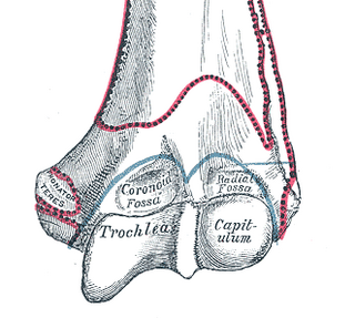
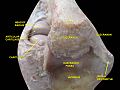
In the human arm, the humeral trochlea is the medial portion of the articular surface of the elbow joint which articulates with the trochlear notch on the ulna in the forearm.

The annular ligament is a strong band of fibers that encircles the head of the radius, and retains it in contact with the radial notch of the ulna.

The radial collateral ligament (RCL), lateral collateral ligament (LCL), or external lateral ligament is a ligament in the elbow on the side of the radius.
The proximal radioulnar articulation, also known as the proximal radioulnar joint (PRUJ), is a synovial pivot joint between the circumference of the head of the radius and the ring formed by the radial notch of the ulna and the annular ligament.
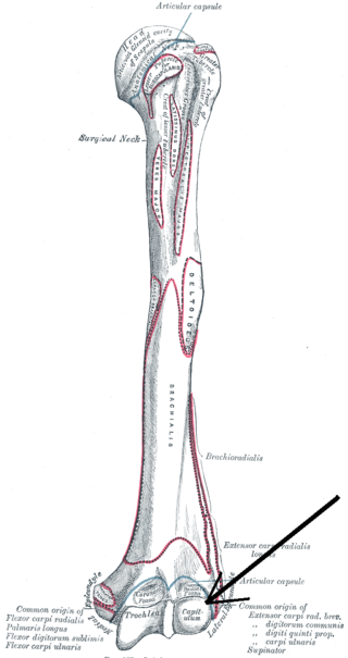
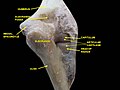
In human anatomy of the arm, the capitulum of the humerus is a smooth, rounded eminence on the lateral portion of the distal articular surface of the humerus. It articulates with the cup-shaped depression on the head of the radius, and is limited to the front and lower part of the bone.

The humeroradial joint is the joint between the head of the radius and the capitulum of the humerus, is a limited ball-and-socket joint, hinge type of synovial joint.
The humeroulnar joint is part of the elbow-joint. It is composed of two bones, the humerus and ulna, and is the junction between the trochlear notch of ulna and the trochlea of humerus. It is classified as a simple hinge-joint, which allows for movements of flexion, extension and circumduction. Owing to the obliquity of the trochlea of the humerus, this movement does not take place in the antero-posterior plane of the body of the humerus.

The elbow is the region between the upper arm and the forearm that surrounds the elbow joint. The elbow includes prominent landmarks such as the olecranon, the cubital fossa, and the lateral and the medial epicondyles of the humerus. The elbow joint is a hinge joint between the arm and the forearm; more specifically between the humerus in the upper arm and the radius and ulna in the forearm which allows the forearm and hand to be moved towards and away from the body. The term elbow is specifically used for humans and other primates, and in other vertebrates it is not used. In those cases, forelimb plus joint is used.
In human anatomy, the quadrate ligament or ligament of Denucé is one of the ligaments of the proximal radioulnar joint in the upper forearm.