High resolution electron energy loss spectroscopy

Last updated

High resolution electron energy loss spectroscopy (HREELS) is a tool used in surface science. The inelastic scattering of electrons from surfaces is utilized to study electronic excitations or vibrational modes of the surface of a material or of molecules adsorbed to a surface. In contrast to other electron energy loss spectroscopies (EELS), HREELS deals with small energy losses in the range of 10−3 eV to 1 eV. It plays an important role in the investigation of surface structure, catalysis, dispersion of surface phonons and the monitoring of epitaxial growth.

Contents

Overview of HREELS

An HREELS spectrum showing surface phonons at 15 and 38 meV. The appearance of peaks on both sides of the elastic scattering peak can be understood by comparison to Raman spectroscopy. HREELS spectrum.gif
An HREELS spectrum showing surface phonons at 15 and 38 meV. The appearance of peaks on both sides of the elastic scattering peak can be understood by comparison to Raman spectroscopy.

In general, electron energy loss spectroscopy is based on the energy losses of electrons when inelastically scattered on matter. An incident beam of electrons with a known energy (Ei) is scattered on a sample. The scattering of these electrons can excite the electronic structure of the sample. If this is the case the scattered electron loses the specific energy (ΔE) needed to cause the excitation. Those scattering processes are called inelastic. It may be easiest to imagine that the energy loss is for example due to an excitation of an electron from an atomic K-shell to the M-shell. The energy for this excitation is taken away from the electron's kinetic energy. The energies of the scattered electrons (Es) are measured and the energy loss can be calculated. From the measured data an intensity versus energy loss diagram is established. In the case of scattering by phonons the so-called energy loss can also be a gain of energy (similar to anti-Stokes Raman spectroscopy). These energy losses allow, using comparison to other experiments or theory, one to draw conclusions about surface properties of a sample.

Excitations of the surface structure are usually very low energy, ranging from 10−3 eV to 10 eV. In HREELS spectra electrons with only small energy losses, like also Raman scattering, the interesting features are all located very close together and especially near to the very strong elastic scattering peak. Hence EELS spectrometers require a high energy resolution. Therefore, this regime of EELS is called High Resolution EELS. In this context resolution shall be defined as the energy difference in which two features in a spectrum are just distinguishable divided by the mean energy of those features:

In the case of EELS the first thing to think of in order to achieve high resolution is using incident electrons of a very precisely defined energy and a high quality analyzer. Further high resolution is only possible when the energies of the incident electrons are not far bigger than the energy losses. For HREELS the energy of the incident electrons is therefore mostly significantly smaller than 102 eV.

Considering that 102 eV electrons have a mean free path of around 1 nm (corresponds to a few monolayers), which decreases with lower energies, this automatically implies that HREELS is a surface sensitive technique. This is the reason why HREELS must be measured in reflection mode and must be implemented in ultra high vacuum (UHV). This is in contrast to Core Level EELS which operates at very high energies and can therefore also be found in transmission electron microscopes (TEM). Instrumental developments have also enabled vibrational spectroscopy to be performed in TEM. [1] [2]

In HREELS not only the electron energy loss can be measured, often the angular distribution of electrons of a certain energy loss in reference to the specular direction gives interesting insight to the structures on a surface.

Physics of HREELS

As mentioned above HREELS involves an inelastic scattering process on a surface. For those processes the conservation of energy as well as the conservation of momentum's projection parallel to the surface hold:

E are energies, k and q are wave vectors and G denotes a reciprocal lattice vector. One should mention at this point that for non perfect surfaces G is not in any case a well defined quantum number, what has to be considered when using the second relation. Variables subscripted with i denote values of incident electrons those subscripted with s values of scattered electrons. "||" denotes parallel to the surface.

For the description of the inelastic scattering processes due to the excitation of vibrational modes of adsorbates different approaches exist. The simplest approach distinguishes between regimes of small and large scattering angles:

Dipole scattering

Figurative interpretation of dipole scattering HREELS dipole scattering.JPG
Figurative interpretation of dipole scattering

The so-called dipole scattering can be applied when the scattered beam is very near to the specular direction. In this case a macroscopic theory can be applied to explain the results. It can be approached using the so-called dielectrical theory introduced by Lucas and Šunjić of which a quantum mechanical treatment was first presented by E. Evans and D.L. Mills in the early 1970s. [3]

Alternatively there is a more unfamiliar model which only holds exactly for perfect conductors: A unit cell at the surface does not have a homogeneous surrounding, hence it is supposed to have an electrical dipole moment. When a molecule is adsorbed to the surface there can be an additional dipole moment and the total dipole moment P is present. This dipole moment causes a long range electronic potential in the vacuum above the surface. On this potential the incident electron can scatter inelastically which means it excites vibrations in the dipole structure. The dipole moment can then be written as . When the adsorbate sticks to a metal surface, imaginary dipoles occur as shown in the figure on the right. Hence for an adsorbed dipole normal to the surface the dipole moment "seen" from the vacuum doubles. Whereas the dipole moment of a parallel to the surface adsorbed dipole vanishes. Hence an incident electron can excite the adsorbed dipole only when it is adsorbed normal to the surface and the vibrational mode can be detected in the energy loss spectrum. If the dipole is adsorbed parallel then no energy losses will be detected and the vibrational modes of the dipole are missing in the energy loss spectrum. When measuring the intensity of the electron energy loss peaks and comparing to other experimental results or to theoretical models it can also be determined whether a molecule is adsorbed normal to the surface or tilted by an angle.

The dielectric model also holds when the material on which the molecule adsorbs is not a metal. The picture shown above is then the limit for where denotes the relative dielectrical constant.

As the incident electron in this model is scattered in the region above the surface, it does not directly impact the surface and because the amount of momentum transferred is small the scattering is mostly in the specular direction.

Impact scattering

Impact scattering is the regime which deals with electrons that are scattered further away from the specular direction. In those cases no macroscopic theory exists and a microscopic theory like, quantum mechanical dispersion theory, has to be applied. Symmetry considerations then also result in certain selection rules (it is also assumed that the energy loss in the inelastic scattering process is negligible):

All those selection rules make it possible to identify the normal coordinates of the adsorbed molecules.

Intermediate negative ion resonance

In intermediate negative ion resonance the electron forms a compound state with an adsorbed molecule during the scattering process. However, the lifetime of those states are so short that this type of scattering is barely observed. All of these regimes can at once be described with the help of the single microscopic theory.

Selection rules for dipole scattering from the perspective of vibrational eigenmodes

A microscopic theory makes it possible to approach the selection rule for dipole scattering in a more exact way. The scattering cross section is only non-vanishing in the case of a non-zero matrix element . Where i denotes the initial and f the final vibrational energy level of the adsorbed molecule and pz the z component of its dipole moment.

As the dipole moment is something like charge times length, pz has the same symmetry properties as z, which is totally symmetric. Hence the product of i and f must also be a totally symmetric function, otherwise the matrix element vanishes. Hence

excitations from the totally symmetrical ground state of a molecule are only possible to a totally symmetric vibrational state.

This is the surface selection rule for dipole scattering. Note that it says nothing about the intensity for scattering or the displacement of the atoms of the adsorbate, but its total dipole moment is the operator in the matrix element. This is important as a vibration of the atoms parallel to the surface can also cause a vibration of the dipole moment normal to the surface. Therefore, the result in the "dipole scattering" section above is not exactly correct.

When trying to gain information from selection rules, one must carefully consider whether a pure dipole or impact scattering region is investigated. Further symmetry-breaking due to strong bindings to the surface must be considered. Another problem is that in cases of larger molecules often many vibrational modes are degenerate, which could again be resolved due to strong molecule-surface interactions. Those interactions can also generate completely new dipole moments which the molecule does not have on its own. But when carefully investigating it is mostly possible to get a very good picture of how the molecule adheres to the surface by analysis of normal dipole modes.[ citation needed ]

High resolution electron energy loss spectrometer

Principle of a HREELS setup HREELS Spectrometer.svg
Principle of a HREELS setup

As the electrons used for HREELS are of low energy they do not only have a very short mean free path length in the sample materials but also under normal atmospheric conditions. Therefore, one has to set up the spectrometer in UHV. The spectrometer is in general a computer simulated design that optimizes the resolution while keeping an acceptable electron flux.

The electrons are generated in an electron source, by heating a tungsten cathode, which is encapsulated by a negatively charged so called repeller that prevents stray electrons from coming into the detector unit. The electrons can leave the source only through a lens system, like e.g. a slot lens system consisting of several slits all on different potential. The purpose of this system is to focus the electrons on the entrance of the monochromator unit, to get a high initial electron flux.

The monochromator is usually a concentric hemispherical analyser (CHA). In more sensitive setups an additional pre-monochromator is used. The task of the monochromator is to reduce the energy of the passing electrons to some eV due to the help of electron lenses. It further lets only those electrons pass which have the chosen initial energy. To achieve a good resolution it is already important to have incident electrons of a well defined energy one normally chooses a resolution of for the monochromator. This means, the electrons leaving the monochromator with e.g. 10 eV have an energy accurate to 10−1 eV. The beam's flux is then in the orders of 10−8 A to 10−10 A. The radii of the CHA are in the order of several 10 mm. And the deflector electrodes have a saw tooth profile to backscatter electrons which are reflected from the walls in order to reduce the background of electrons with the wrong Ei. The electrons are then focused by a lens system onto the sample. These lenses are, in contrary to those of the emitter system very flexible, as it is important is to get a good focus on the sample. To enable measurements of angular distributions all those elements are mounted on a rotate able table with the axis cantered at the sample.Its negative charge causes the electron beam to broaden. What can be prevented by charging the top and bottom plates of the CHA deflectors negative. What again causes a change in the deflection angle and has to be considered when designing the experiment.

In the scattering process at the sample the electrons can lose energies from several 10−2 eV up to a few electron volt. The scattered electron beam which is of around 10−3 lower flux than the incident beam then enters, the analyzer, another CHA.

The analyzer CHA again allows only electrons of certain energies to pass to the analyzing unit, a channel electron multiplier (CEM). For this analyzing CHA the same facts are valid as for the monochromator. Except that a higher resolution as in the monochromator is wanted. Hence the radial dimensions of this CHA are mostly bigger by like a factor 2. Due to aberrations of the lens systems the beam has also broadened. To sustain a high enough electron flux to the analyzer the apertures are also about a factor 2 bigger. To make the analysis more accurate, especially to reduce the background of in the deflector scattered electrons often two analyzers are used, or additional apertures are added behind the analyzers as scattered electrons of the wrong energy normally leave the CHAs under large angles. In this way energy losses of 10−2 eV to 10 eV can be detected with accuracies of about 10−2 eV.

General problems of HREEL spectrometers

Due to the electron flux the apertures can become negatively charged, which makes them effectively smaller for the passing electrons. This has to be considered when doing the design of the setup as it is anyway difficult to keep different potentials, of repeller, lenses, screening elements, and the reflector, constant. Unstable potentials on lenses or CHA deflectors would cause fluctuations in the measured signal. Similar problems are caused by external electric or magnetic fields, either they cause fluctuations in the signal, or add a constant offset. That is why the sample is normally shielded by equipotential, metal electrodes to keep the region of the sample field free so that neither the probe electrons nor the sample is affected by external electric fields. Further a cylinder of a material with a high magnetic permeability, e.g. Mu-metal, built around the whole spectrometer to keep magnetic fields or field inhomogeneities at the experiment down to 10 mG or 1mG/cm. Because of the same reason the whole experiment, except the lenses which are normally made of coated copper, is designed in stainless antimagnetic steel and insulating parts are avoided wherever possible.

See also

Related Research Articles

<span class="mw-page-title-main">Infrared spectroscopy</span> Measurement of infrared radiations interaction with matter

Infrared spectroscopy is the measurement of the interaction of infrared radiation with matter by absorption, emission, or reflection. It is used to study and identify chemical substances or functional groups in solid, liquid, or gaseous forms. It can be used to characterize new materials or identify and verify known and unknown samples. The method or technique of infrared spectroscopy is conducted with an instrument called an infrared spectrometer which produces an infrared spectrum. An IR spectrum can be visualized in a graph of infrared light absorbance on the vertical axis vs. frequency, wavenumber or wavelength on the horizontal axis. Typical units of wavenumber used in IR spectra are reciprocal centimeters, with the symbol cm−1. Units of IR wavelength are commonly given in micrometers, symbol μm, which are related to the wavenumber in a reciprocal way. A common laboratory instrument that uses this technique is a Fourier transform infrared (FTIR) spectrometer. Two-dimensional IR is also possible as discussed below.

<span class="mw-page-title-main">Spectroscopy</span> Study involving matter and electromagnetic radiation

Spectroscopy is the field of study that measures and interprets the electromagnetic spectra that result from the interaction between electromagnetic radiation and matter as a function of the wavelength or frequency of the radiation. In simpler terms, spectroscopy is the precise study of color as generalized from visible light to all bands of the electromagnetic spectrum.

<span class="mw-page-title-main">Raman spectroscopy</span> Spectroscopic technique

Raman spectroscopy is a spectroscopic technique typically used to determine vibrational modes of molecules, although rotational and other low-frequency modes of systems may also be observed. Raman spectroscopy is commonly used in chemistry to provide a structural fingerprint by which molecules can be identified.

<span class="mw-page-title-main">X-ray photoelectron spectroscopy</span> Spectroscopic technique

X-ray photoelectron spectroscopy (XPS) is a surface-sensitive quantitative spectroscopic technique based on the photoelectric effect that can identify the elements that exist within a material or are covering its surface, as well as their chemical state, and the overall electronic structure and density of the electronic states in the material. XPS is a powerful measurement technique because it not only shows what elements are present, but also what other elements they are bonded to. The technique can be used in line profiling of the elemental composition across the surface, or in depth profiling when paired with ion-beam etching. It is often applied to study chemical processes in the materials in their as-received state or after cleavage, scraping, exposure to heat, reactive gasses or solutions, ultraviolet light, or during ion implantation.

<span class="mw-page-title-main">Electron energy loss spectroscopy</span> Form of microscopy using an electron beam

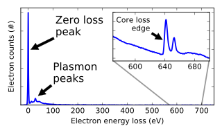
Electron energy loss spectroscopy (EELS) is a form of electron microscopy in which a material is exposed to a beam of electrons with a known, narrow range of kinetic energies. Some of the electrons will undergo inelastic scattering, which means that they lose energy and have their paths slightly and randomly deflected. The amount of energy loss can be measured via an electron spectrometer and interpreted in terms of what caused the energy loss. Inelastic interactions include phonon excitations, inter- and intra-band transitions, plasmon excitations, inner shell ionizations, and Cherenkov radiation. The inner-shell ionizations are particularly useful for detecting the elemental components of a material. For example, one might find that a larger-than-expected number of electrons comes through the material with 285 eV less energy than they had when they entered the material. This is approximately the amount of energy needed to remove an inner-shell electron from a carbon atom, which can be taken as evidence that there is a significant amount of carbon present in the sample. With some care, and looking at a wide range of energy losses, one can determine the types of atoms, and the numbers of atoms of each type, being struck by the beam. The scattering angle can also be measured, giving information about the dispersion relation of whatever material excitation caused the inelastic scattering.

<span class="mw-page-title-main">Fluorescence spectroscopy</span> Type of electromagnetic spectroscopy

Fluorescence spectroscopy is a type of electromagnetic spectroscopy that analyzes fluorescence from a sample. It involves using a beam of light, usually ultraviolet light, that excites the electrons in molecules of certain compounds and causes them to emit light; typically, but not necessarily, visible light. A complementary technique is absorption spectroscopy. In the special case of single molecule fluorescence spectroscopy, intensity fluctuations from the emitted light are measured from either single fluorophores, or pairs of fluorophores.

<span class="mw-page-title-main">Raman scattering</span> Inelastic scattering of photons

Raman scattering or the Raman effect is the inelastic scattering of photons by matter, meaning that there is both an exchange of energy and a change in the light's direction. Typically this effect involves vibrational energy being gained by a molecule as incident photons from a visible laser are shifted to lower energy. This is called normal Stokes Raman scattering. The effect is exploited by chemists and physicists to gain information about materials for a variety of purposes by performing various forms of Raman spectroscopy. Many other variants of Raman spectroscopy allow rotational energy to be examined and electronic energy levels may be examined if an X-ray source is used in addition to other possibilities. More complex techniques involving pulsed lasers, multiple laser beams and so on are known.

<span class="mw-page-title-main">Neutron scattering</span> Physical phenomenon

Neutron scattering, the irregular dispersal of free neutrons by matter, can refer to either the naturally occurring physical process itself or to the man-made experimental techniques that use the natural process for investigating materials. The natural/physical phenomenon is of elemental importance in nuclear engineering and the nuclear sciences. Regarding the experimental technique, understanding and manipulating neutron scattering is fundamental to the applications used in crystallography, physics, physical chemistry, biophysics, and materials research.

<span class="mw-page-title-main">Resonance Raman spectroscopy</span> Raman spectroscopy technique

Resonance Raman spectroscopy is a variant of Raman spectroscopy in which the incident photon energy is close in energy to an electronic transition of a compound or material under examination. This similarity in energy (resonance) leads to greatly increased intensity of the Raman scattering of certain vibrational modes, compared to ordinary Raman spectroscopy.

In physics and chemistry, a selection rule, or transition rule, formally constrains the possible transitions of a system from one quantum state to another. Selection rules have been derived for electromagnetic transitions in molecules, in atoms, in atomic nuclei, and so on. The selection rules may differ according to the technique used to observe the transition. The selection rule also plays a role in chemical reactions, where some are formally spin-forbidden reactions, that is, reactions where the spin state changes at least once from reactants to products.

<span class="mw-page-title-main">X-ray absorption spectroscopy</span> Panel of different types of X-ray absorption spectroscopy requiring a synchrotron radiation facility

X-ray absorption spectroscopy (XAS) is a widely used technique for determining the local geometric and/or electronic structure of matter. The experiment is usually performed at synchrotron radiation facilities, which provide intense and tunable X-ray beams. Samples can be in the gas phase, solutions, or solids.

Helium atom scattering (HAS) is a surface analysis technique used in materials science. It provides information about the surface structure and lattice dynamics of a material by measuring the diffracted atoms from a monochromatic helium beam incident on the sample.

<span class="mw-page-title-main">Low-energy ion scattering</span>

Low-energy ion scattering spectroscopy (LEIS), sometimes referred to simply as ion scattering spectroscopy (ISS), is a surface-sensitive analytical technique used to characterize the chemical and structural makeup of materials. LEIS involves directing a stream of charged particles known as ions at a surface and making observations of the positions, velocities, and energies of the ions that have interacted with the surface. Data that is thus collected can be used to deduce information about the material such as the relative positions of atoms in a surface lattice and the elemental identity of those atoms. LEIS is closely related to both medium-energy ion scattering (MEIS) and high-energy ion scattering, differing primarily in the energy range of the ion beam used to probe the surface. While much of the information collected using LEIS can be obtained using other surface science techniques, LEIS is unique in its sensitivity to both structure and composition of surfaces. Additionally, LEIS is one of a very few surface-sensitive techniques capable of directly observing hydrogen atoms, an aspect that may make it an increasingly more important technique as the hydrogen economy is being explored.

<span class="mw-page-title-main">Surface phonon</span>

In solid state physics, a surface phonon is the quantum of a lattice vibration mode associated with a solid surface. Similar to the ordinary lattice vibrations in a bulk solid, the nature of surface vibrations depends on details of periodicity and symmetry of a crystal structure. Surface vibrations are however distinct from the bulk vibrations, as they arise from the abrupt termination of a crystal structure at the surface of a solid. Knowledge of surface phonon dispersion gives important information related to the amount of surface relaxation, the existence and distance between an adsorbate and the surface, and information regarding presence, quantity, and type of defects existing on the surface.

Inelastic electron tunneling spectroscopy (IETS) is an experimental tool for studying the vibrations of molecular adsorbates on metal oxides. It yields vibrational spectra of the adsorbates with high resolution (< 0.5 meV) and high sensitivity (< 1013 molecules are required to provide a spectrum). An additional advantage is the fact that optically forbidden transitions may be observed as well. Within IETS, an oxide layer with molecules adsorbed on it is put between two metal plates. A bias voltage is applied between the two contacts. An energy diagram of the metal-oxide-metal device under bias is shown in the top figure. The metal contacts are characterized by a constant density of states, filled up to the Fermi energy. The metals are assumed to be equal. The adsorbates are situated on the oxide material. They are represented by a single bridge electronic level, which is the upper dashed line. If the insulator is thin enough, there is a finite probability that the incident electron tunnels through the barrier. Since the energy of the electron is not changed by this process, it is an elastic process. This is shown in the left figure.

X-ray Raman scattering (XRS) is non-resonant inelastic scattering of X-rays from core electrons. It is analogous to vibrational Raman scattering, which is a widely used tool in optical spectroscopy, with the difference being that the wavelengths of the exciting photons fall in the X-ray regime and the corresponding excitations are from deep core electrons.

<span class="mw-page-title-main">Resonant inelastic X-ray scattering</span> Advanced X-ray spectroscopy technique

Resonant inelastic X-ray scattering (RIXS) is an advanced X-ray spectroscopy technique.

The technique of vibrational analysis with scanning probe microscopy allows probing vibrational properties of materials at the submicrometer scale, and even of individual molecules. This is accomplished by integrating scanning probe microscopy (SPM) and vibrational spectroscopy. This combination allows for much higher spatial resolution than can be achieved with conventional Raman/FTIR instrumentation. The technique is also nondestructive, requires non-extensive sample preparation, and provides more contrast such as intensity contrast, polarization contrast and wavelength contrast, as well as providing specific chemical information and topography images simultaneously.

Electron orbital imaging is an X-ray synchrotron technique used to produce images of electron orbitals in real space. It utilizes the technique of X-ray Raman scattering (XRS), also known as Non-resonant Inelastic X-Ray Scattering (NIXS) to inelastically scatter electrons off a single crystal. It is an element specific spectroscopic technique for studying the valence electrons of transition metals.

Raman spectroelectrochemistry (Raman-SEC) is a technique that studies the inelastic scattering or Raman scattering of monochromatic light related to chemical compounds involved in an electrode process. This technique provides information about vibrational energy transitions of molecules, using a monochromatic light source, usually from a laser that belongs to the UV, Vis or NIR region. Raman spectroelectrochemistry provides specific information about structural changes, composition and orientation of the molecules on the electrode surface involved in an electrochemical reaction, being the Raman spectra registered a real fingerprint of the compounds.

References

  1. Krivanek, Ondrej L.; Lovejoy, Tracy C.; Dellby, Niklas; Aoki, Toshihiro; Carpenter, R. W.; Rez, Peter; Soignard, Emmanuel; Zhu, Jiangtao; Batson, Philip E.; Lagos, Maureen J.; Egerton, Ray F. (2014). "Vibrational spectroscopy in the electron microscope". Nature. 514 (7521): 209–212. doi:10.1038/nature13870. ISSN   0028-0836. PMID   25297434. S2CID   4467249.
  2. Venkatraman, Kartik; Levin, Barnaby D.A.; March, Katia; Rez, Peter; Crozier, Peter A. (2019). "Vibrational spectroscopy at atomic resolution with electron impact scattering". Nature Physics. 15 (12): 1237–1241. arXiv: 1812.08895 . doi:10.1038/s41567-019-0675-5. S2CID   119452520.
  3. E.Evans; D.L. Mills (1972). "Theory of Inelastic Scattering of Slow Electrons by Long-Wavelength Surface Optical Phonons". Phys. Rev. B. 5 (10): 4126–4139. doi:10.1103/PhysRevB.5.4126.

Bibliography