
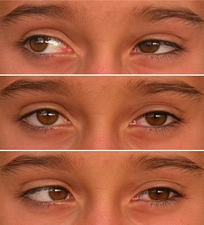
Esotropia is a form of strabismus in which one or both eyes turns inward. The condition can be constantly present, or occur intermittently, and can give the affected individual a "cross-eyed" appearance. It is the opposite of exotropia and usually involves more severe axis deviation than esophoria. Esotropia is sometimes erroneously called "lazy eye", which describes the condition of amblyopia; a reduction in vision of one or both eyes that is not the result of any pathology of the eye and cannot be resolved by the use of corrective lenses. Amblyopia can, however, arise as a result of esotropia occurring in childhood: In order to relieve symptoms of diplopia or double vision, the child's brain will ignore or "suppress" the image from the esotropic eye, which when allowed to continue untreated will lead to the development of amblyopia. Treatment options for esotropia include glasses to correct refractive errors, the use of prisms and/or orthoptic exercises and/or eye muscle surgery. The term is from Greek eso meaning "inward" and trope meaning "a turning".

Strabismus is a condition in which the eyes do not properly align with each other when looking at an object. The eye that is focused on an object can alternate. The condition may be present occasionally or constantly. If present during a large part of childhood, it may result in amblyopia or lazy eyes and loss of depth perception. If onset is during adulthood, it is more likely to result in double vision.

An eye examination is a series of tests performed to assess vision and ability to focus on and discern objects. It also includes other tests and examinations pertaining to the eyes. Eye examinations are primarily performed by an optometrist, ophthalmologist, orthoptist, or an optician. Health care professionals often recommend that all people should have periodic and thorough eye examinations as part of routine primary care, especially since many eye diseases are asymptomatic.

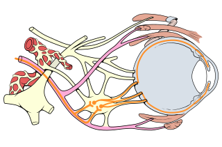
The accommodation reflex is a reflex action of the eye, in response to focusing on a near object, then looking at a distant object, comprising coordinated changes in vergence, lens shape (accommodation) and pupil size. It is dependent on cranial nerve II, superior centers (interneuron) and cranial nerve III. The change in the shape of the lens is controlled by ciliary muscles inside the eye. Changes in contraction of the ciliary muscles alters the focal distance of the eye, causing nearer or farther images to come into focus on the retina; this process is known as accommodation. The reflex, controlled by the parasympathetic nervous system, involves three responses: pupil constriction, lens accommodation, and convergence.
Duane syndrome is a congenital rare type of strabismus most commonly characterized by the inability of the eye to move outward. The syndrome was first described by ophthalmologists Jakob Stilling (1887) and Siegmund Türk (1896), and subsequently named after Alexander Duane, who discussed the disorder in more detail in 1905.

The medial rectus muscle is a muscle in the orbit near the eye. It is one of the extraocular muscles. It originates from the annulus of Zinn, and inserts into the anteromedial surface of the eye. It is supplied by the inferior division of the oculomotor nerve (III). It rotates the eye medially (adduction).
The inferior oblique muscle or obliquus oculi inferior is a thin, narrow muscle placed near the anterior margin of the floor of the orbit. The inferior oblique is an extraocular muscle, and is attached to the maxillary bone (origin) and the posterior, inferior, lateral surface of the eye (insertion). The inferior oblique is innervated by the inferior branch of the oculomotor nerve.

The extraocular muscles, are the seven extrinsic muscles of the human eye. Six of the extraocular muscles control movement of the eye and the other muscle, the levator palpebrae, controls eyelid elevation. The actions of the six muscles responsible for eye movement depend on the position of the eye at the time of muscle contraction.

Sixth nerve palsy, or abducens nerve palsy, is a disorder associated with dysfunction of cranial nerve VI, which is responsible for causing contraction of the lateral rectus muscle to abduct the eye. The inability of an eye to turn outward, results in a convergent strabismus or esotropia of which the primary symptom is diplopia in which the two images appear side-by-side. Thus, the diplopia is horizontal and worse in the distance. Diplopia is also increased on looking to the affected side and is partly caused by overaction of the medial rectus on the unaffected side as it tries to provide the extra innervation to the affected lateral rectus. These two muscles are synergists or "yoke muscles" as both attempt to move the eye over to the left or right. The condition is commonly unilateral but can also occur bilaterally.

Congenital fourth nerve palsy is a condition present at birth characterized by a vertical misalignment of the eyes due to a weakness or paralysis of the superior oblique muscle.

Hypertropia is a condition of misalignment of the eyes (strabismus), whereby the visual axis of one eye is higher than the fellow fixating eye. Hypotropia is the similar condition, focus being on the eye with the visual axis lower than the fellow fixating eye. Dissociated vertical deviation is a special type of hypertropia leading to slow upward drift of one or rarely both eyes, usually when the patient is inattentive.

Strabismus surgery is surgery on the extraocular muscles to correct strabismus, the misalignment of the eyes. Strabismus surgery is a one-day procedure that is usually performed under general anesthesia most commonly by either a neuro- or pediatric ophthalmologist. The patient spends only a few hours in the hospital with minimal preoperative preparation. After surgery, the patient should expect soreness and redness but is generally free to return home.
Oculomotor nerve palsy or oculomotor neuropathy is an eye condition resulting from damage to the third cranial nerve or a branch thereof. As the name suggests, the oculomotor nerve supplies the majority of the muscles controlling eye movements. Damage to this nerve will result in an inability to move the eye normally. The nerve also supplies the upper eyelid muscle and is accompanied by parasympathetic fibers innervating the muscles responsible for pupil constriction. The limitations of eye movement resulting from the condition are generally so severe that patients are often unable to maintain normal eye alignment when gazing straight ahead, leading to strabismus and, as a consequence, double vision (diplopia).
Chronic progressive external ophthalmoplegia (CPEO), is a type of eye disorder characterized by slowly progressive inability to move the eyes and eyebrows. It is often the only feature of mitochondrial disease, in which case the term CPEO may be given as the diagnosis. In other people suffering from mitochondrial disease, CPEO occurs as part of a syndrome involving more than one part of the body, such as Kearns–Sayre syndrome. Occasionally CPEO may be caused by conditions other than mitochondrial diseases.
Dissociated vertical deviation (DVD) is an eye condition which occurs in association with a squint, typically infantile esotropia. The exact cause is unknown, although it is logical to assume it is from faulty innervation of eye muscles.
The Parks–Bielschowsky three-step test, also known as Park's three-step test or Bielschowsky head tilt test, is a method used to isolate the paretic extraocular muscle, particularly superior oblique muscle and trochlear nerve, in acquired vertical double vision. It was originally described by Marshall M. Parks.
Botulinum toxin therapy of strabismus is a medical technique used sometimes in the management of strabismus, in which botulinum toxin is injected into selected extraocular muscles in order to reduce the misalignment of the eyes. The injection of the toxin to treat strabismus, reported upon in 1981, is considered to be the first ever use of botulinum toxin for therapeutic purposes. Today, the injection of botulinum toxin into the muscles that surround the eyes is one of the available options in the management of strabismus. Other options for strabismus management are vision therapy and occlusion therapy, corrective glasses and prism glasses, and strabismus surgery.

The Maddox rod test can be used to subjectively detect and measure a latent, manifest, horizontal or vertical strabismus for near and distance. The test is based on the principle of diplopic projection. Dissociation of the deviation is brought about by presenting a red line image to one eye and a white light to the other, while prisms are used to superimpose these and effectively measure the angle of deviation. The strength of the prism is increased until the streak of the light passes through the centre of the prism, as the strength of the prism indicates the amount of deviation present. The Maddox rod is a handheld instrument composed of red parallel plano convex cylinder lens, which refracts light rays so that a point source of light is seen as a line or streak of light. Due to the optical properties, the streak of light is seen perpendicular to the axis of the cylinder.
Bagolini striated glasses test, or BSGT, is a subjective clinical test to detect the presence or extent of binocular functions and is generally performed by an Optometrist(O.D) or orthoptist or ophthalmologist. It is mainly used in strabismus clinics. Through this test, suppression, microtropia, diplopia and manifest deviations can be noted. However this test should always be used in conjunction with other clinical tests, such as Worth 4 dot test, Cover test, Prism cover test and Maddox rod to come to a diagnosis.
The management of strabismus may include the use of drugs or surgery to correct the strabismus. Agents used include paralytic agents such as botox used on extraocular muscles, topical autonomic nervous system agents to alter the refractive index in the eyes, and agents that act in the central nervous system to correct amblyopia.

